Revision 8

#69994

Store at -20C

CST Logo
Orders:

877-616-CELL (2355)

[email protected]

Support:

877-678-TECH (8324)

3 Trask Lane | Danvers | Massachusetts | 01923 | USA

For Research Use Only. Not for Use in Diagnostic Procedures.

Applications:
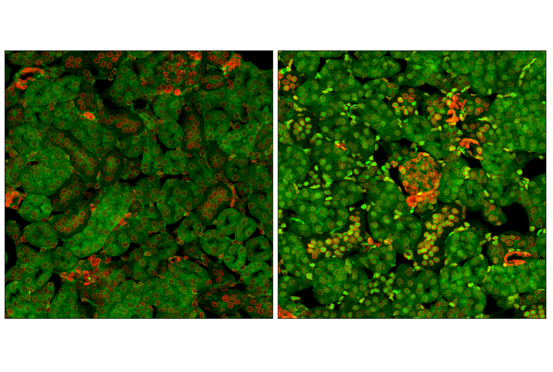
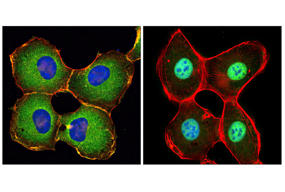
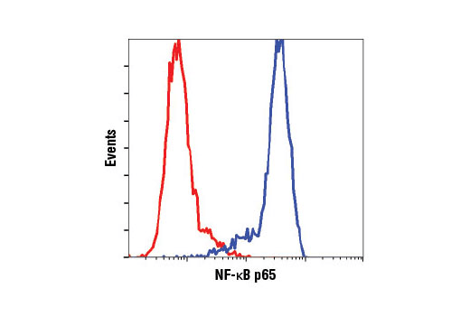
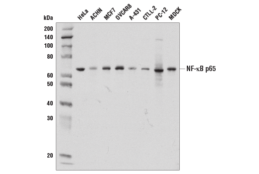
W, IHC-P, IF-F, IF-IC, FC-FP

Reactivity:
H M R Hm Mk Dg

Sensitivity:
Endogenous

MW (kDa):
65

Source/Isotype:
Rabbit IgG

UniProt ID:
#Q04206

Entrez-Gene Id:
5970

Product Usage Information

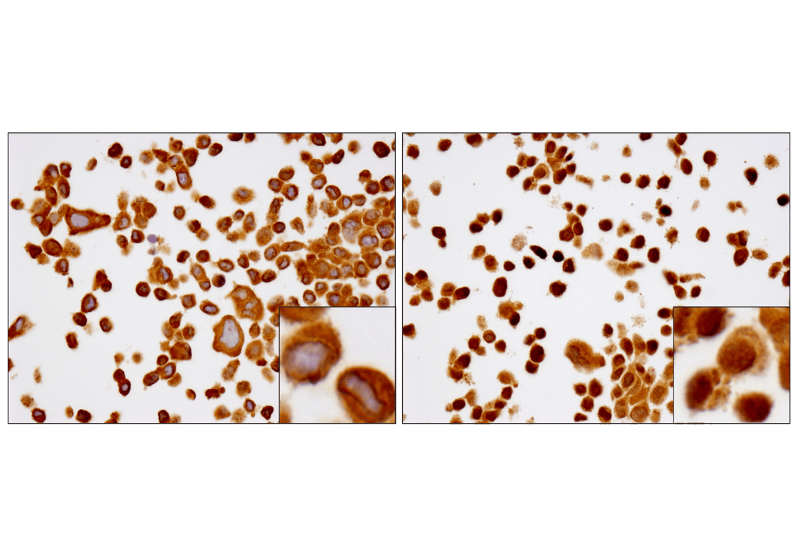
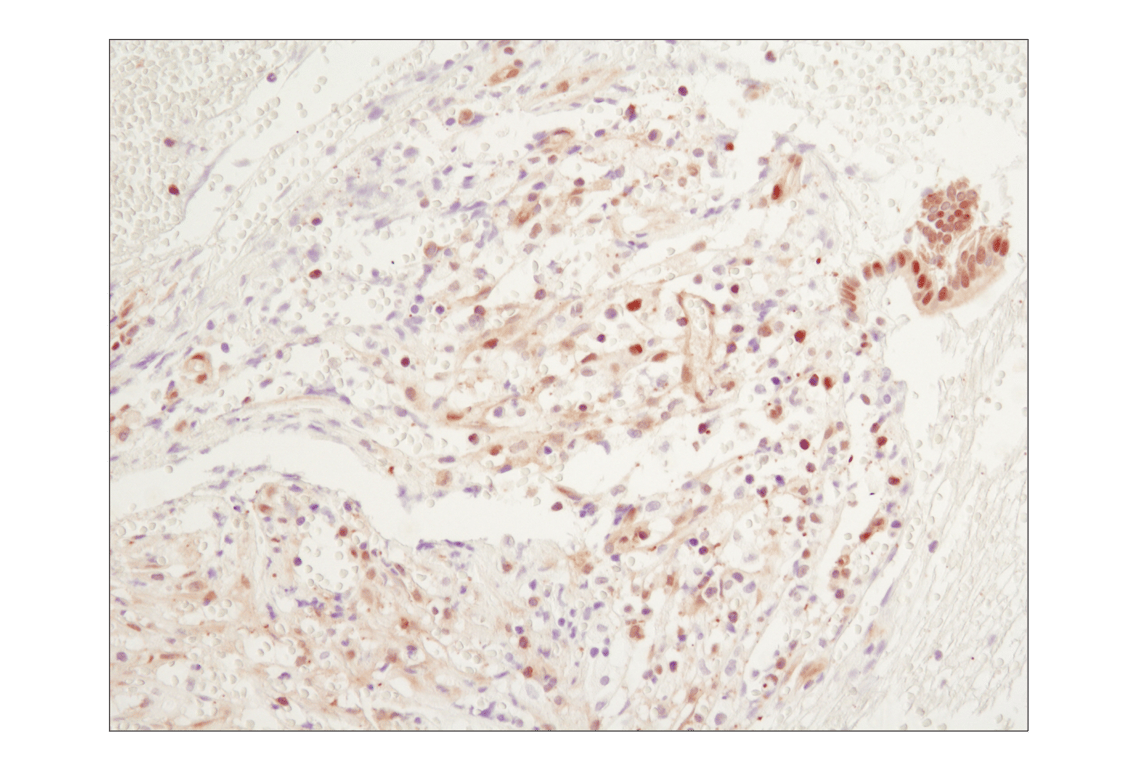
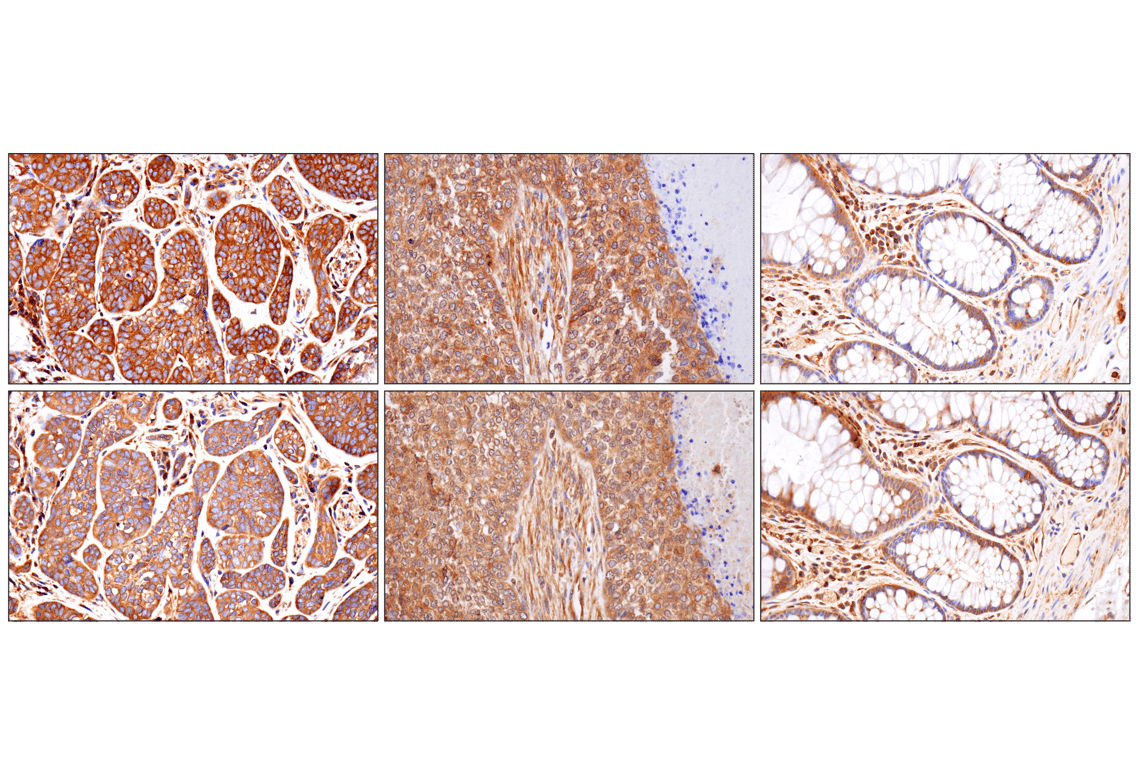
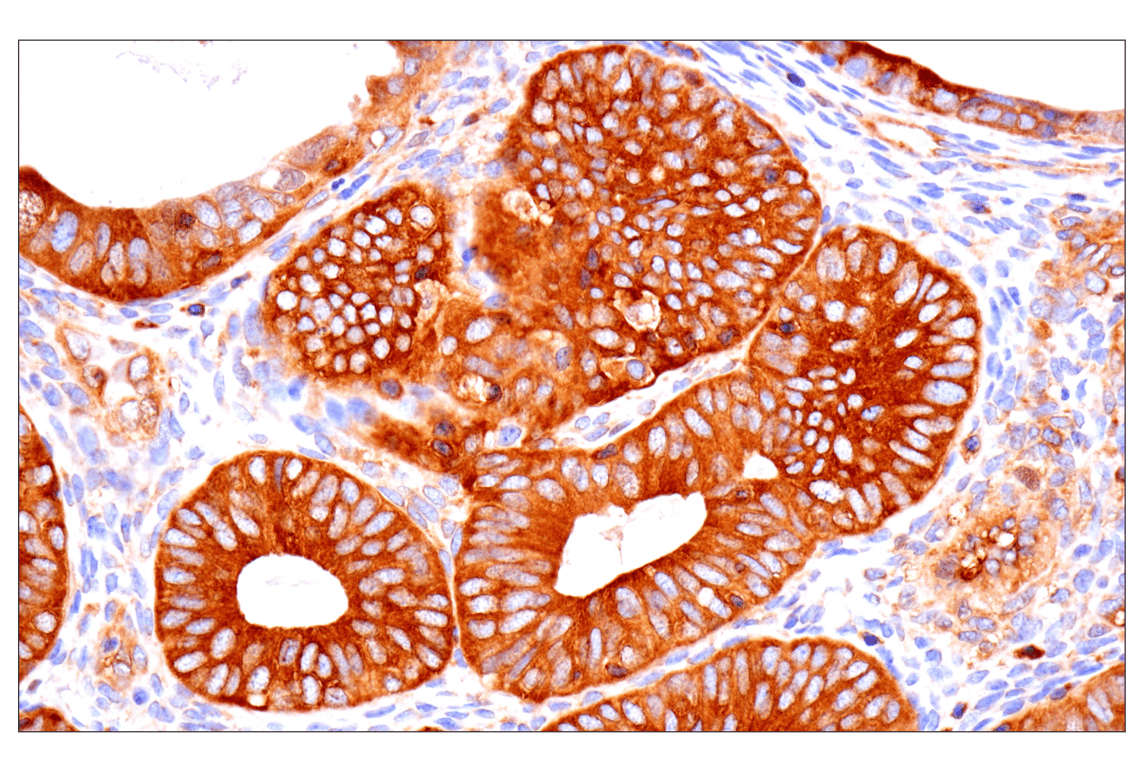
This product is the carrier free version of product #8242. All data were generated using the same antibody clone in the standard formulation which contains BSA and glycerol.

This formulation is ideal for use with technologies requiring specialized or custom antibody labeling, including fluorophores, metals, lanthanides, and oligonucleotides. It is not recommended for ChIP, ChIP-seq, CUT&RUN or CUT&Tag assays. If you require a carrier free formulation for chromatin profiling, please contact us. Optimal dilutions/concentrations should be determined by the end user.

BSA and Azide Free antibodies are quality control tested by size exclusion chromatography (SEC) to determine antibody integrity.

Formulation

Supplied in 1X Borate (20 mM H3BO3, 150 mM NaCl (pH 9.0)). BSA and Azide Free.

For standard formulation of this product see product #8242.

Storage

Store at -20°C. This product will freeze at -20°C so it is recommended to aliquot into single-use vials to avoid multiple freeze/thaw cycles. A slight precipitate may be present and can be dissolved by gently vortexing. This will not interfere with antibody performance.

Specificity/Sensitivity

NF-kappaB p65 (D14E12) Rabbit Monoclonal Antibody (BSA and Azide Free) recognizes endogenous levels of total NF-κB p65/RelA protein. This antibody does not cross react with other NF-κB/Rel family members. Non-specific staining was observed in pancreatic islets by immunohistochemistry.

Source / Purification

Monoclonal antibody is produced by immunizing animals with a synthetic peptide corresponding to residues surrounding Glu498 of human NF-κB p65/RelA protein.

Background

Transcription factors of the nuclear factor κB (NF-κB)/Rel family play a pivotal role in inflammatory and immune responses (1,2). There are five family members in mammals: RelA, c-Rel, RelB, NF-κB1 (p105/p50), and NF-κB2 (p100/p52). Both p105 and p100 are proteolytically processed by the proteasome to produce p50 and p52, respectively. Rel proteins bind p50 and p52 to form dimeric complexes that bind DNA and regulate transcription. In unstimulated cells, NF-κB is sequestered in the cytoplasm by IκB inhibitory proteins (3-5). NF-κB-activating agents can induce the phosphorylation of IκB proteins, targeting them for rapid degradation through the ubiquitin-proteasome pathway and releasing NF-κB to enter the nucleus where it regulates gene expression (6-8). NIK and IKKα (IKK1) regulate the phosphorylation and processing of NF-κB2 (p100) to produce p52, which translocates to the nucleus (9-11).

Species Reactivity

Species reactivity is determined by testing in at least one approved application (e.g., western blot).

Applications Key

W: Western Blotting IHC-P: Immunohistochemistry (Paraffin) IF-F: Immunofluorescence (Frozen) IF-IC: Immunofluorescence (Immunocytochemistry) FC-FP: Flow Cytometry (Fixed/Permeabilized)

Cross-Reactivity Key

H: Human M: Mouse R: Rat Hm: Hamster Mk: Monkey Dg: Dog

Trademarks and Patents

Cell Signaling Technology is a trademark of Cell Signaling Technology, Inc.

All other trademarks are the property of their respective owners. Visit cellsignal.com/trademarks for more information.

使用に関する制限

使用に関する制限

法的な権限を与えられたCSTの担当者が署名した書面によって別途明示的に合意された場合を除き、CST、その関連会社または代理店が提供する製品には以下の条件が適用されます。お客様が定める条件でここに定められた条件に含まれるものを超えるもの、または、ここに定められた条件と異なるものは、法的な権限を与えられたCSTの担当者が別途書面にて受諾した場合を除き、拒絶され、いかなる効力も効果も有しません。

研究専用 (For Research Use Only) またはこれに類似する表示がされた製品は、 いかなる目的についても FDA または外国もしくは国内のその他の規制機関により承認、認可または許可を受けていません。 お客様は製品を診断もしくは治療目的で使用してはならず、また、製品に表示された内容に違反する方法で使用してはなりません。 CST が販売または使用許諾する製品は、エンドユーザーであるお客様に対し、使途を研究および開発のみに限定して提供されるものです。 診断、予防もしくは治療目的で製品を使用することまたは製品を再販売 (単独であるか他の製品等の一部であるかを問いません) もしくはその他の商業的利用の目的で購入することについては、CST から別途許諾を得る必要があります。 お客様は以下の事項を遵守しなければなりません。(a) CST の製品 (単独であるか他の資材と一緒であるかを問いません) を販売、使用許諾、貸与、寄付もしくはその他の態様で第三者に譲渡したり使用させたりしてはなりません。また、商用の製品を製造するために CST の製品を使用してはなりません。(b) 複製、改変、リバースエンジニアリング、逆コンパイル、 分解または他の方法により製品の構造または技術を解明しようとしてはなりません。また、 CST の製品またはサービスと競合する製品またはサービスを開発する目的で CST の製品を使用してはなりません。(c) CST の製品の商標、商号、ロゴ、特許または著作権に関する通知または表示を除去したり改変したりしてはなりません。(d) CST の製品をCST 製品販売条件(CST’s Product Terms of Sale) および該当する書面のみに従って使用しなければなりません。(e) CST の製品に関連してお客様が使用する第三者の製品またはサービスに関する使用許諾条件、 サービス提供条件またはこれに類する合意事項を遵守しなければなりません。

Orders: 877-616-CELL (2355) [email protected] Support: 877-678-TECH (8324) [email protected] Web: cellsignal.com
For Research Use Only. Not for Use in Diagnostic Procedures.

Revision 8

Cell Signaling Technologyロゴ
Western blot analysis of extracts from various cell lines using NF-κB p65 (D14E12) XP® Rabbit mAb. Data were generated using the standard formulation of this product.
Western Blotting Image 1: NF-kappaB p65 (D14E12) Rabbit Monoclonal Antibody (BSA and Azide Free)
Immunohistochemical analysis using NF-κB p65 (D14E12) XP® Rabbit mAb on SignalSlide® NF-κB p65 IHC Controls #12873 (paraffin-embedded HCT116 cells, untreated (left) or treated with hTNF-α (right). Data were generated using the standard formulation of this product.
Immunohistochemistry Image 1: NF-kappaB p65 (D14E12) Rabbit Monoclonal Antibody (BSA and Azide Free)
Immunohistochemical analysis of paraffin-embedded human chronic cholecystitis using NF-κB p65 (D14E12) XP® Rabbit mAb. Data were generated using the standard formulation of this product.
Immunohistochemistry Image 2: NF-kappaB p65 (D14E12) Rabbit Monoclonal Antibody (BSA and Azide Free)
Orders: 877-616-CELL (2355) [email protected] Support: 877-678-TECH (8324) [email protected] Web: cellsignal.com
For Research Use Only. Not for Use in Diagnostic Procedures.

Revision 8

Cell Signaling Technologyロゴ
Immunohistochemical analysis of paraffin-embedded human adenoid cystic carcinoma of the salivary gland (left), squamous cell lung carcinoma (middle) or normal colon (right) using NF-κB p65 (D14E12) XP® Rabbit mAb (top) or an NF-κB p65 Mouse mAb (bottom). These two antibodies detect unique, non-overlapping epitopes on human NF-κB p65. The similar staining patterns obtained with both antibodies help to confirm the specificity of the staining. Data were generated using the standard formulation of this product.
Immunohistochemistry Image 3: NF-kappaB p65 (D14E12) Rabbit Monoclonal Antibody (BSA and Azide Free)
Immunohistochemical analysis of paraffin-embedded human endometrioid carcinoma using NF-κB p65 (D14E12) XP® Rabbit mAb. Data were generated using the standard formulation of this product.
Immunohistochemistry Image 4: NF-kappaB p65 (D14E12) Rabbit Monoclonal Antibody (BSA and Azide Free)
Immunohistochemical analysis of paraffin-embedded human thymoma using NF-κB p65 (D14E12) XP® Rabbit mAb. Data were generated using the standard formulation of this product.
Immunohistochemistry Image 5: NF-kappaB p65 (D14E12) Rabbit Monoclonal Antibody (BSA and Azide Free)
Orders: 877-616-CELL (2355) [email protected] Support: 877-678-TECH (8324) [email protected] Web: cellsignal.com
For Research Use Only. Not for Use in Diagnostic Procedures.

Revision 8

Cell Signaling Technologyロゴ
Immunohistochemical analysis of paraffin-embedded human gastric carcinoma using NF-κB p65 (D14E12) XP® Rabbit mAb. Data were generated using the standard formulation of this product.
Immunohistochemistry Image 6: NF-kappaB p65 (D14E12) Rabbit Monoclonal Antibody (BSA and Azide Free)
Immunohistochemical analysis of paraffin-embedded normal human small intestine using NF-κB p65 (D14E12) XP® Rabbit mAb. Data were generated using the standard formulation of this product.
Immunohistochemistry Image 7: NF-kappaB p65 (D14E12) Rabbit Monoclonal Antibody (BSA and Azide Free)
Immunohistochemical analysis of paraffin-embedded human lung adenocarcinoma using NF-κB p65 (D14E12) XP® Rabbit mAb. Data were generated using the standard formulation of this product.
Immunohistochemistry Image 8: NF-kappaB p65 (D14E12) Rabbit Monoclonal Antibody (BSA and Azide Free)
Orders: 877-616-CELL (2355) [email protected] Support: 877-678-TECH (8324) [email protected] Web: cellsignal.com
For Research Use Only. Not for Use in Diagnostic Procedures.

Revision 8

Cell Signaling Technologyロゴ
Confocal immunofluorescent analysis of fixed frozen kidney from untreated (left) or LPS-treated mice (2 hrs, 5 mg/kg) (right) using NF-kappaB p65 (D14E12) Rabbit Monoclonal Antibody (green) and Lamin A/C (4C11) Mouse Monoclonal Antibody (Alexa Fluor® 647 Conjugate) #41357 (red). Data were generated using the standard formulation of this product.
Immunofluorescence Image 1: NF-kappaB p65 (D14E12) Rabbit Monoclonal Antibody (BSA and Azide Free)
Confocal immunofluorescent analysis of HT-1080 cells, untreated (left) or treated with hTNF-alpha (20 ng/ml, 20 min; right), using NF-kappaB p65 (D14E12) Rabbit Monoclonal Antibody (green), DyLight® 554 Phalloidin #13054 (red), and DRAQ5® #4084 (blue). Data were generated using the standard formulation of this product.
Immunofluorescence Image 1: NF-kappaB p65 (D14E12) Rabbit Monoclonal Antibody (BSA and Azide Free)
Flow cytometric analysis of fixed and permeabilized Jurkat cells using NF-κB p65 (D14E12) Rabbit Monoclonal Antibody (solid line) or concentration-matched Rabbit (DA1E) Monoclonal Antibody IgG Isotype Control #3900 (dashed line). Anti-rabbit IgG (H+L), F(ab')2 Fragment (Alexa Fluor® 488 Conjugate) #4412 was used as a secondary antibody.
Flow Cytometry Image 1: NF-kappaB p65 (D14E12) Rabbit Monoclonal Antibody (BSA and Azide Free)
Orders: 877-616-CELL (2355) [email protected] Support: 877-678-TECH (8324) [email protected] Web: cellsignal.com
For Research Use Only. Not for Use in Diagnostic Procedures.