Revision 17
#8480
Store at -20C
877-616-CELL (2355)
877-678-TECH (8324)
3 Trask Lane | Danvers | Massachusetts | 01923 | USA
For Research Use Only. Not for Use in Diagnostic Procedures.
Applications:
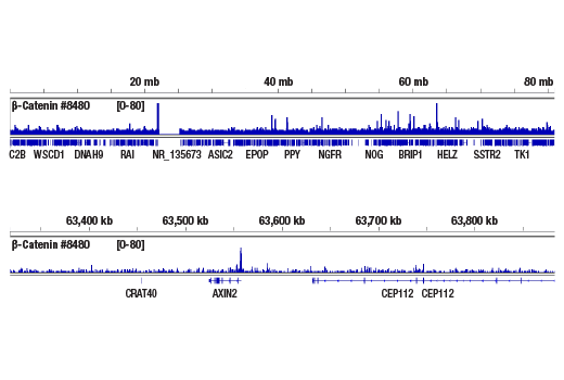
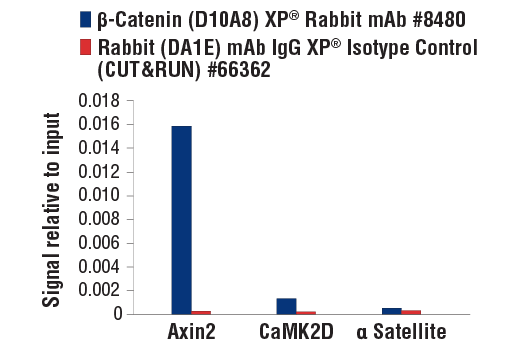
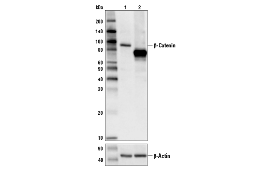
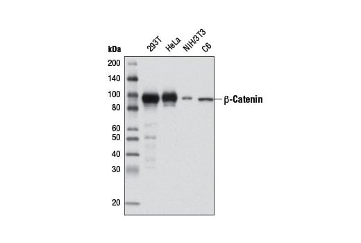
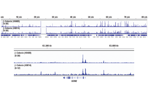
W, W-S, IP, IHC-Bond, IHC-P, IF-F, IF-IC, FC-FP, ChIP, ChIP-seq, C&R
Reactivity:
H M R Mk
Sensitivity:
Endogenous
MW (kDa):
92
Source/Isotype:
Rabbit IgG
UniProt ID:
#P35222
Entrez-Gene Id:
1499
Product Usage Information
The CUT&RUN dilution was determined using CUT&RUN Assay Kit #86652.
| Application | Dilution |
|---|---|
| Western Blotting | 1:1000 |
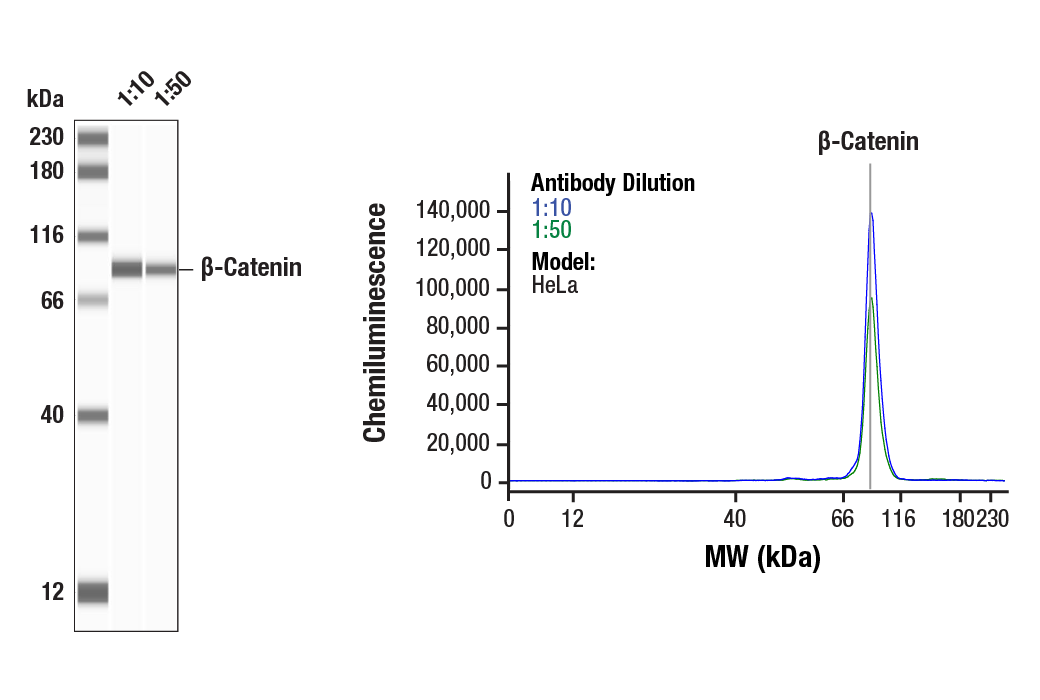
| Simple Western™ | 1:10 - 1:50 |
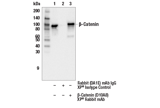
| Immunoprecipitation | 1:50 |
| IHC Leica Bond | 1:50 - 1:200 |
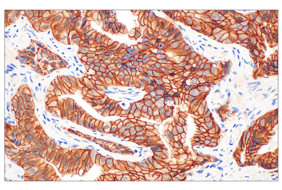
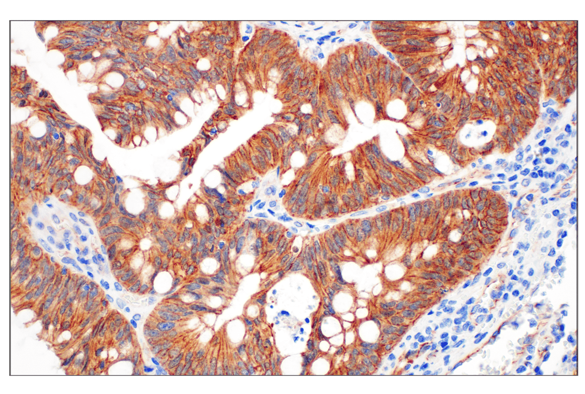
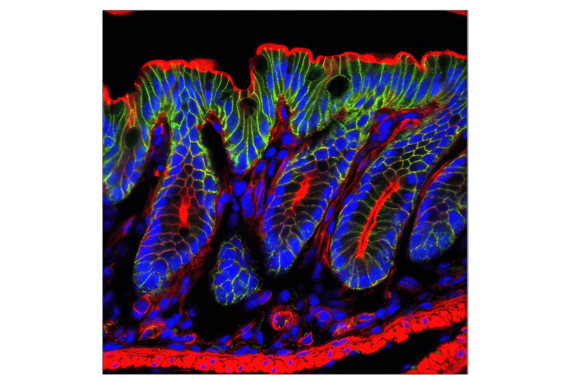
| Immunohistochemistry (Paraffin) | 1:50 - 1:200 |
| Immunofluorescence (Frozen) | 1:50 - 1:100 |
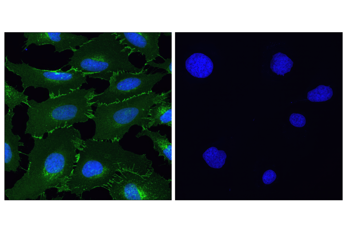
| Immunofluorescence (Immunocytochemistry) | 1:100 - 1:400 |
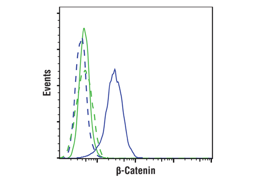
| Flow Cytometry (Fixed/Permeabilized) | 1:50 - 1:200 |
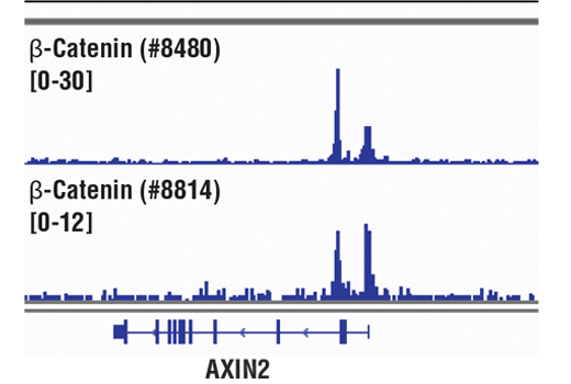
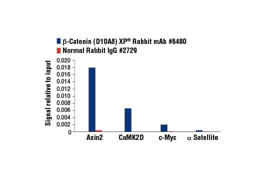
| Chromatin IP | 1:25 |
| Chromatin IP-seq | 1:25 |
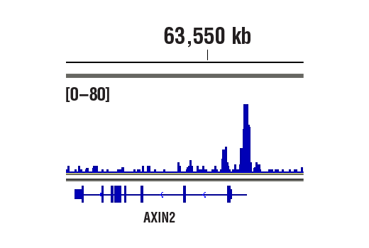
| CUT&RUN | 1:25 |
Storage
For a carrier free (BSA and azide free) version of this product see product #84441.
Specificity/Sensitivity
Species predicted to react based on 100% sequence homology
Source / Purification
Background
Background References
- Cadigan, K.M. and Nusse, R. (1997) Genes Dev 11, 3286-3305.
- Wodarz, A. and Nusse, R. (1998) Annu Rev Cell Dev Biol 14, 59-88.
- Polakis, P. (1999) Curr Opin Genet Dev 9, 15-21.
- Amit, S. et al. (2002) Genes Dev 16, 1066-76.
- Liu, C. et al. (2002) Cell 108, 837-47.
- Yanagawa, S. et al. (2002) EMBO J 21, 1733-42.
- Yost, C. et al. (1996) Genes Dev 10, 1443-54.
- Morin, P.J. et al. (1997) Science 275, 1787-90.
Species Reactivity
Species reactivity is determined by testing in at least one approved application (e.g., western blot).
Western Blot Buffer
IMPORTANT: For western blots, incubate membrane with diluted primary antibody in 5% w/v nonfat dry milk, 1X TBS, 0.1% Tween® 20 at 4°C with gentle shaking, overnight.
Applications Key
W: Western Blotting W-S: Simple Western™ IP: Immunoprecipitation IHC-Bond: IHC Leica Bond IHC-P: Immunohistochemistry (Paraffin) IF-F: Immunofluorescence (Frozen) IF-IC: Immunofluorescence (Immunocytochemistry) FC-FP: Flow Cytometry (Fixed/Permeabilized) ChIP: Chromatin IP ChIP-seq: Chromatin IP-seq C&R: CUT&RUN
Cross-Reactivity Key
H: Human M: Mouse R: Rat Mk: Monkey
Trademarks and Patents
Cell Signaling Technology is a trademark of Cell Signaling Technology, Inc.
SignalStain is a registered trademark of Cell Signaling Technology, Inc.
SimpleChIP is a registered trademark of Cell Signaling Technology, Inc.
All other trademarks are the property of their respective owners. Visit cellsignal.com/trademarks for more information.
使用に関する制限
使用に関する制限
法的な権限を与えられたCSTの担当者が署名した書面によって別途明示的に合意された場合を除き、CST、その関連会社または代理店が提供する製品には以下の条件が適用されます。お客様が定める条件でここに定められた条件に含まれるものを超えるもの、または、ここに定められた条件と異なるものは、法的な権限を与えられたCSTの担当者が別途書面にて受諾した場合を除き、拒絶され、いかなる効力も効果も有しません。
研究専用 (For Research Use Only) またはこれに類似する表示がされた製品は、 いかなる目的についても FDA または外国もしくは国内のその他の規制機関により承認、認可または許可を受けていません。 お客様は製品を診断もしくは治療目的で使用してはならず、また、製品に表示された内容に違反する方法で使用してはなりません。 CST が販売または使用許諾する製品は、エンドユーザーであるお客様に対し、使途を研究および開発のみに限定して提供されるものです。 診断、予防もしくは治療目的で製品を使用することまたは製品を再販売 (単独であるか他の製品等の一部であるかを問いません) もしくはその他の商業的利用の目的で購入することについては、CST から別途許諾を得る必要があります。 お客様は以下の事項を遵守しなければなりません。(a) CST の製品 (単独であるか他の資材と一緒であるかを問いません) を販売、使用許諾、貸与、寄付もしくはその他の態様で第三者に譲渡したり使用させたりしてはなりません。また、商用の製品を製造するために CST の製品を使用してはなりません。(b) 複製、改変、リバースエンジニアリング、逆コンパイル、 分解または他の方法により製品の構造または技術を解明しようとしてはなりません。また、 CST の製品またはサービスと競合する製品またはサービスを開発する目的で CST の製品を使用してはなりません。(c) CST の製品の商標、商号、ロゴ、特許または著作権に関する通知または表示を除去したり改変したりしてはなりません。(d) CST の製品をCST 製品販売条件(CST’s Product Terms of Sale) および該当する書面のみに従って使用しなければなりません。(e) CST の製品に関連してお客様が使用する第三者の製品またはサービスに関する使用許諾条件、 サービス提供条件またはこれに類する合意事項を遵守しなければなりません。
Revision 17
Revision 17
Revision 17
Revision 17
Revision 17
Revision 17
Revision 17
Revision 17