Revision 3
#89348
Store at -20C
877-616-CELL (2355)
877-678-TECH (8324)
3 Trask Lane | Danvers | Massachusetts | 01923 | USA
For Research Use Only. Not for Use in Diagnostic Procedures.
Applications:
W, W-S, IHC-P
Reactivity:
H
Sensitivity:
Endogenous
MW (kDa):
51-60
Source/Isotype:
Rabbit IgG
UniProt ID:
#P19544
Entrez-Gene Id:
7490
Product Usage Information
| Application | Dilution |
|---|---|
| Western Blotting | 1:1000 |
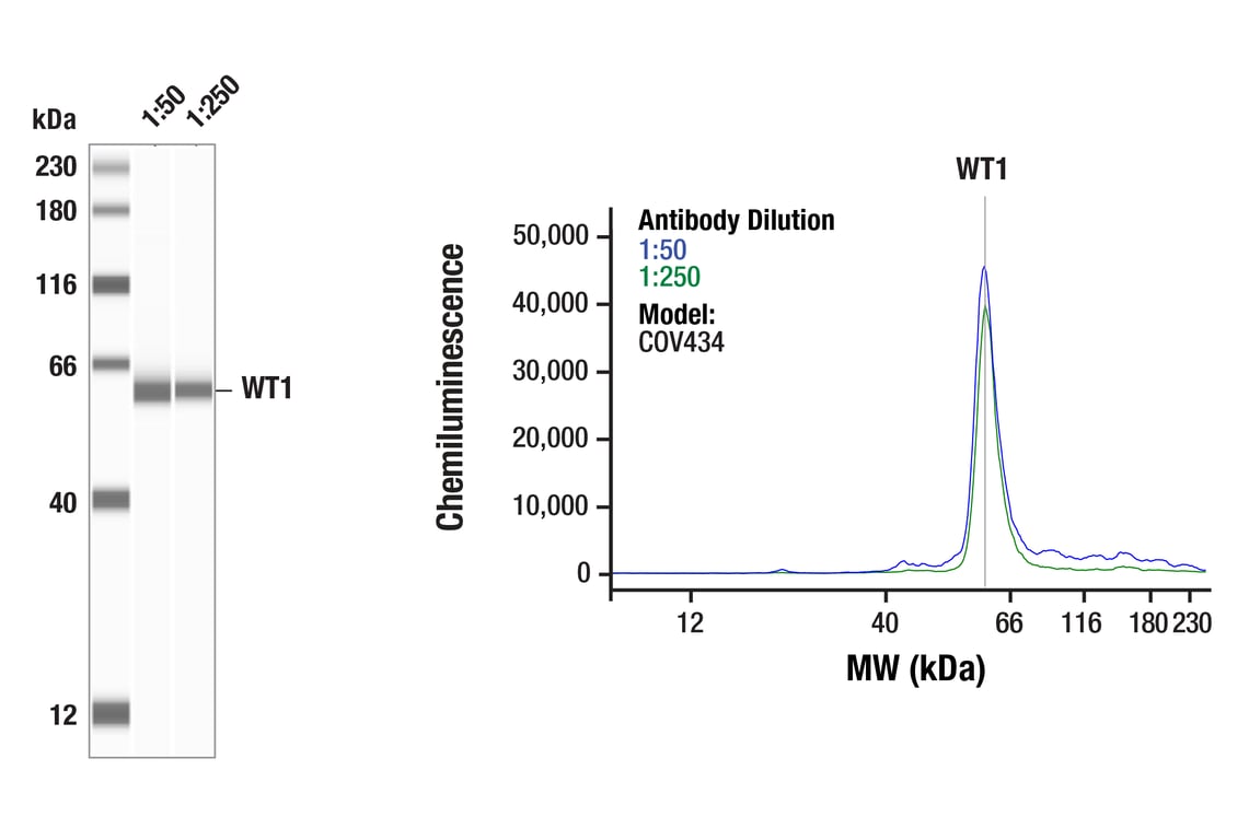
| Simple Western™ | 1:50 - 1:200 |
| Immunohistochemistry (Paraffin) | 1:400 - 1:1600 |
Storage
Specificity/Sensitivity
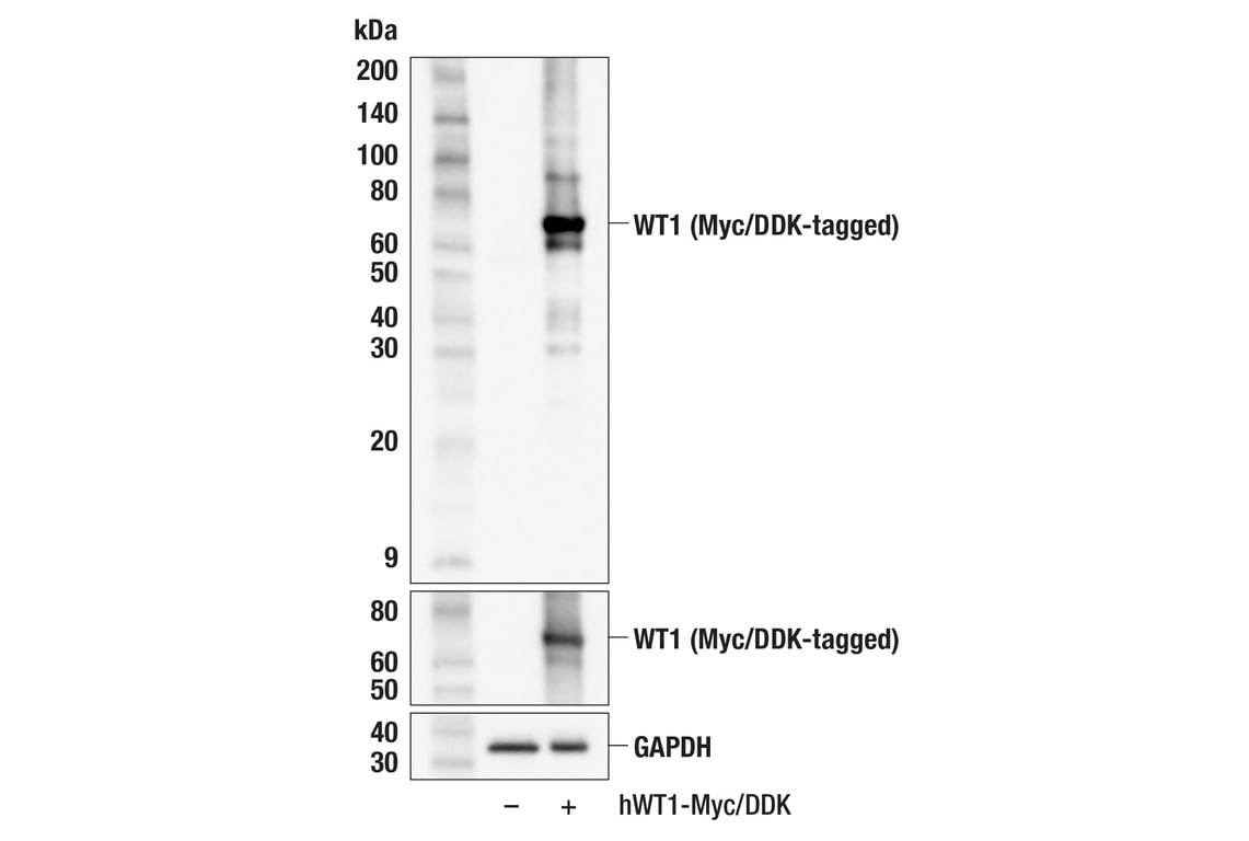
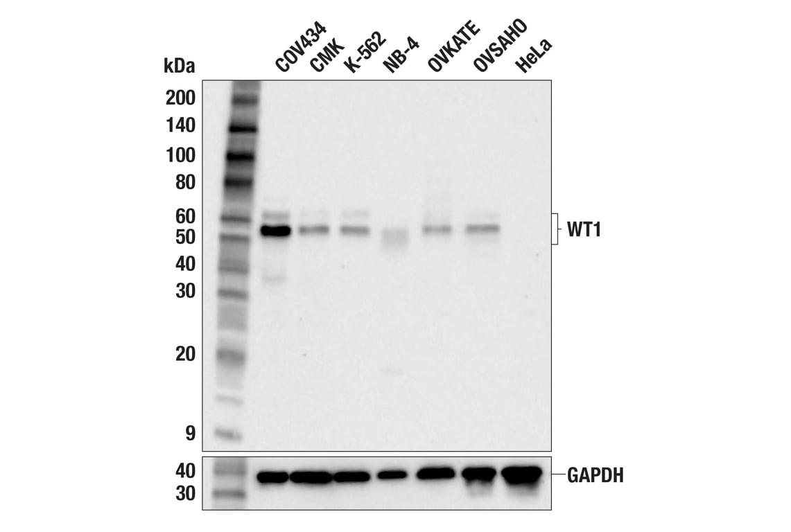
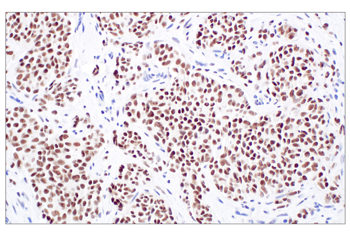
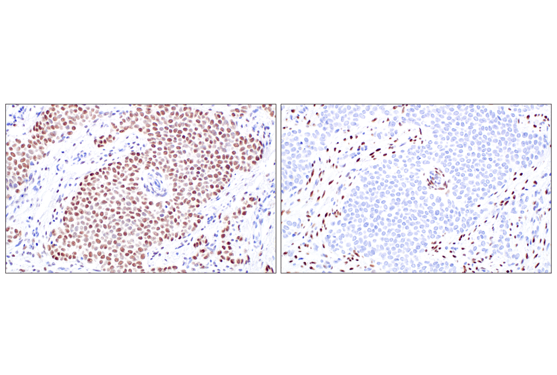
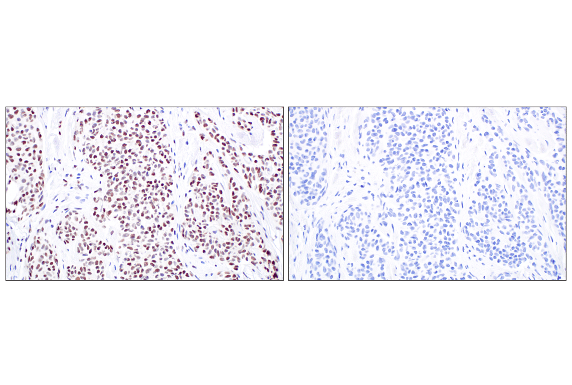
WT1 (F3T7B) Rabbit mAb (Carboxy-terminal Antigen) recognizes endogenous levels of total WT1 protein.
This antibody is recommended for the detection of fusion proteins containing the C-terminus region of WT1 found in desmoplastic small round cell tumors by immunohistochemistry. For the detection of wild-type WT1, it is recommended to use WT1 (D8I7F) XP® Rabbit mAb #83535.
Non-specific staining was observed in kidney, liver, and small intestine by immunohistochemistry.
Species predicted to react based on 100% sequence homology
Source / Purification
Background
WT1 has a myriad of biological functions and a host of interacting partners and target genes (6). It can behave as a transcriptional activator, or a repressor, and can act as an oncogene or a tumor suppressor (7). It exerts influence over the epigenetic landscape, and also has post-translational influence of gene expression through RNA interactions (8). The diverse biological roles of WT1 have been attributed to the existence of multiple isoforms and post-translational modifications of the protein (9).
Background References
- Royer-Pokora, B. et al. (2004) Am J Med Genet A 127A, 249-57.
- Kohsaka, T. et al. (1999) Hum Mutat 14, 466-70.
- Little, M. et al. (2000) Hum Mutat 15, 389.
- Takata, A. et al. (2000) J Med Genet 37, 698-701.
- Suri, M. et al. (2007) Am J Med Genet A 143A, 2312-20.
- Toska, E. and Roberts, S.G. (2014) Biochem J 461, 15-32.
- Yang, L. et al. (2007) Leukemia 21, 868-76.
- Weiss, T.C. and Romaniuk, P.J. (2009) Biochemistry 48, 148-55.
- Haber, D.A. et al. (1991) Proc Natl Acad Sci USA 88, 9618-22.
Species Reactivity
Species reactivity is determined by testing in at least one approved application (e.g., western blot).
Western Blot Buffer
IMPORTANT: For western blots, incubate membrane with diluted primary antibody in 5% w/v BSA, 1X TBS, 0.1% Tween® 20 at 4°C with gentle shaking, overnight.
Applications Key
W: Western Blotting W-S: Simple Western™ IHC-P: Immunohistochemistry (Paraffin)
Cross-Reactivity Key
H: Human
Trademarks and Patents
Cell Signaling Technology is a trademark of Cell Signaling Technology, Inc.
All other trademarks are the property of their respective owners. Visit cellsignal.com/trademarks for more information.
使用に関する制限
使用に関する制限
法的な権限を与えられたCSTの担当者が署名した書面によって別途明示的に合意された場合を除き、CST、その関連会社または代理店が提供する製品には以下の条件が適用されます。お客様が定める条件でここに定められた条件に含まれるものを超えるもの、または、ここに定められた条件と異なるものは、法的な権限を与えられたCSTの担当者が別途書面にて受諾した場合を除き、拒絶され、いかなる効力も効果も有しません。
研究専用 (For Research Use Only) またはこれに類似する表示がされた製品は、 いかなる目的についても FDA または外国もしくは国内のその他の規制機関により承認、認可または許可を受けていません。 お客様は製品を診断もしくは治療目的で使用してはならず、また、製品に表示された内容に違反する方法で使用してはなりません。 CST が販売または使用許諾する製品は、エンドユーザーであるお客様に対し、使途を研究および開発のみに限定して提供されるものです。 診断、予防もしくは治療目的で製品を使用することまたは製品を再販売 (単独であるか他の製品等の一部であるかを問いません) もしくはその他の商業的利用の目的で購入することについては、CST から別途許諾を得る必要があります。 お客様は以下の事項を遵守しなければなりません。(a) CST の製品 (単独であるか他の資材と一緒であるかを問いません) を販売、使用許諾、貸与、寄付もしくはその他の態様で第三者に譲渡したり使用させたりしてはなりません。また、商用の製品を製造するために CST の製品を使用してはなりません。(b) 複製、改変、リバースエンジニアリング、逆コンパイル、 分解または他の方法により製品の構造または技術を解明しようとしてはなりません。また、 CST の製品またはサービスと競合する製品またはサービスを開発する目的で CST の製品を使用してはなりません。(c) CST の製品の商標、商号、ロゴ、特許または著作権に関する通知または表示を除去したり改変したりしてはなりません。(d) CST の製品をCST 製品販売条件(CST’s Product Terms of Sale) および該当する書面のみに従って使用しなければなりません。(e) CST の製品に関連してお客様が使用する第三者の製品またはサービスに関する使用許諾条件、 サービス提供条件またはこれに類する合意事項を遵守しなければなりません。
Revision 3
Revision 3
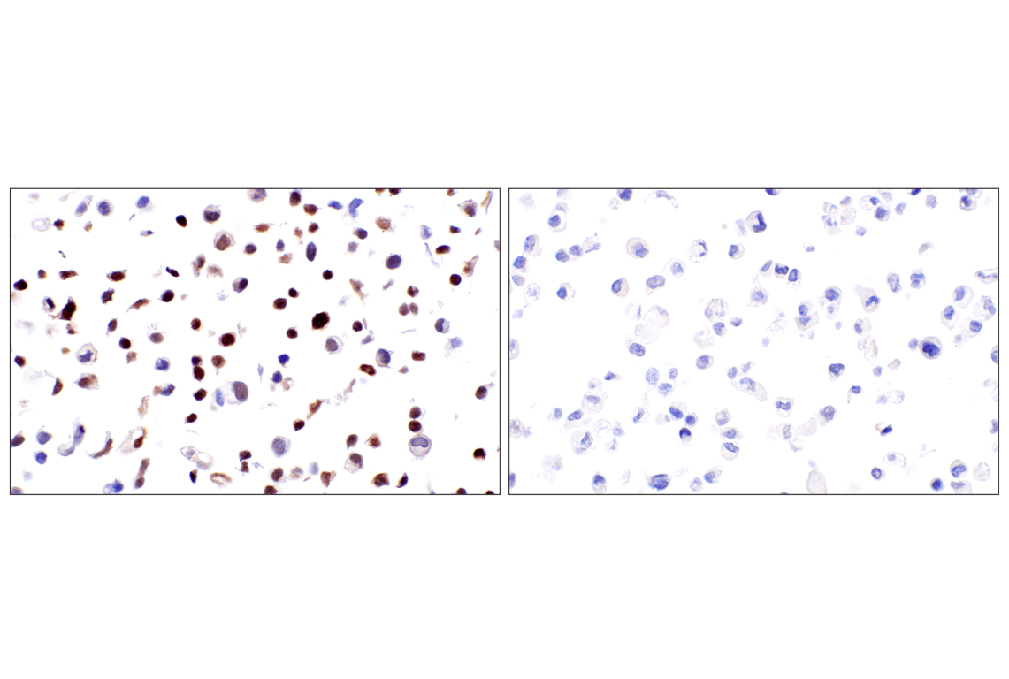
(metastatic site) using WT1 (F3T7B) Rabbit mAb.
Revision 3