Revision 4

#99618

Store at -20C

CST Logo
Orders:

877-616-CELL (2355)

[email protected]

Support:

877-678-TECH (8324)

3 Trask Lane | Danvers | Massachusetts | 01923 | USA

For Research Use Only. Not for Use in Diagnostic Procedures.

Applications:
W, IHC-P, FC-L

Reactivity:
H

Sensitivity:
Endogenous

MW (kDa):
200-220

Source/Isotype:
Mouse IgG1 kappa

UniProt ID:
#P16070

Entrez-Gene Id:
960

Product Usage Information

Application Dilution
Western Blotting 1:1000
Immunohistochemistry (Paraffin) 1:50 - 1:200
Flow Cytometry (Live) 1:100 - 1:400

Storage

Supplied in 10 mM sodium HEPES (pH 7.5), 150 mM NaCl, 100 µg/mL BSA, 50% glycerol, and less than 0.02% sodium azide. Store at –20°C. Do not aliquot the antibody.

Specificity/Sensitivity

CD44 v6 (C44Mab-9) Mouse Monoclonal Antibody recognizes endogenous levels of total CD44 protein containing the variant v6 exon.

Source / Purification

Monoclonal antibody is produced by immunizing animals with recombinant protein specific to the human CD44 variant v3-v10. This antigen has been further characterized as corresponding to residues surrounding Phe404 of human CD44 protein, within the CD44 variant v6 exon.

Background

CD44 is a type I transmembrane glycoprotein that mediates cell-cell and cell-matrix interaction through its affinity for hyaluronic acid (HA) and possibly through other parts of the extracellular matrix (ECM). CD44 is highly polymorphic, possesses a number of alternative splice variants and undergoes extensive post-translational modifications (1,2). Increased surface levels of CD44 are characteristic of T cell activation, and expression of the protein is upregulated during the inflammatory response. Research studies have shown that interactions between CD44 and HER2 are linked to an increase in ovarian carcinoma cell growth (1-3). CD44 interacts with ezrin, radixin, and moesin (ERM), linking the actin cytoskeleton to the plasma membrane and the ECM (4-6). CD44 is constitutively phosphorylated at Ser325 in resting cells. Activation of PKC results in phosphorylation of Ser291, dephosphorylation of Ser325, disassociation of ezrin from CD44, and directional motility (4).

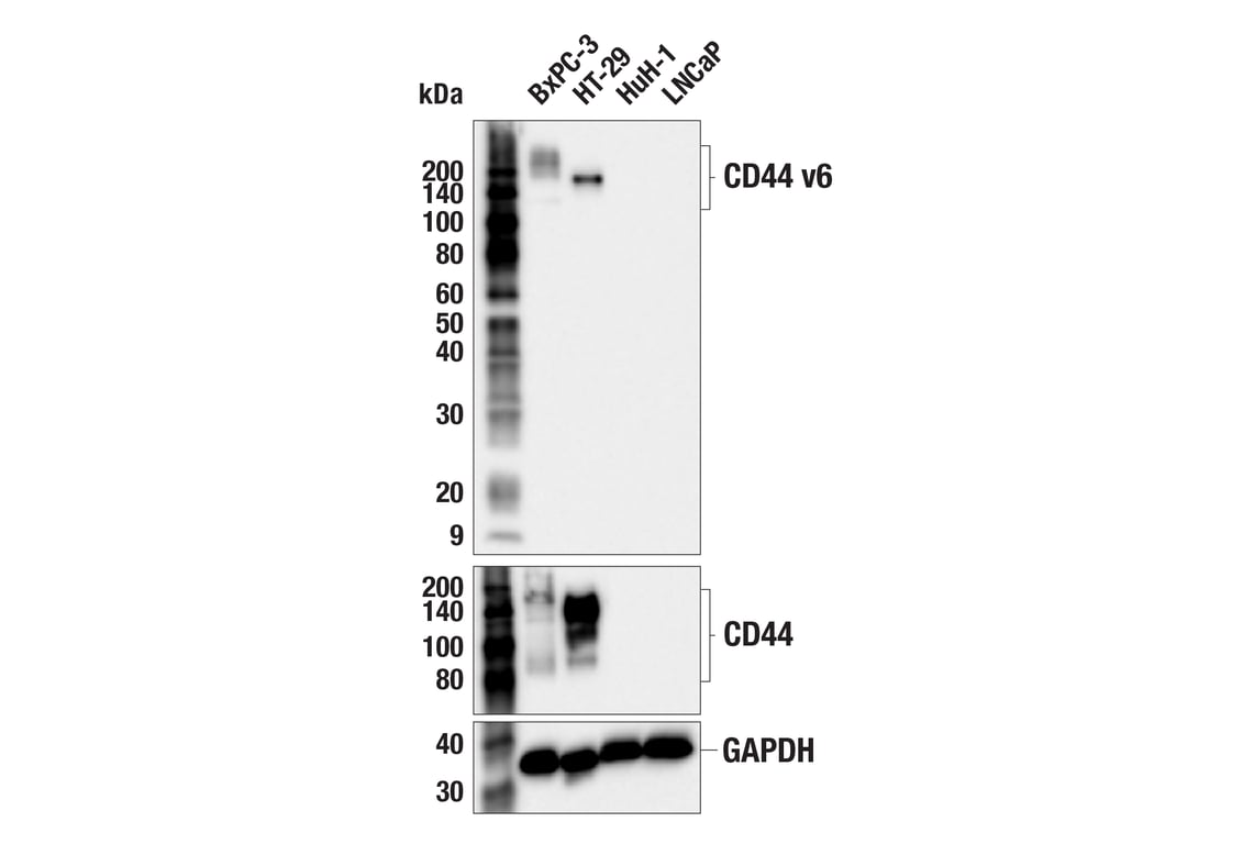
Human CD44 consists of 19 exons, of which 10 are expressed in the standard isoform (CD44s) and all other isoforms. The nine variant exons (v2-v10) inserted between the constant regions via alternative splicing are the source of CD44 heterogeneity, and can dramatically alter the cell-adhesion properties of CD44-expressing cells (7-10). Expression of CD44 isoforms containing exon v6 is associated with metastasis and poor clinical outcomes in colorectal cancer, osteosarcoma, breast cancer, head and neck squamous cell carcinoma, endometriosis, and pancreatic carcinoma (11-16).

Among pancreatic ductal adenocarcinomas (PDAC) cell lines, those that highly express CD44v, including CD44 v6, exhibit an epithelial or MET phenotype, express E-cadherin, and have an increased growth rate (9). Conversely, PDAC cells that highly express CD44s exhibit a mesenchymal phenotype, have high gemcitabine resistance, and co-express proteins associated with EMT transition, including vimentin and ZEB-1 (9). In vivo, PDAC cells have the ability to switch between expression of these CD44 isoforms in response to chemotherapy, demonstrating the importance of CD44-targeted therapies for treatment of some cancers (9).

Species Reactivity

Species reactivity is determined by testing in at least one approved application (e.g., western blot).

Western Blot Buffer

IMPORTANT: For western blots, incubate membrane with diluted primary antibody in 5% w/v nonfat dry milk, 1X TBS, 0.1% Tween® 20 at 4°C with gentle shaking, overnight.

Applications Key

W: Western Blotting IHC-P: Immunohistochemistry (Paraffin) FC-L: Flow Cytometry (Live)

Cross-Reactivity Key

H: Human

Trademarks and Patents

Cell Signaling Technology is a trademark of Cell Signaling Technology, Inc.

All other trademarks are the property of their respective owners. Visit cellsignal.com/trademarks for more information.

使用に関する制限

使用に関する制限

法的な権限を与えられたCSTの担当者が署名した書面によって別途明示的に合意された場合を除き、CST、その関連会社または代理店が提供する製品には以下の条件が適用されます。お客様が定める条件でここに定められた条件に含まれるものを超えるもの、または、ここに定められた条件と異なるものは、法的な権限を与えられたCSTの担当者が別途書面にて受諾した場合を除き、拒絶され、いかなる効力も効果も有しません。

研究専用 (For Research Use Only) またはこれに類似する表示がされた製品は、 いかなる目的についても FDA または外国もしくは国内のその他の規制機関により承認、認可または許可を受けていません。 お客様は製品を診断もしくは治療目的で使用してはならず、また、製品に表示された内容に違反する方法で使用してはなりません。 CST が販売または使用許諾する製品は、エンドユーザーであるお客様に対し、使途を研究および開発のみに限定して提供されるものです。 診断、予防もしくは治療目的で製品を使用することまたは製品を再販売 (単独であるか他の製品等の一部であるかを問いません) もしくはその他の商業的利用の目的で購入することについては、CST から別途許諾を得る必要があります。 お客様は以下の事項を遵守しなければなりません。(a) CST の製品 (単独であるか他の資材と一緒であるかを問いません) を販売、使用許諾、貸与、寄付もしくはその他の態様で第三者に譲渡したり使用させたりしてはなりません。また、商用の製品を製造するために CST の製品を使用してはなりません。(b) 複製、改変、リバースエンジニアリング、逆コンパイル、 分解または他の方法により製品の構造または技術を解明しようとしてはなりません。また、 CST の製品またはサービスと競合する製品またはサービスを開発する目的で CST の製品を使用してはなりません。(c) CST の製品の商標、商号、ロゴ、特許または著作権に関する通知または表示を除去したり改変したりしてはなりません。(d) CST の製品をCST 製品販売条件(CST’s Product Terms of Sale) および該当する書面のみに従って使用しなければなりません。(e) CST の製品に関連してお客様が使用する第三者の製品またはサービスに関する使用許諾条件、 サービス提供条件またはこれに類する合意事項を遵守しなければなりません。

Orders: 877-616-CELL (2355) [email protected] Support: 877-678-TECH (8324) [email protected] Web: cellsignal.com
For Research Use Only. Not for Use in Diagnostic Procedures.

Revision 4

Cell Signaling Technologyロゴ
Western blot analysis of extracts from 293T cells, mock transfected (-) or transfected with constructs expressing Myc/DDK-tagged human CD44 variant v6 (hCD44 v6-Myc/DDK; +), Myc/DDK-tagged full-length human CD44 (hCD44-Myc/DDK; +), or Myc/DDK-tagged human CD44 isoform s (hCD44s-Myc/DDK), using CD44 v6 (C44Mab-9) Mouse mAb (upper), CD44 (156-3C11) Mouse mAb #3570 (middle), or GAPDH (D16H11) XP® Rabbit mAb #5174 (lower). The full-length CD44 construct contains all the CD44 variant v2-v10 exons, while the CD44 isoform s construct lacks the CD44 variant v2-v10 exons.
Western Blotting Image 1: CD44 v6 (C44Mab-9) Mouse Monoclonal Antibody
Western blot analysis of extracts from various cell lines using CD44 v6 (C44Mab-9) Mouse mAb (upper), CD44 (E7K2Y) XP® Rabbit mAb #37259 (middle), or GAPDH (D16H11) XP® Rabbit mAb #5174 (lower). Negative expression of CD44 v6 and CD44 protein in HuH-1 and LNCaP cells is consistent with the predicted expression pattern.
Western Blotting Image 2: CD44 v6 (C44Mab-9) Mouse Monoclonal Antibody
Immunohistochemical analysis of paraffin-embedded human urothelial carcinoma using CD44 v6 (C44Mab-9) Mouse mAb.
Immunohistochemistry Image 1: CD44 v6 (C44Mab-9) Mouse Monoclonal Antibody
Orders: 877-616-CELL (2355) [email protected] Support: 877-678-TECH (8324) [email protected] Web: cellsignal.com
For Research Use Only. Not for Use in Diagnostic Procedures.

Revision 4

Cell Signaling Technologyロゴ
Immunohistochemical analysis of paraffin-embedded human prostate adenocarcinoma using CD44 v6 (C44Mab-9) Mouse mAb.
Immunohistochemistry Image 2: CD44 v6 (C44Mab-9) Mouse Monoclonal Antibody
Immunohistochemical analysis of paraffin-embedded human esophageal carcinoma using CD44 v6 (C44Mab-9) Mouse mAb.
Immunohistochemistry Image 3: CD44 v6 (C44Mab-9) Mouse Monoclonal Antibody

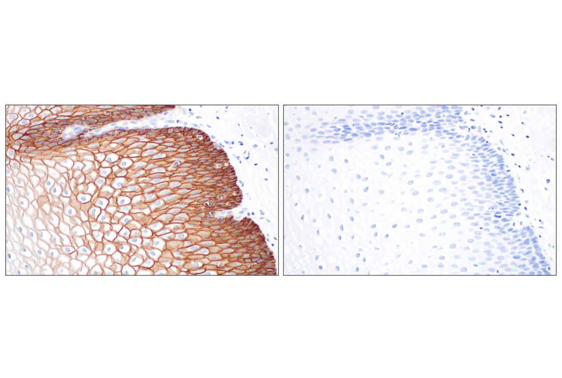
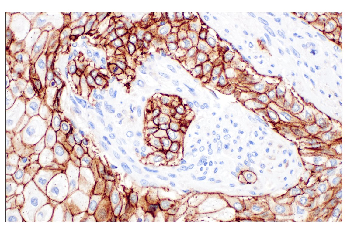
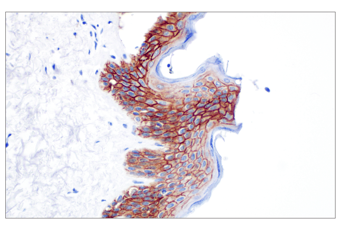
Immunohistochemical analysis of paraffin-embedded normal human skin using CD44 v6 (C44Mab-9) Mouse mAb.

Immunohistochemistry Image 4: CD44 v6 (C44Mab-9) Mouse Monoclonal Antibody
Orders: 877-616-CELL (2355) [email protected] Support: 877-678-TECH (8324) [email protected] Web: cellsignal.com
For Research Use Only. Not for Use in Diagnostic Procedures.

Revision 4

Cell Signaling Technologyロゴ

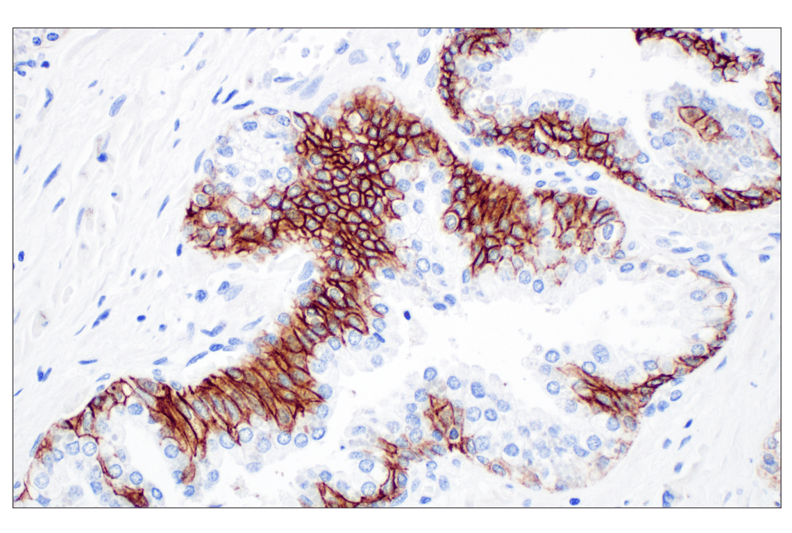
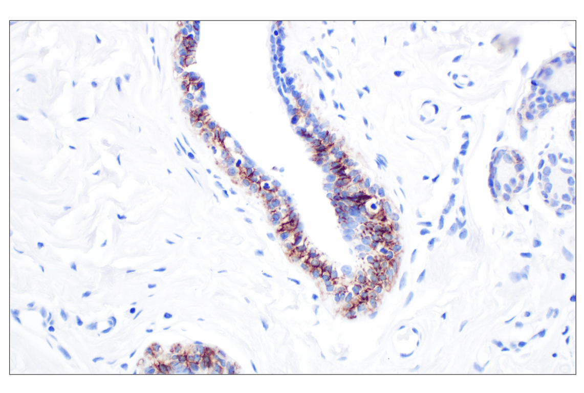
Immunohistochemical analysis of paraffin-embedded normal human breast using CD44 v6 (C44Mab-9) Mouse mAb.

Immunohistochemistry Image 5: CD44 v6 (C44Mab-9) Mouse Monoclonal Antibody

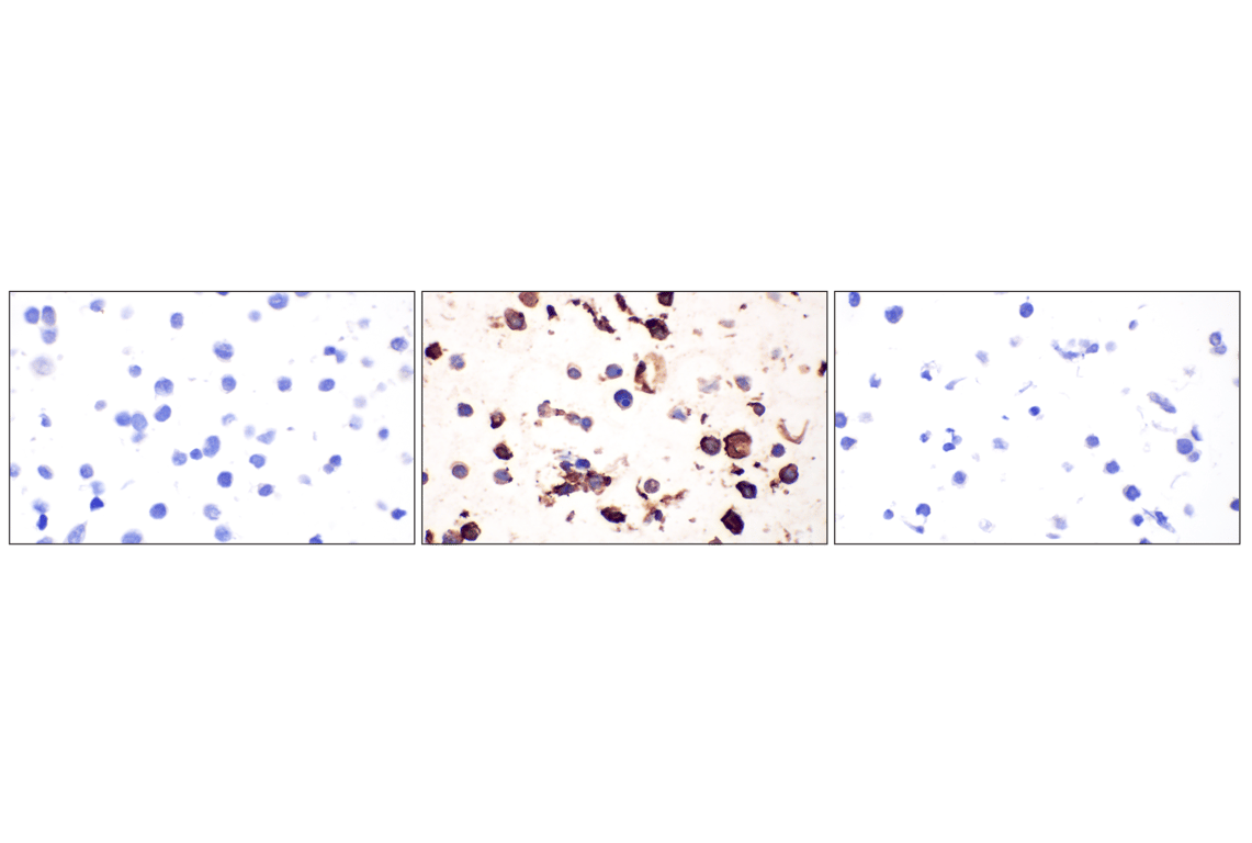
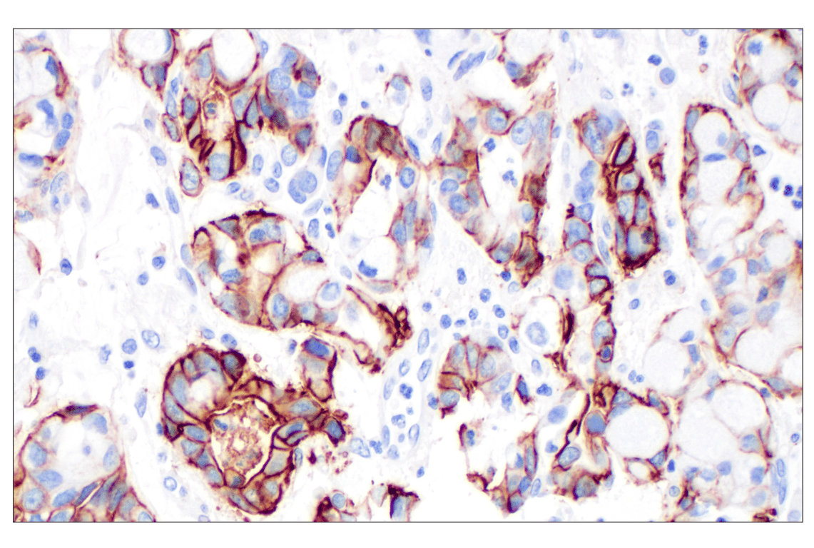
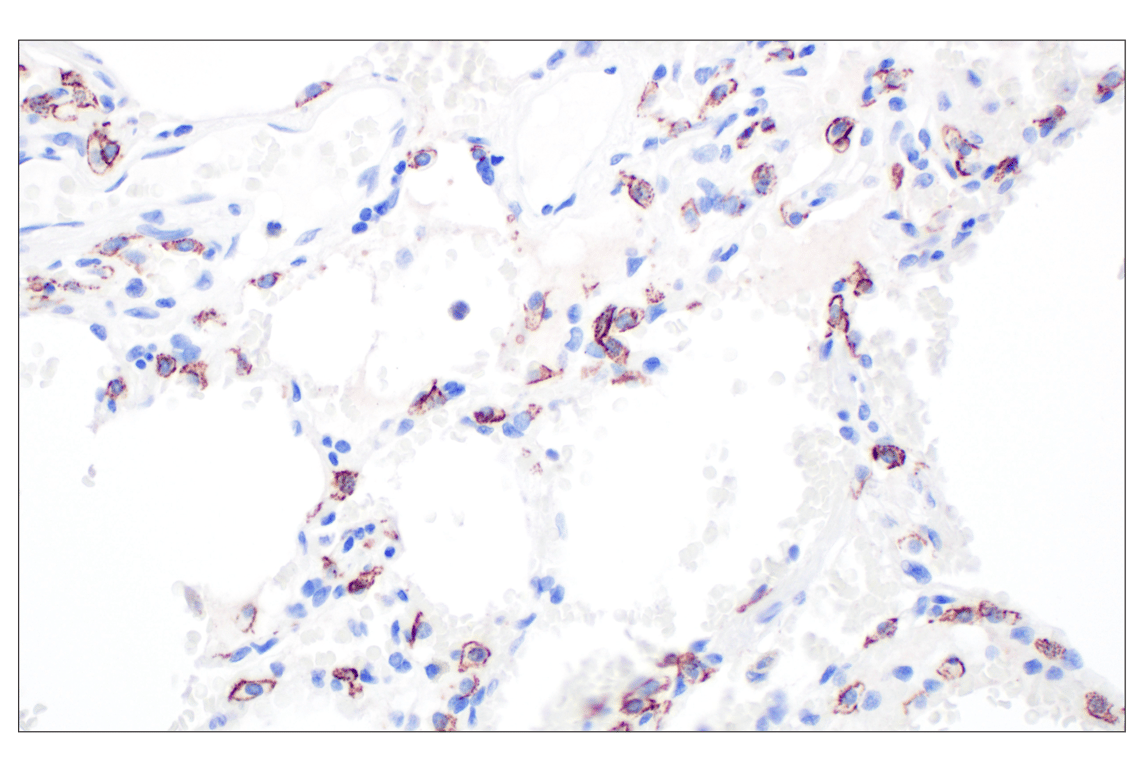
Immunohistochemical analysis of paraffin-embedded normal human lung using CD44 v6 (C44Mab-9) Mouse mAb.

Immunohistochemistry Image 6: CD44 v6 (C44Mab-9) Mouse Monoclonal Antibody
Immunohistochemical analysis of paraffin-embedded normal human esophagus (left), urothelial carcinoma (middle), and squamous cell lung carcinoma (right) using CD44 v6 (C44Mab-9) Mouse mAb (top) or a CD44 v6 Mouse mAb (bottom). These two antibodies detect unique, non-overlapping epitopes on human CD44 v6. The similar staining patterns obtained with both antibodies help to confirm the specificity of the staining.
Immunohistochemistry Image 7: CD44 v6 (C44Mab-9) Mouse Monoclonal Antibody
Orders: 877-616-CELL (2355) [email protected] Support: 877-678-TECH (8324) [email protected] Web: cellsignal.com
For Research Use Only. Not for Use in Diagnostic Procedures.

Revision 4

Cell Signaling Technologyロゴ
Immunohistochemical analysis of paraffin-embedded normal human esophagus using CD44 v6 (C44Mab-9) Mouse mAb (left) compared to concentration-matched Mouse (G3A1) mAb IgG1 Isotype Control #5415 (right).
Immunohistochemistry Image 8: CD44 v6 (C44Mab-9) Mouse Monoclonal Antibody
Immunohistochemical analysis of paraffin-embedded 293T cell pellet, untransfected (left), CD44 variant v6-transfected (middle), or CD44-transfected (right), using CD44 v6 (C44Mab-9) Mouse mAb.
Immunohistochemistry Image 9: CD44 v6 (C44Mab-9) Mouse Monoclonal Antibody
Flow cytometric analysis of HuH-1 cells (blue, negative) and HT-29 cells (green, positive) using CD44 v6 (C44Mab-9) Mouse mAb (solid lines) or concentration-matched Mouse (G3A1) mAb IgG Isotype Control #5415 (dashed lines). Anti-mouse IgG (H+L), F(ab')2 Fragment (Alexa Fluor® 488 Conjugate) #4408 was used as a secondary antibody.
Flow Cytometry Image 1: CD44 v6 (C44Mab-9) Mouse Monoclonal Antibody
Orders: 877-616-CELL (2355) [email protected] Support: 877-678-TECH (8324) [email protected] Web: cellsignal.com
For Research Use Only. Not for Use in Diagnostic Procedures.