Product Information
Storage
Specificity / Sensitivity
Species Reactivity:
Human, Mouse
Product Description
Background
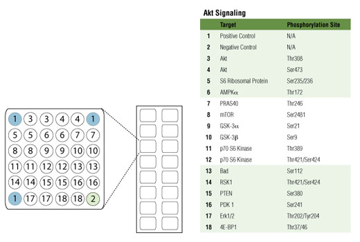
The Akt signaling module is typically activated in response to growth factor stimulation of receptor tyrosine kinases transmitting primarily anabolic growth and survival signals. Akt1/2 are ubiquitously expressed protein kinases having a multitude of cellular substrates and are involved in the regulation of a wide range of cellular processes. Akt is activated by phosphorylation at two distinct sites: Ser473 by the mTORC2 complex and Thr308 by the plasma membrane residing kinase PDK1.
PI3 kinase is a lipid kinase that phosphorylates inositol phospholipids at position three to generate docking sites for Akt at the plasma membrane where Akt is activated. PTEN is a lipid phosphatase that negates the action of PI3 kinase to downregulate the signal emanating from this module.
mTOR integrates growth factor signaling and nutrient availability and is a core component of two macromolecular complexes, mTORC1 and mTORC2. The autophosphorylation of mTOR at Ser2481 correlates with the levels of its activation. mTORC1 phosphorylation of p70 S6 kinase leads to kinase activation, which in turn activates protein synthesis. The S6 ribosomal protein is found downstream of p70 S6 kinase and its phoshporylation at Ser235/236 reflects mTOR pathway activation. The mTORC2 complex activates Akt by phosphorylating it at Ser473. Phosphorylation of PRAS40 at Thr246 by Akt relieves PRAS40 inhibition of mTORC1.
4E-BP1 is a repressor of translation and inhibits cap-dependent translation initiation. Hyperphosphorylation of 4E-BP1 by mTORC1 leads to derepression of this blockade, which results in activation of cap-dependent translation.
Phosphorylation of the pro-apoptotic protein Bad at Ser112 and the multifunctional kinases GSK-3α and GSK-3β at Ser21 and Ser9, respectively, by Akt inhibits their activity and promotes cell survival.
AMPK is an energy sensor that is activated by phosphorylation at Thr172 in response to elevated AMP levels. Under conditions of low energy and elevated levels of AMP, AMPK helps to ensure that anabolic processes, such as those triggered by Akt, are decreased until energy levels are restored.
Although not a component of the Akt signaling network, Erk1 and Erk2 kinases are a central component of the Ras/MAP kinase signaling module. Erk1/2 regulate multiple cellular functions and are involved in a broad range of cellular processes, such as proliferation, differentiation, and motility. Erk and Akt signaling modules cross regulate each other at multiple points and through a variety of mechanisms. Erk is activated by a wide range of extracellular signals including growth factors, cytokines, hormones, and neurotransmitters, leading to dual phosphorylation at Thr202 and Tyr204.
The 90 kDa ribosomal S6 kinase 1 (RSK1) is activated primarily by Erk1/2 in response to many growth factors, polypeptide hormones, and neurotransmitters. p90RSK1 phosphorylates a wide range of substrates including ribosomal protein S6, and positively regulates protein translation and cellular growth. p90RSK1 can also be activated by kinases that regulate the response to cellular stress
- Lawlor, M.A. and Alessi, D.R. (2001) J Cell Sci 114, 2903-10.
- Brazil, D.P. and Hemmings, B.A. (2001) Trends Biochem Sci 26, 657-64.
- Brazil, D.P. et al. (2002) Cell 111, 293-303.
- Luo, J. et al. (2003) Cancer Cell 4, 257-62.
- Manning, B.D. and Cantley, L.C. (2007) Cell 129, 1261-74.
- Franke, T.F. (2008) Sci Signal 1, pe29.
- Huang, J. and Manning, B.D. (2009) Biochem Soc Trans 37, 217-22.
- Hers, I. et al. (2011) Cell Signal 23, 1515-27.
- Dufner, A. and Thomas, G. (1999) Exp Cell Res 253, 100-9.
- Rubinfeld, H. and Seger, R. (2005) Mol Biotechnol 31, 151-74.
- Mihaylova, M.M. and Shaw, R.J. (2011) Nat Cell Biol 13, 1016-23.
- Hardie, D.G. et al. (2012) Nat Rev Mol Cell Biol 13, 251-62.
Species Reactivity
Species reactivity is determined by testing in at least one approved application (e.g., western blot).
Cross-Reactivity Key
H: human M: mouse R: rat Hm: hamster Mk: monkey Vir: virus Mi: mink C: chicken Dm: D. melanogaster X: Xenopus Z: zebrafish B: bovine Dg: dog Pg: pig Sc: S. cerevisiae Ce: C. elegans Hr: horse GP: Guinea Pig Rab: rabbit All: all species expected
Trademarks and Patents
使用に関する制限
法的な権限を与えられたCSTの担当者が署名した書面によって別途明示的に合意された場合を除き、 CST、その関連会社または代理店が提供する製品には以下の条件が適用されます。お客様が定める条件でここに定められた条件に含まれるものを超えるもの、 または、ここに定められた条件と異なるものは、法的な権限を与えられたCSTの担当者が別途書面にて受諾した場合を除き、拒絶され、 いかなる効力も効果も有しません。
研究専用 (For Research Use Only) またはこれに類似する表示がされた製品は、 いかなる目的についても FDA または外国もしくは国内のその他の規制機関により承認、認可または許可を受けていません。 お客様は製品を診断もしくは治療目的で使用してはならず、また、製品に表示された内容に違反する方法で使用してはなりません。 CST が販売または使用許諾する製品は、エンドユーザーであるお客様に対し、使途を研究および開発のみに限定して提供されるものです。 診断、予防もしくは治療目的で製品を使用することまたは製品を再販売 (単独であるか他の製品等の一部であるかを問いません) もしくはその他の商業的利用の目的で購入することについては、CST から別途許諾を得る必要があります。 お客様は以下の事項を遵守しなければなりません。(a) CST の製品 (単独であるか他の資材と一緒であるかを問いません) を販売、使用許諾、貸与、寄付もしくはその他の態様で第三者に譲渡したり使用させたりしてはなりません。また、商用の製品を製造するために CST の製品を使用してはなりません。(b) 複製、改変、リバースエンジニアリング、逆コンパイル、 分解または他の方法により製品の構造または技術を解明しようとしてはなりません。また、 CST の製品またはサービスと競合する製品またはサービスを開発する目的で CST の製品を使用してはなりません。(c) CST の製品の商標、商号、ロゴ、特許または著作権に関する通知または表示を除去したり改変したりしてはなりません。(d) CST の製品をCST 製品販売条件(CST’s Product Terms of Sale) および該当する書面のみに従って使用しなければなりません。(e) CST の製品に関連してお客様が使用する第三者の製品またはサービスに関する使用許諾条件、 サービス提供条件またはこれに類する合意事項を遵守しなければなりません。