#Q16539, #P45983, #P40763, #P42226, #P27361, #O43318, #O15264, #P25963, #P31751, #Q14653, #Q9Y243, #P53778, #P43403, #P07947, #P07948, #P43405, #P42229, #P51692, #P28482, #P06239, #P55210, #O95786, #P31749, #P08631, #P42224, #P12931, #P06241, #Q15759
1432, 5599, 6774, 6778, 5595, 6885, 5603, 4792, 208, 3661, 10000, 6300, 7535, 7525, 4067, 6850, 6776, 6777, 5594, 3932, 840, 23586, 207, 3055, 6772, 6714, 2534, 5600
Product Information
Storage
Specificity / Sensitivity
Species Reactivity:
Human
Product Description
Background
The vertebrate immune response relies on both a non-specific, innate immune system and an antigen-specific, adaptive immune response. Organisms rely on cell-mediated and antibody-mediated responses to detect pathogens and protect against infection and disease. Inflammation is an immune response in which circulating immune system cells localize to sites of injury or infection. Regulation of inflammation is essential, as unchecked inflammation can result in permanent damage to tissues and organs. A variety of signaling proteins are known to regulate the immune and inflammatory response.
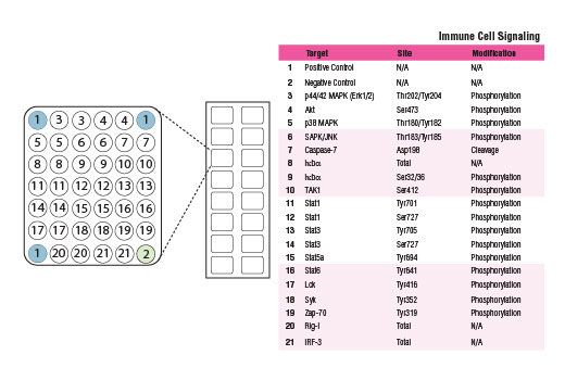
Akt and p44/42 MAPK (Erk1/2) kinases are key regulators of cell fate that generally transmit growth promoting and survival signals. These two kinases serve as major signal integration hubs and very few cellular processes occur without their involvement. Both p38 MAPK and SAPK/JNK kinases are activated through a dual phosphorylation mechanism in response to pro-inflammatory cytokines, stressful conditions, or genotoxic stress. Caspase-7 is an intracellular protease involved in apoptosis and is part of a cellular sub-organelle called the inflammasome. Caspase-7 is activated by cleavage at Asp198. Activation of the essential immune system regulator NF-κB is triggered by a diverse group of extracellular signals promoted by inflammatory cytokines, growth factors, and chemokines. The proteasome-mediated degradation of the NF-κB/Rel inhibitor IκBα results from phosphorylation of IκBα at Ser32 and Ser36 and targeting of IκBα to the proteasome. The TAK1 kinase responds to a variety of cytokines to regulate cellular kinases and activate the NF-κB pathway; phosphorylation of TAK1 at Ser412 by PKA regulates kinase activity.
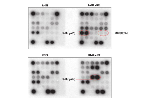
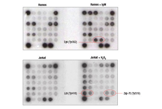
Stat family transcription factors are activated by a variety of cytokines, chemokines, or growth factors to regulate the immune response. Phosphorylation of Stat proteins at specific tyrosine or serine residues leads to Stat protein dimerization, nuclear translocation, and binding of the transcription factor to genes that regulate the immune response. The range and specificity of responses regulated by Stat proteins is determined in part by the tissue-specific expression of different cytokine receptors and by the combinatorial coupling of various Stat members to different receptors. Lck, Syk, and Zap-70 are Src family tyrosine kinases that couple the activated B cells and T cell immunoreceptors to downstream signaling events that mediate the immune response. Rig-1 is a Toll-receptor signaling complex component that plays a role in the antiviral innate immune response. The transcription factor IRF-3 plays a role in regulating interferon (IFN) and IFN-inducible gene expression in response to viral infection.
- Sojka, D.K. et al. (2009) Immunol Res 45, 239-50.
- Si-Tahar, M. et al. (2009) Clin Exp Immunol 156, 194-8.
- Girardin, S.E. and Philpott, D.J. (2009) Semin Immunol 21, 173-4.
- Lukic, M.L. et al. (2009) Mol Immunol 47, 1-2.
- Gaestel, M. et al. (2009) Nat Rev Drug Discov 8, 480-99.
- Nag, K. and Chaudhary, A. (2009) Curr Signal Transduct Ther 4, 76-81.
- Müller-Hübenthal, B. et al. (2009) Anticancer Res 29, 4795-805.
- Yu, H. et al. (2009) Nat Rev Cancer 9, 798-809.
- Grivennikov, S.I. et al. (2010) Cell 140, 883-99.
- Maletzki, C. and Emmrich, J. (2010) Dig Dis 28, 574-8.
- Davis, B.K. et al. (2011) Annu Rev Immunol 29, 707-35.
- Newton, K. and Dixit, V.M. (2012) Cold Spring Harb Perspect Biol 4.
- Mantovani, A. and Garlanda, C. (2013) Nat Immunol 14, 768-70.
Species Reactivity
Species reactivity is determined by testing in at least one approved application (e.g., western blot).
Cross-Reactivity Key
H: human M: mouse R: rat Hm: hamster Mk: monkey Vir: virus Mi: mink C: chicken Dm: D. melanogaster X: Xenopus Z: zebrafish B: bovine Dg: dog Pg: pig Sc: S. cerevisiae Ce: C. elegans Hr: horse GP: Guinea Pig Rab: rabbit All: all species expected
Trademarks and Patents
使用に関する制限
法的な権限を与えられたCSTの担当者が署名した書面によって別途明示的に合意された場合を除き、 CST、その関連会社または代理店が提供する製品には以下の条件が適用されます。お客様が定める条件でここに定められた条件に含まれるものを超えるもの、 または、ここに定められた条件と異なるものは、法的な権限を与えられたCSTの担当者が別途書面にて受諾した場合を除き、拒絶され、 いかなる効力も効果も有しません。
研究専用 (For Research Use Only) またはこれに類似する表示がされた製品は、 いかなる目的についても FDA または外国もしくは国内のその他の規制機関により承認、認可または許可を受けていません。 お客様は製品を診断もしくは治療目的で使用してはならず、また、製品に表示された内容に違反する方法で使用してはなりません。 CST が販売または使用許諾する製品は、エンドユーザーであるお客様に対し、使途を研究および開発のみに限定して提供されるものです。 診断、予防もしくは治療目的で製品を使用することまたは製品を再販売 (単独であるか他の製品等の一部であるかを問いません) もしくはその他の商業的利用の目的で購入することについては、CST から別途許諾を得る必要があります。 お客様は以下の事項を遵守しなければなりません。(a) CST の製品 (単独であるか他の資材と一緒であるかを問いません) を販売、使用許諾、貸与、寄付もしくはその他の態様で第三者に譲渡したり使用させたりしてはなりません。また、商用の製品を製造するために CST の製品を使用してはなりません。(b) 複製、改変、リバースエンジニアリング、逆コンパイル、 分解または他の方法により製品の構造または技術を解明しようとしてはなりません。また、 CST の製品またはサービスと競合する製品またはサービスを開発する目的で CST の製品を使用してはなりません。(c) CST の製品の商標、商号、ロゴ、特許または著作権に関する通知または表示を除去したり改変したりしてはなりません。(d) CST の製品をCST 製品販売条件(CST’s Product Terms of Sale) および該当する書面のみに従って使用しなければなりません。(e) CST の製品に関連してお客様が使用する第三者の製品またはサービスに関する使用許諾条件、 サービス提供条件またはこれに類する合意事項を遵守しなければなりません。