#Q16539, #P27361, #O15264, #P31751, #Q9Y243, #P53778, #P62753, #P28482, #P31749, #Q15759
1432, 5595, 5603, 208, 10000, 6300, 6194, 5594, 207, 5600
Product Information
Storage
Specificity / Sensitivity
Product Description
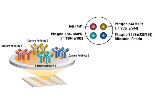
Briefly, 4 capture antibodies with distinct target specificity have been spotted onto the bottom of each well of a microwell plate. After incubation with cell lysates, the spotted antibodies capture the target proteins. Following extensive washing, a mixture of detection antibodies is added to detect the captured target proteins. An HRP-linked secondary solution is then used to recognize the bound detection antibodies. Chemiluminescent HRP substrate is used to produce luminescent signal.
Background
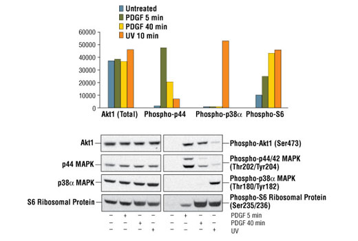
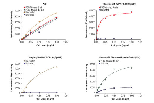
Cells must respond in an appropriate fashion to many complex signaling events. Extracellular signaling cues are organized into well defined signal transduction modules that control fundamental cellular behavior. Two prominent signaling modules that are among the best characterized are the p44/42 MAP kinase (ERK MAPK) and p38 MAPK. The p44/42 MAPK is activated by a wide variety of extracellular signals including growth and neurotrophic factors, cytokines, hormones and neurotransmitters. p44/42 MAPK activation occurs through phosphorylation of threonine and tyrosine at the sequence T*EY* by a dual specificity kinase called MAP kinase kinase (MEK). p38 MAPK is dually phosphorylated in response to pro-inflammatory cytokines or cellular stress. These signaling modules are central controllers of fundamental cellular behavior such as growth, proliferation, movement, and response to stress. Growth factor stimulation results in phosphorylation of S6 ribosomal protein on Ser235/236, leading to an increase in protein synthesis and cell cycle progression. Phosphorylation levels of critical molecular switches such as MAPKs therefore serve as a reliable indicator of the activation state of the entire signaling module. The profiling of phosphorylation events using phospho-specific antibodies is now widely used to investigate diagnostic pathology (1,2). While profiling of protein phosphorylation events was shown to predict the progression of a tumor to a more invasive stage (3), it has been observed that the ratio between p44/42 MAPK and p38 MAPK may predict whether tumor cells will proliferate or enter a state of dormancy in vivo (4). PathScan® Signaling Nodes I 4-Plex Array Kit provides the researcher with means to profile numerous chemical compounds and obtain in-cell relative potency (5).
Species Reactivity
Species reactivity is determined by testing in at least one approved application (e.g., western blot).
Cross-Reactivity Key
H: human M: mouse R: rat Hm: hamster Mk: monkey Vir: virus Mi: mink C: chicken Dm: D. melanogaster X: Xenopus Z: zebrafish B: bovine Dg: dog Pg: pig Sc: S. cerevisiae Ce: C. elegans Hr: horse GP: Guinea Pig Rab: rabbit All: all species expected
Trademarks and Patents
使用に関する制限
法的な権限を与えられたCSTの担当者が署名した書面によって別途明示的に合意された場合を除き、 CST、その関連会社または代理店が提供する製品には以下の条件が適用されます。お客様が定める条件でここに定められた条件に含まれるものを超えるもの、 または、ここに定められた条件と異なるものは、法的な権限を与えられたCSTの担当者が別途書面にて受諾した場合を除き、拒絶され、 いかなる効力も効果も有しません。
研究専用 (For Research Use Only) またはこれに類似する表示がされた製品は、 いかなる目的についても FDA または外国もしくは国内のその他の規制機関により承認、認可または許可を受けていません。 お客様は製品を診断もしくは治療目的で使用してはならず、また、製品に表示された内容に違反する方法で使用してはなりません。 CST が販売または使用許諾する製品は、エンドユーザーであるお客様に対し、使途を研究および開発のみに限定して提供されるものです。 診断、予防もしくは治療目的で製品を使用することまたは製品を再販売 (単独であるか他の製品等の一部であるかを問いません) もしくはその他の商業的利用の目的で購入することについては、CST から別途許諾を得る必要があります。 お客様は以下の事項を遵守しなければなりません。(a) CST の製品 (単独であるか他の資材と一緒であるかを問いません) を販売、使用許諾、貸与、寄付もしくはその他の態様で第三者に譲渡したり使用させたりしてはなりません。また、商用の製品を製造するために CST の製品を使用してはなりません。(b) 複製、改変、リバースエンジニアリング、逆コンパイル、 分解または他の方法により製品の構造または技術を解明しようとしてはなりません。また、 CST の製品またはサービスと競合する製品またはサービスを開発する目的で CST の製品を使用してはなりません。(c) CST の製品の商標、商号、ロゴ、特許または著作権に関する通知または表示を除去したり改変したりしてはなりません。(d) CST の製品をCST 製品販売条件(CST’s Product Terms of Sale) および該当する書面のみに従って使用しなければなりません。(e) CST の製品に関連してお客様が使用する第三者の製品またはサービスに関する使用許諾条件、 サービス提供条件またはこれに類する合意事項を遵守しなければなりません。