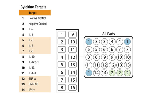
#P40763, #P01579, #P29459, #P22301, #P35225, #P05112, #P05231, #Q16552, #P60568, #P04141, #P05113, #P10145, #P01375
6774, 3458, 3592, 3586, 3596, 3565, 3569, 3605, 3558, 1437, 3567, 3576, 7124
Product Information
Storage
Specificity / Sensitivity
Species Reactivity:
Human
Product Description
Background
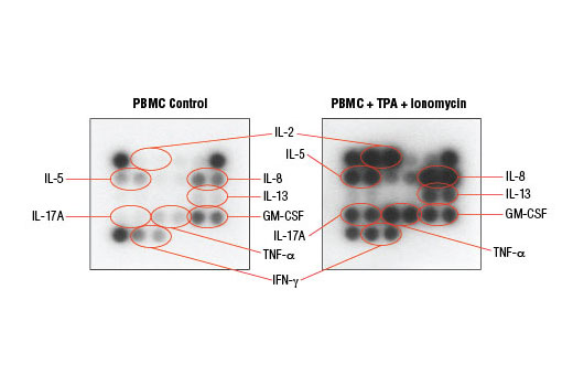
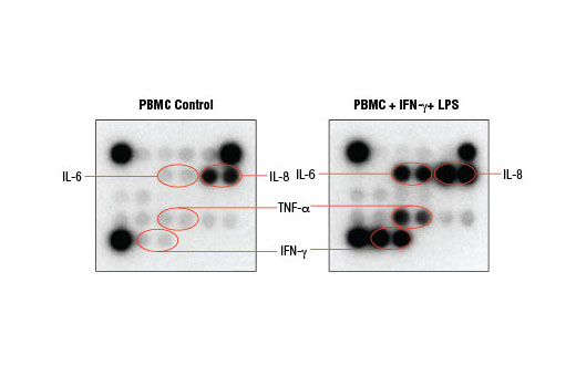
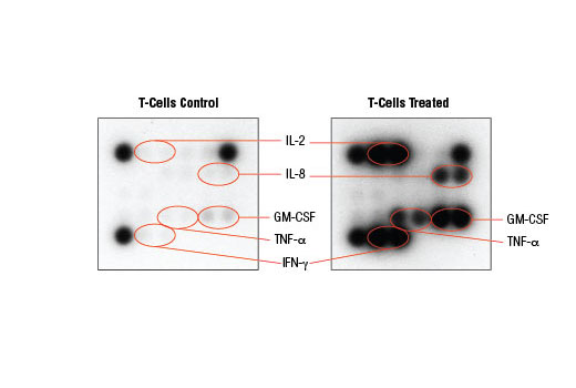
Cytokines are secreted intercellular signaling molecules that regulate many biological processes including inflammation, host defense, and cell differentiation. Cytokine profiles may provide insight into the molecular mechanisms that distinguish between healthy and diseased states. The PathScan® Th1/Th2/Th17 Cytokine Antibody Array Kit offers an antibody panel against a broad array of cytokines to enable measurement of their relative changes in cell culture supernatants.
Upon activation, naive CD4+ helper T cells differentiate into distinct functional subsets. The development of these subsets is driven, in part, by the cytokine milieu. Type 1 (Th1) cells help drive cellular immunity against intracellular pathogens. IL-12 and IFN-γ induce Th1 cell development. Th1 cells produce IFN-γ and IL-2, which provide a positive feedback loop to enhance Th1 cell differentiation and NK cell and CD8+ T cell cytolytic activity.
Th2 cells play a crucial role in the humoral immune response against extracellular pathogens. IL-4 drives development of Th2 cells, which subsequently produce IL-4, IL-5, and IL-13. These cytokines induce B cell proliferation, antibody production, IgE class switching, and activate eosinophils respectively.
Another distinct helper T cell lineage, Th17, is important for mucosal immunity. Dysregulation of Th17 may significantly contribute to the development of autoimmunity. IL-17 produced by Th17 cells induces secretion of pro-inflammatory cytokines IL-6, IL-8, GM-CSF, and TNF-α. Many of these molecules link innate and adaptive immunity through the recruitment and activation of innate immune cells.
Effective immune responses require finely tuned coordination between pro and anti-inflammatory signals. Pro-inflammatory molecules play important roles in activating key immune players to fight infection. IL-8 induces granulocyte migration and activates neutrophil phagocytic activity. GM-CSF mobilizes monocytes into infected tissue and activates macrophage and neutrophils. TNF-α is a multifunctional pro-inflammatory cytokine involved with a number of processes including cell proliferation, differentiation, and apoptosis.
Uncontrolled inflammation may damage surrounding host tissue. IL-10 is a prototypical anti-inflammatory cytokine that serves to terminate the acute inflammatory response by inhibiting Th1 cell function and pro-inflammatory cytokine production.
- Romagnani, S. (1999) Inflamm Bowel Dis 5, 285-94.
- Bradley, L.M. et al. (2000) Immunol Res 21, 149-58.
- O'Garra, A. and Arai, N. (2000) Trends Cell Biol 10, 542-50.
- Harrington, L.E. et al. (2005) Nat Immunol 6, 1123-32.
- Stockinger, B. and Veldhoen, M. (2007) Curr Opin Immunol 19, 281-6.
- Köhidai, L. and Csaba, G. (1998) Cytokine 10, 481-6.
- Carey, A.J. et al. (2012) JAKSTAT 1, 159-167.
Species Reactivity
Species reactivity is determined by testing in at least one approved application (e.g., western blot).
Cross-Reactivity Key
H: human M: mouse R: rat Hm: hamster Mk: monkey Vir: virus Mi: mink C: chicken Dm: D. melanogaster X: Xenopus Z: zebrafish B: bovine Dg: dog Pg: pig Sc: S. cerevisiae Ce: C. elegans Hr: horse GP: Guinea Pig Rab: rabbit All: all species expected
Trademarks and Patents
使用に関する制限
法的な権限を与えられたCSTの担当者が署名した書面によって別途明示的に合意された場合を除き、 CST、その関連会社または代理店が提供する製品には以下の条件が適用されます。お客様が定める条件でここに定められた条件に含まれるものを超えるもの、 または、ここに定められた条件と異なるものは、法的な権限を与えられたCSTの担当者が別途書面にて受諾した場合を除き、拒絶され、 いかなる効力も効果も有しません。
研究専用 (For Research Use Only) またはこれに類似する表示がされた製品は、 いかなる目的についても FDA または外国もしくは国内のその他の規制機関により承認、認可または許可を受けていません。 お客様は製品を診断もしくは治療目的で使用してはならず、また、製品に表示された内容に違反する方法で使用してはなりません。 CST が販売または使用許諾する製品は、エンドユーザーであるお客様に対し、使途を研究および開発のみに限定して提供されるものです。 診断、予防もしくは治療目的で製品を使用することまたは製品を再販売 (単独であるか他の製品等の一部であるかを問いません) もしくはその他の商業的利用の目的で購入することについては、CST から別途許諾を得る必要があります。 お客様は以下の事項を遵守しなければなりません。(a) CST の製品 (単独であるか他の資材と一緒であるかを問いません) を販売、使用許諾、貸与、寄付もしくはその他の態様で第三者に譲渡したり使用させたりしてはなりません。また、商用の製品を製造するために CST の製品を使用してはなりません。(b) 複製、改変、リバースエンジニアリング、逆コンパイル、 分解または他の方法により製品の構造または技術を解明しようとしてはなりません。また、 CST の製品またはサービスと競合する製品またはサービスを開発する目的で CST の製品を使用してはなりません。(c) CST の製品の商標、商号、ロゴ、特許または著作権に関する通知または表示を除去したり改変したりしてはなりません。(d) CST の製品をCST 製品販売条件(CST’s Product Terms of Sale) および該当する書面のみに従って使用しなければなりません。(e) CST の製品に関連してお客様が使用する第三者の製品またはサービスに関する使用許諾条件、 サービス提供条件またはこれに類する合意事項を遵守しなければなりません。