Revision 2

#5723Store at -20C

Cell Signaling Technology

Orders: 877-616-CELL (2355) [email protected]

Support: 877-678-TECH (8324)

Web: [email protected] cellsignal.com

3 Trask LaneDanversMassachusetts01923USA
For Research Use Only. Not for Use in Diagnostic Procedures.
:

:

:

:

UniProt ID:

#P42574

Entrez-Gene Id:

836

Product Information

Storage

This kit contains mixed storage components. Please store this entire kit at -20°C for long term storage. Upon first use, please allow components to thaw and then store each component as indicated on individual component labels.

Specificity / Sensitivity

Caspase-3 Activity Assay Kit detects fluorescent AMC dye produced from cleavage of Ac-DEVD-AMC by activated caspase-3 in apoptotic cells. This kit is expected to work in most species. Depending on the cell type and the incubation time applied in the assay, 0.5 - 2x10⁵ cells/well (or 100 μg/well of total lysate protein) is sufficient for most experimental setups. For best results, cell number or lysate concentration titrations are recommended (see Figures 1 and 2). Because caspase-7 shares the same susbtrate sequence as caspase-3, this kit also detects caspase-7 activity.

Species Reactivity:

All Species Expected

Product Description

The Caspase-3 Activity Assay Kit is a fluorescent assay that detects the activity of caspase-3 in cell lysates. It contains a fluorogenic substrate (N-Acetyl-Asp-Glu-Val-Asp-7-amino-4-methylcoumarin or Ac-DEVD-AMC) for caspase-3. During the assay, activated caspase-3 cleaves this substrate between DEVD and AMC, generating highly fluorescent AMC that can be detected using a fluorescence reader with excitation at 380 nm and emission between 420 - 460 nm. Cleavage of the substrate only occurs in lysates of apoptotic cells; therefore, the amount of AMC produced is proportional to the number of apoptotic cells in the sample.

Background

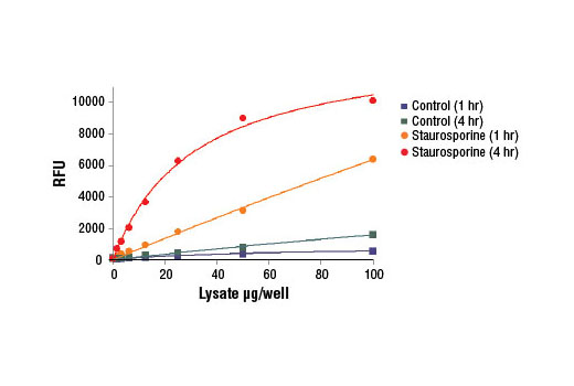
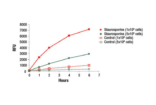
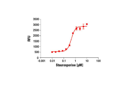
Caspase-3 (CPP-32, Apopain, Yama, SCA-1) is a critical executioner of apoptosis, as it is either partially or totally responsible for the proteolytic cleavage of many key proteins, such as the nuclear enzyme poly (ADP-ribose) polymerase (PARP) (1). Activation of caspase-3 requires proteolytic processing of its inactive zymogen into activated p17 and p12 fragments. Cleavage of caspase-3 requires the aspartic acid residue at the P1 position (2).

Caspase-7 (CMH-1, Mch3, ICE-LAP3) has been identified as a major contributor to the execution of apoptosis (3-6). Caspase-7, like caspase-3, is an effector caspase that is responsible for cleaving downstream substrates, such as PARP (3,5). During apoptosis, caspase-7 is activated by upstream caspases through proteolytic processsing at Asp23, Asp198, and Asp206, thereby producing the mature subunits (3,5). Similar to caspases-2 and -3, caspase-7 preferentially cleaves substrates following the recognition sequence DEVD (7).

  1. Fernandes-Alnemri, T. et al. (1994) J Biol Chem 269, 30761-4.
  2. Nicholson, D.W. et al. (1995) Nature 376, 37-43.
  3. Fernandes-Alnemri, T. et al. (1995) Cancer Res 55, 6045-52.
  4. Duan, H. et al. (1996) J Biol Chem 271, 1621-5.
  5. Lippke, J.A. et al. (1996) J Biol Chem 271, 1825-8.
  6. Cohen, G.M. (1997) Biochem J 326 ( Pt 1), 1-16.
  7. Thornberry, N.A. et al. (1997) J Biol Chem 272, 17907-11.

Species Reactivity

Species reactivity is determined by testing in at least one approved application (e.g., western blot).

Cross-Reactivity Key

H: human M: mouse R: rat Hm: hamster Mk: monkey Vir: virus Mi: mink C: chicken Dm: D. melanogaster X: Xenopus Z: zebrafish B: bovine Dg: dog Pg: pig Sc: S. cerevisiae Ce: C. elegans Hr: horse GP: Guinea Pig Rab: rabbit All: all species expected

Trademarks and Patents

Cell Signaling Technology is a trademark of Cell Signaling Technology, Inc.
PathScan is a registered trademark of Cell Signaling Technology, Inc.
All other trademarks are the property of their respective owners. Visit cellsignal.com/trademarks for more information.

使用に関する制限

法的な権限を与えられたCSTの担当者が署名した書面によって別途明示的に合意された場合を除き、 CST、その関連会社または代理店が提供する製品には以下の条件が適用されます。お客様が定める条件でここに定められた条件に含まれるものを超えるもの、 または、ここに定められた条件と異なるものは、法的な権限を与えられたCSTの担当者が別途書面にて受諾した場合を除き、拒絶され、 いかなる効力も効果も有しません。

研究専用 (For Research Use Only) またはこれに類似する表示がされた製品は、 いかなる目的についても FDA または外国もしくは国内のその他の規制機関により承認、認可または許可を受けていません。 お客様は製品を診断もしくは治療目的で使用してはならず、また、製品に表示された内容に違反する方法で使用してはなりません。 CST が販売または使用許諾する製品は、エンドユーザーであるお客様に対し、使途を研究および開発のみに限定して提供されるものです。 診断、予防もしくは治療目的で製品を使用することまたは製品を再販売 (単独であるか他の製品等の一部であるかを問いません) もしくはその他の商業的利用の目的で購入することについては、CST から別途許諾を得る必要があります。 お客様は以下の事項を遵守しなければなりません。(a) CST の製品 (単独であるか他の資材と一緒であるかを問いません) を販売、使用許諾、貸与、寄付もしくはその他の態様で第三者に譲渡したり使用させたりしてはなりません。また、商用の製品を製造するために CST の製品を使用してはなりません。(b) 複製、改変、リバースエンジニアリング、逆コンパイル、 分解または他の方法により製品の構造または技術を解明しようとしてはなりません。また、 CST の製品またはサービスと競合する製品またはサービスを開発する目的で CST の製品を使用してはなりません。(c) CST の製品の商標、商号、ロゴ、特許または著作権に関する通知または表示を除去したり改変したりしてはなりません。(d) CST の製品をCST 製品販売条件(CST’s Product Terms of Sale) および該当する書面のみに従って使用しなければなりません。(e) CST の製品に関連してお客様が使用する第三者の製品またはサービスに関する使用許諾条件、 サービス提供条件またはこれに類する合意事項を遵守しなければなりません。

Revision 2
#5723

Caspase-3 Activity Assay Kit

Caspase-3 Activity Assay Kit: Image 1 Expand Image
Figure 1. NIH/3T3 cells were treated with Staurosporine #9953 (5 μM, 5 hr) and then lysed in PathScan® Sandwich ELISA Lysis Buffer (1X) #7018 (supplied with kit). Various amounts of cell lysate were added to assay plates containing the substrate solution, and plates were incubated at 37ºC in the dark. Relative fluorescent units (RFUs) were acquired at 1 and 4 hr.
Caspase-3 Activity Assay Kit: Image 2 Expand Image
Figure 2. NIH/3T3 cells were seeded in a 96-well plate at 1x10⁵ cells/well or 5x10⁴ cells/well, and then treated with Staurosporine #9953 (5 μM, 5 hr) and then lysed in 30 μl PathScan® Sandwich ELISA Lysis Buffer (1X) #7018 (supplied with kit). Cell lysate was added to assay plates containing the substrate solution, and plates were incubated at 37ºC in the dark. Relative fluorescent units (RFUs) were acquired at 0, 1, 2, 4, and 6 hr.
Caspase-3 Activity Assay Kit: Image 3 Expand Image
Figure 3. HeLa cells were seeded at 1x10⁵ cells/well in a 96-well plate and incubated overnight. Cells were treated with various concentrations of Staurosporine #9953 (5 hr) and then lysed in 30 μL of PathScan® Sandwich ELISA Lysis Buffer (1X) #7018 (supplied with kit). Cell lysate was mixed with substrate solution and incubated at 37ºC in the dark for 2 hr and relative fluorescent units (RFUs) were acquired.