Revision 1

#13837Store at -20C

Cell Signaling Technology

Orders: 877-616-CELL (2355) [email protected]

Support: 877-678-TECH (8324)

Web: [email protected] cellsignal.com

3 Trask LaneDanversMassachusetts01923USA
For Research Use Only. Not for Use in Diagnostic Procedures.
:

:

:

:

Product Information

Storage

Calcein-AM stock solution in DMSO is stable for up to 12 months at -20ºC if protected from light and moisture. Aliquot the Calcein-AM stock solution and store aliquots at the recommended condition. Close the cap tightly after each use. Any aqueous dilution of the Calcein-AM stock should be used within the same day. Propidium Iodide is relatively stable and can be stored at 4ºC and protected from light for up to 12 months.

Specificity / Sensitivity

The Cell Health Assay Kit will detect cell viability/toxicity in most eukaryotic cells. Between 500 and 50,000 cells/well can be used in this assay. A cell number titration is recommended when using a plate-reader with 96-well plate. For optimal conditions, titrations of Calcein-AM and Propidium Iodide are recommended for each cell line.

Product Description

The Cell Health Assay Kit is a fluorescence-based assay that determines cell viability by measuring intracellular esterase activity and plasma membrane integrity. The kit contains Calcein-AM, which stains viable cells, and Propidium Iodide (PI), an indicator of dead cells. Calcein-AM is the acetomethoxy form of calcein, a highly lipophilic, cell membrane permeable dye. Intracellular esterase converts the non-fluorescent Calcein-AM to the highly fluorescent calcein, which is retained within live cells and produces an intense green fluorescence. The DNA-binding agent Propidium Iodide is cell membrane impermeable and only enters dead cells or those with damaged cell membranes. Intracellular PI binds DNA and undergoes an approximate 40-fold enhancement in fluorescence intensity. As a result, live cells will produce a strong green fluorescence resulting from the conversion of Calcein-AM to calcein, while dead cells produce a strong red fluorescence due to the presence of Propidium Iodide.

At the recommended reagent concentrations and volumes, one kit will provide for 1000 assays (96-well plate format) or 200 flow cytometry assays.

Background

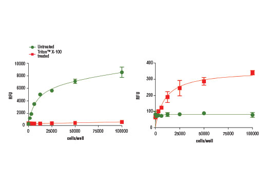
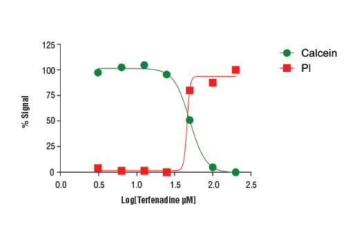
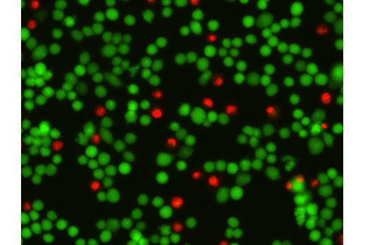
Measures of cell viability and cytotoxicity are broadly used to study the effects of growth factors and cytokines, inhibitors and activators, and immune response signals. Many viability and toxicity assay kits (such as XTT, resazurin, and BrdU cell kits) monitor the presence or viability of live cells by measuring the metabolic conversion or incorporation of a substrate into cellular molecules. Assay kits that measure dead or dying cells usually rely on the activity of apoptotic enzymes (i.e. caspase-3) or the intracellular release of an enzyme or molecule indicative of cell death (i.e. cytochrome c, LDH). The Cell Health Assay Kit is able to stain both live and dead cells simultaneously by using two fluorescent probes. After treating cells with a combination of Calcein-AM and Propidium Iodide, live cells will produce strong green fluorescence while dead cells produce strong red fluorescence (1,2). These fluorescent signals can be detected using multiple methods, including a plate reader or scanner, flow cytometer, or fluorescent microscope. This dual assay is specifically designed to assess viability and toxicity for eukaryotic cells (1,2), but may also be used to assay some bacteria or yeasts (3,4).

  1. Papadopoulos, N.G. et al. (1994) J Immunol Methods 177, 101-11.
  2. Decherchi, P. et al. (1997) J Neurosci Methods 71, 205-13.
  3. Cárdenas, W. et al. (2004) Fish Shellfish Immunol 17, 223-33.
  4. Hiraoka, Y. and Kimbara, K. (2002) Appl Environ Microbiol 68, 2031-5.

Species Reactivity

Species reactivity is determined by testing in at least one approved application (e.g., western blot).

Cross-Reactivity Key

H: human M: mouse R: rat Hm: hamster Mk: monkey Vir: virus Mi: mink C: chicken Dm: D. melanogaster X: Xenopus Z: zebrafish B: bovine Dg: dog Pg: pig Sc: S. cerevisiae Ce: C. elegans Hr: horse GP: Guinea Pig Rab: rabbit All: all species expected

Trademarks and Patents

Cell Signaling Technology is a trademark of Cell Signaling Technology, Inc.
All other trademarks are the property of their respective owners. Visit cellsignal.com/trademarks for more information.

使用に関する制限

法的な権限を与えられたCSTの担当者が署名した書面によって別途明示的に合意された場合を除き、 CST、その関連会社または代理店が提供する製品には以下の条件が適用されます。お客様が定める条件でここに定められた条件に含まれるものを超えるもの、 または、ここに定められた条件と異なるものは、法的な権限を与えられたCSTの担当者が別途書面にて受諾した場合を除き、拒絶され、 いかなる効力も効果も有しません。

研究専用 (For Research Use Only) またはこれに類似する表示がされた製品は、 いかなる目的についても FDA または外国もしくは国内のその他の規制機関により承認、認可または許可を受けていません。 お客様は製品を診断もしくは治療目的で使用してはならず、また、製品に表示された内容に違反する方法で使用してはなりません。 CST が販売または使用許諾する製品は、エンドユーザーであるお客様に対し、使途を研究および開発のみに限定して提供されるものです。 診断、予防もしくは治療目的で製品を使用することまたは製品を再販売 (単独であるか他の製品等の一部であるかを問いません) もしくはその他の商業的利用の目的で購入することについては、CST から別途許諾を得る必要があります。 お客様は以下の事項を遵守しなければなりません。(a) CST の製品 (単独であるか他の資材と一緒であるかを問いません) を販売、使用許諾、貸与、寄付もしくはその他の態様で第三者に譲渡したり使用させたりしてはなりません。また、商用の製品を製造するために CST の製品を使用してはなりません。(b) 複製、改変、リバースエンジニアリング、逆コンパイル、 分解または他の方法により製品の構造または技術を解明しようとしてはなりません。また、 CST の製品またはサービスと競合する製品またはサービスを開発する目的で CST の製品を使用してはなりません。(c) CST の製品の商標、商号、ロゴ、特許または著作権に関する通知または表示を除去したり改変したりしてはなりません。(d) CST の製品をCST 製品販売条件(CST’s Product Terms of Sale) および該当する書面のみに従って使用しなければなりません。(e) CST の製品に関連してお客様が使用する第三者の製品またはサービスに関する使用許諾条件、 サービス提供条件またはこれに類する合意事項を遵守しなければなりません。

Revision 1
#13837

Cell Health Assay Kit

Cell Health Assay Kit: Image 1 Expand Image
Figure 1. HeLa cells were seeded at 1,500 to 100,000 cells/well in a 96-well black plate with clear bottom and incubated overnight. Cells were exposed to Triton X-100 (0.05%, 10 min) and cell viability/toxicity was measured using the Cell Health Assay Kit #13837. Relative fluorescence for Calcein-AM (left) and Propidium Iodide (right) are shown.
Cell Health Assay Kit: Image 2 Expand Image
Figure 2. Treatment of HeLa cells (4 x10⁴ cells/well) with increasing concentrations of terfenadine (4 hr) results in reduced cell viability as detected by the Cell Health Assay Kit.
Cell Health Assay Kit: Image 3 Expand Image
Figure 4. Fluorescent analysis of HeLa cells (4x10⁴ cells/well) treated with terfenadine (12.5 μM, 4 hr) and stained with Calcein-AM (1 μM, 30 min; green) and Propidium Iodide (3 μM, 30 min; red).