#P00533
1956
Product Information
Specificity / Sensitivity
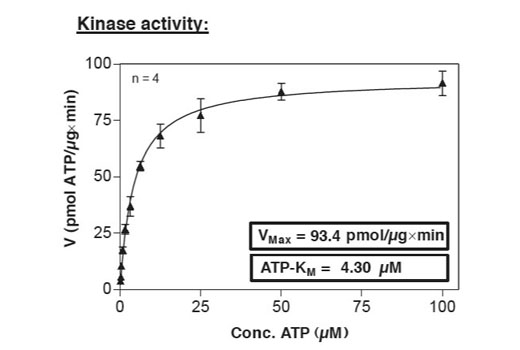
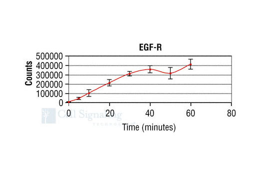
Purified EGF-R kinase was quality controlled for purity using silver stain SDS-PAGE and Western blot. EGF-R kinase Vmax and Km values were measured to determine specific enzymatic activity (fig.5).
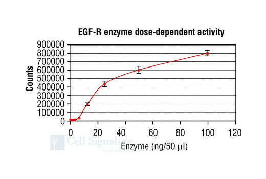
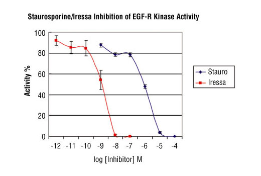
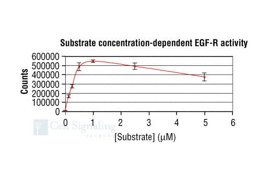
Assay conditions (time course [fig.1], kinase dose-dependence [fig.3], substrate dose-dependence [fig.4] and inhibitor sensitivity [fig.6]) for EGFR kinase activity were verified using the EGF-R substrate peptide provided in this kit.
Source / Purification
Source/Purification: The GST-Kinase fusion protein was produced using a baculovirus expression system from a construct containing a human EGF-R cDNA kinase domain fragment amino-terminally fused to a GST-HIS6-Thrombin cleavage site. The protein was then purified by one-step affinity purification using glutathione-agarose.
The detection antibody, Phospho-Tyrosine Monoclonal Antibody (P-Tyr-100) #9411, was corresponding to mice immunized with phospho-tyrosine-containing peptides .
Background
The epidermal growth factor (EGF) receptor is a transmembrane tyrosine kinase that belongs to the HER/ErbB protein family. Ligand binding results in receptor dimerization, autophosphorylation, activation of downstream signaling, internalization, and lysosomal degradation (1,2). Phosphorylation of EGF receptor (EGFR) at Tyr845 in the kinase domain is implicated in stabilizing the activation loop, maintaining the active state enzyme, and providing a binding surface for substrate proteins (3,4). c-Src is involved in phosphorylation of EGFR at Tyr845 (5). The SH2 domain of PLCγ binds at phospho-Tyr992, resulting in activation of PLCγ-mediated downstream signaling (6). Phosphorylation of EGFR at Tyr1045 creates a major docking site for the adaptor protein c-Cbl, leading to receptor ubiquitination and degradation following EGFR activation (7,8). The GRB2 adaptor protein binds activated EGFR at phospho-Tyr1068 (9). A pair of phosphorylated EGFR residues (Tyr1148 and Tyr1173) provide a docking site for the Shc scaffold protein, with both sites involved in MAP kinase signaling activation (2). Phosphorylation of EGFR at specific serine and threonine residues attenuates EGFR kinase activity. EGFR carboxy-terminal residues Ser1046 and Ser1047 are phosphorylated by CaM kinase II; mutation of either of these serines results in upregulated EGFR tyrosine autophosphorylation (10).
Description: The kit provides a means of performing enzymatic assays with active human EGF-R kinase. It includes active EGF-R kinase (supplied as a GST fusion protein), a biotinylated substrate peptide and a phospho-tyrosine monoclonal antibody for detection of the phosphorylated form of the substrate peptide.
Unit Definition: 10 Units is defined as the amount of EGF-R kinase required to maximally phosphorylate 75 pmol of PTP1B (Tyr66) #C03-1727 biotinylated substrate peptide in 30 minutes at 25ºC in a total reaction volume of 50 µl quantified by DELFIA®.
Peptide Core Sequence: NDY*IN
Molecular Weights: Peptide Substrate, Biotin-PTP1B (Tyr66): 2141 Daltons, GST-EGF-R Kinase domain: 91,163 Daltons
Storage: Antibodies are supplied in in 10 mM sodium HEPES (pH 7.5), 150 mM NaCl, 100 µg/ml BSA and 50% glycerol. Do not aliquot the antibodies. Peptides are supplied at 6 µM in 0.001% DMSO. Enzymes are supplied in 50 mM Tris-HCL (pH 8.0), 100 mM NaCl, 5 mM DTT, 15 mM reduced glutathion and 20% glycerol. Store at -80ºC.
Keep enzymes on ice during use.
Avoid repeated freeze-thaw cycles.
- Hackel, P.O. et al. (1999) Curr Opin Cell Biol 11, 184-9.
- Zwick, E. et al. (1999) Trends Pharmacol Sci 20, 408-12.
- Cooper, J.A. and Howell, B. (1993) Cell 73, 1051-4.
- Hubbard, S.R. et al. (1994) Nature 372, 746-54.
- Biscardi, J.S. et al. (1999) J Biol Chem 274, 8335-43.
- Emlet, D.R. et al. (1997) J Biol Chem 272, 4079-86.
- Levkowitz, G. et al. (1999) Mol Cell 4, 1029-40.
- Ettenberg, S.A. et al. (1999) Oncogene 18, 1855-66.
- Rojas, M. et al. (1996) J Biol Chem 271, 27456-61.
- Feinmesser, R.L. et al. (1999) J Biol Chem 274, 16168-73.
Species Reactivity
Species reactivity is determined by testing in at least one approved application (e.g., western blot).
Cross-Reactivity Key
H: human M: mouse R: rat Hm: hamster Mk: monkey Vir: virus Mi: mink C: chicken Dm: D. melanogaster X: Xenopus Z: zebrafish B: bovine Dg: dog Pg: pig Sc: S. cerevisiae Ce: C. elegans Hr: horse GP: Guinea Pig Rab: rabbit All: all species expected
Trademarks and Patents
使用に関する制限
法的な権限を与えられたCSTの担当者が署名した書面によって別途明示的に合意された場合を除き、 CST、その関連会社または代理店が提供する製品には以下の条件が適用されます。お客様が定める条件でここに定められた条件に含まれるものを超えるもの、 または、ここに定められた条件と異なるものは、法的な権限を与えられたCSTの担当者が別途書面にて受諾した場合を除き、拒絶され、 いかなる効力も効果も有しません。
研究専用 (For Research Use Only) またはこれに類似する表示がされた製品は、 いかなる目的についても FDA または外国もしくは国内のその他の規制機関により承認、認可または許可を受けていません。 お客様は製品を診断もしくは治療目的で使用してはならず、また、製品に表示された内容に違反する方法で使用してはなりません。 CST が販売または使用許諾する製品は、エンドユーザーであるお客様に対し、使途を研究および開発のみに限定して提供されるものです。 診断、予防もしくは治療目的で製品を使用することまたは製品を再販売 (単独であるか他の製品等の一部であるかを問いません) もしくはその他の商業的利用の目的で購入することについては、CST から別途許諾を得る必要があります。 お客様は以下の事項を遵守しなければなりません。(a) CST の製品 (単独であるか他の資材と一緒であるかを問いません) を販売、使用許諾、貸与、寄付もしくはその他の態様で第三者に譲渡したり使用させたりしてはなりません。また、商用の製品を製造するために CST の製品を使用してはなりません。(b) 複製、改変、リバースエンジニアリング、逆コンパイル、 分解または他の方法により製品の構造または技術を解明しようとしてはなりません。また、 CST の製品またはサービスと競合する製品またはサービスを開発する目的で CST の製品を使用してはなりません。(c) CST の製品の商標、商号、ロゴ、特許または著作権に関する通知または表示を除去したり改変したりしてはなりません。(d) CST の製品をCST 製品販売条件(CST’s Product Terms of Sale) および該当する書面のみに従って使用しなければなりません。(e) CST の製品に関連してお客様が使用する第三者の製品またはサービスに関する使用許諾条件、 サービス提供条件またはこれに類する合意事項を遵守しなければなりません。