| Product Includes | Product # | Quantity | Color | Storage Temp |
|---|---|---|---|---|
| MAP Kinase Multi-Target | 79155 | 96 tests |
|
+4C |
| TMB Substrate | 7004 | 11 ml |
|
+4C |
| STOP Solution | 7002 | 11 ml |
|
+4C |
| Sealing Tape | 54503 | 2 ea |
|
+4C |
| ELISA Wash Buffer (20X) | 9801 | 25 ml |
|
+4C |
| ELISA Sample Diluent | 11083 | 25 ml |
|
+4C |
| Cell Lysis Buffer (10X) | 9803 | 15 ml |
|
-20C |
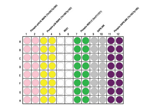
*The microwell plate is supplied as 12 8-well modules - Each module is designed to break apart for 8 tests.
Description
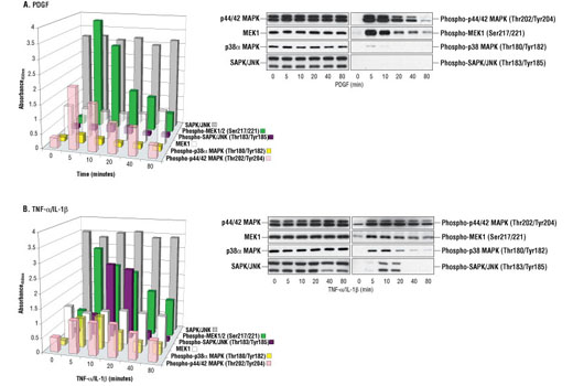
CST's PathScan® MAP Kinase Multi-Target Sandwich ELISA Kit is a solid phase sandwich enzyme-linked immunosorbent assay (ELISA) that combines the reagents necessary to detect endogenous levels of phospho-p44/42 MAPK (Thr202/Tyr204), phospho-p38 MAPK (Thr180/Tyr182), MEK1, phospho-MEK1 (Ser217/221), SAPK/JNK and phospho-SAPK/JNK (Thr183/Tyr185). These molecules represent convergence points and key regulatory proteins in signaling pathways controlling cellular events such as growth, differentiation and the response to stress and inflammation. Sixteen tests are provided for each target protein. Specific assay formulations for the indicated target proteins can be found in the datasheets associated with the individual PathScan® Sandwich ELISA Kits**. Briefly, a capture antibody has been coated onto the microwells. After incubation with cell lysates, the target protein is captured by the coated antibody. Following extensive washing, a detection antibody is added to detect the captured target protein. An HRP-linked secondary antibody is then used to recognize the bound detection antibody. HRP substrate, TMB, is added to develop color. The magnitude of absorbance for this developed color is proportional to the quantity of bound target protein. *Antibodies in kit are custom formulations specific to kit. **See companion products.
Specificity/Sensitivity
This kit detects proteins from the indicated species, as determined through in-house testing, but may also detect homologous proteins from other species.
Background
Both p44 and p42 MAP kinases (Erk1 and Erk2) function in a protein kinase cascade that plays a critical role in the regulation of cell growth and differentiation (1-6). MAP kinases are activated by a wide variety of extracellular signals including growth and neurotrophic factors, cytokines, hormones and neurotransmitters. Activation of MAP kinases occurs through phosphorylation of threonine and tyrosine (Thr202 and Tyrr204 of human MAP kinase or Thr183 and Tyr185 of rat MAP kinase) at the sequence T*EY* by a single upstream MAP kinase kinase (MEK) (7,8).
MEK1 and MEK2 are dual-specificity protein kinases that function in a mitogen activated protein kinase cascade controlling cell growth and differentiation. Activation of MEK1 and MEK2 occurs through phosphorylation of Ser217 and Ser221 by Raf-like molecules. MEK activates p44 and p42 MAP kinase (1,9,10).
p38 MAP kinase (MAPK) participates in a signaling cascade controlling the cellular response to pro-inflammatory cytokines and a variety of cellular stresses. MKK3, MKK6 and SEK (MKK4) activate p38 MAP kinase by phosphorylation at Thr180 and Tyr182 (11-14).
The stress-activated protein kinase/Jun-amino-terminal kinase SAPK/JNK is activated by a variety of environmental stresses, including UV and gamma radiation, ceramides, inflammatory cytokines and in some instances, by growth factors and GPCR agonists (15-20). As with the other MAPKs, the core-signaling unit is composed of a MAPKKK, typically MEKK1-4, or by a mixed lineage kinase (MLK), which phosphorylates and activates MKK4-7, which then phosphorylates Thr183 and Tyr185 to activate the SAPK/JNK kinase (16). Stress signals are delivered to this cascade by small GTPases of the Rho family (Rac, Rho, cdc42) (17). Both Rac1 and cdc42 mediate the stimulation of MEKKs and MLKs (17). Alternatively, MKK4-7 can be activated by a pathway independent of small GTPases via stimulation of a member of the germinal center kinase (GCK) family (18).
- McKay, M.M. and Morrison, D.K. (2007) Oncogene 26, 3113-21.
- Pearson, G. et al. (2001) Endocr Rev 22, 153-83.
- Marshall, C.J. (1995) Cell 80, 179-85.
- Hunter, T. (1995) Cell 80, 225-36.
- Hill, C.S. and Treisman, R. (1995) Cell 80, 199-211.
- Cowley, S. et al. (1994) Cell 77, 841-52.
- Sturgill, T.W. et al. (1988) Nature 334, 715-8.
- Payne, D.M. et al. (1991) EMBO J 10, 885-92.
- Alessi, D.R. et al. (1994) EMBO J 13, 1610-9.
- Pearson, G. et al. (2001) Endocr Rev 22, 153-83.
- Raingeaud, J. et al. (1995) J Biol Chem 270, 7420-6.
- Raman, M. et al. (2007) Oncogene 26, 3100-12.
- Zarubin, T. and Han, J. (2005) Cell Res 15, 11-8.
- Roux, P.P. and Blenis, J. (2004) Microbiol Mol Biol Rev 68, 320-44.
- Davis, R.J. (1999) Biochem Soc Symp 64, 1-12.
- Ichijo, H. (1999) Oncogene 18, 6087-93.
- Kyriakis, J.M. and Avruch, J. (2001) Physiol Rev 81, 807-69.
- Kyriakis, J.M. (1999) J Biol Chem 274, 5259-62.
- Leppä, S. and Bohmann, D. (1999) Oncogene 18, 6158-62.
- Whitmarsh, A.J. and Davis, R.J. (1998) Trends Biochem Sci 23, 481-5.
Background References
Cross-Reactivity Key
H: human M: mouse R: rat Hm: hamster Mk: monkey Vir: virus Mi: mink C: chicken Dm: D. melanogaster X: Xenopus Z: zebrafish B: bovine Dg: dog Pg: pig Sc: S. cerevisiae Ce: C. elegans Hr: horse GP: Guinea Pig Rab: rabbit All: all species expected
Trademarks and Patents
使用に関する制限
法的な権限を与えられたCSTの担当者が署名した書面によって別途明示的に合意された場合を除き、 CST、その関連会社または代理店が提供する製品には以下の条件が適用されます。お客様が定める条件でここに定められた条件に含まれるものを超えるもの、 または、ここに定められた条件と異なるものは、法的な権限を与えられたCSTの担当者が別途書面にて受諾した場合を除き、拒絶され、 いかなる効力も効果も有しません。
研究専用 (For Research Use Only) またはこれに類似する表示がされた製品は、 いかなる目的についても FDA または外国もしくは国内のその他の規制機関により承認、認可または許可を受けていません。 お客様は製品を診断もしくは治療目的で使用してはならず、また、製品に表示された内容に違反する方法で使用してはなりません。 CST が販売または使用許諾する製品は、エンドユーザーであるお客様に対し、使途を研究および開発のみに限定して提供されるものです。 診断、予防もしくは治療目的で製品を使用することまたは製品を再販売 (単独であるか他の製品等の一部であるかを問いません) もしくはその他の商業的利用の目的で購入することについては、CST から別途許諾を得る必要があります。 お客様は以下の事項を遵守しなければなりません。(a) CST の製品 (単独であるか他の資材と一緒であるかを問いません) を販売、使用許諾、貸与、寄付もしくはその他の態様で第三者に譲渡したり使用させたりしてはなりません。また、商用の製品を製造するために CST の製品を使用してはなりません。(b) 複製、改変、リバースエンジニアリング、逆コンパイル、 分解または他の方法により製品の構造または技術を解明しようとしてはなりません。また、 CST の製品またはサービスと競合する製品またはサービスを開発する目的で CST の製品を使用してはなりません。(c) CST の製品の商標、商号、ロゴ、特許または著作権に関する通知または表示を除去したり改変したりしてはなりません。(d) CST の製品をCST 製品販売条件(CST’s Product Terms of Sale) および該当する書面のみに従って使用しなければなりません。(e) CST の製品に関連してお客様が使用する第三者の製品またはサービスに関する使用許諾条件、 サービス提供条件またはこれに類する合意事項を遵守しなければなりません。