Revision 2
Cell Signaling Technology

Orders: 877-616-CELL (2355) [email protected]

Support: 877-678-TECH (8324)

Web: [email protected] cellsignal.com

3 Trask LaneDanversMassachusetts01923USA
For Research Use Only. Not for Use in Diagnostic Procedures.
Applications:

FC-FP

:

:

:

:

UniProt ID:

#P06332, #P10144, #P22646, #Q9BZS1, #P11942, #P01731

Entrez-Gene Id:

12504, 3002, 12501, 50943, 12502, 12525

Product Information

Product Usage Information

All antibodies in this kit are compatible with the FoxP3/Transcription Factor Fixation/Permeabilization Kit #43481 and can be used in a single staining mix on fixed and permeabilized cells. Prior to fixation and antibody incubation, we recommend adding a fixable viability dye such as the Ghost Dye Violet 510 Viability Dye to enable identification and exclusion of dead cells from analysis.

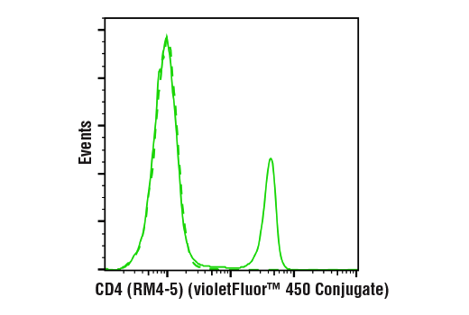
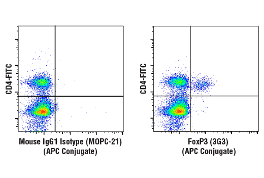
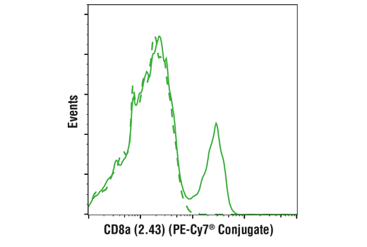
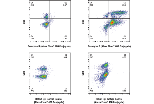
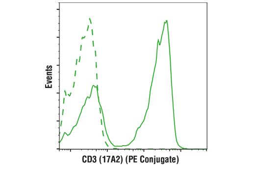
Gating strategy for identifying essential T cell subsets:
If a fixable viability dye was used, first gate on viable cells. Next, gate on lymphocytes based on forward scatter and side scatter. Conventional helper T cells are the CD3+CD4+ cells within the lymphocyte gate. Regulatory CD3+CD4+ T cells are positive for expression of FoxP3. Conventional cytotoxic T cells are the CD3+CD8+ cells within the lymphocyte gate. Both CD3+CD4+ and CD3+CD8+ cells can be positive for expression of the cytotoxic molecule Granzyme B.

Storage

CD3 (17A2) Rat mAb (PE Conjugate), CD4 (RM4-5) Rat mAb (violetFluor 450 Conjugate), FoxP3 (3G3) Mouse mAb (APC Conjugate), and CD8α (2.43) Rat mAb (PE-Cy7® Conjugate) are supplied in 10 mM NaH2PO4, 150 mM NaCl, 0.09% NaN3, 0.1% gelatin, pH7.2. Granzyme B (D2H2F) Rabbit mAb (Alexa Fluor® 488 Conjugate) is supplied in PBS (pH 7.2), less than 0.1% sodium azide and 2 mg/ml BSA. Store at 4oC. Do not aliquot the antibodies. Protect from light. Do not freeze.

All components in this kit are stable in accordance with the date printed on the outer packaging label when stored at the recommended temperature. Please refer to product labels, datasheets, or web pages for specific “Best By” dates for each individual component.

Specificity / Sensitivity

Each antibody in the Mouse Essential T Cell Markers Flow Cytometry Panel detects endogenous levels of its target protein. CD3 (17A2) Rat mAb (PE Conjugate), CD4 (RM4-5) Rat mAb (violetFluor 450 Conjugate), and CD8α (2.43) Rat mAb (PE-Cy7® Conjugate) detect epitopes within the extracellular domains. FoxP3 (3G3) Mouse mAb (APC Conjugate) and Granzyme B (D2H2F) Rabbit mAb (Alexa Fluor® 488 Conjugate) detect epitopes within the intracellular domains.

Species Reactivity:

Mouse

Source / Purification

Monoclonal antibodies were purified from tissue culture supernatant via affinity chromatography. The purified antibodies were conjugated under optimal conditions, with unreacted dye removed from the preparation.

Product Description

The Mouse Essential T Cell Markers Flow Cytometry Panel can be used to identify major subsets of mouse T cells.

T cells are identified by expression of CD3. There are two major subsets of conventional T cells: helper T cells which express CD4, and cytotoxic T cells which express CD8. Regulatory CD4+ T cells express FoxP3. Upon antigen stimulation, T cells upregulate expression of Granzyme B, a serine protease that is secreted by these cells to mediate target cell cytotoxicity.

Species Reactivity

Species reactivity is determined by testing in at least one approved application (e.g., western blot).

Applications Key

FC-FP: Flow Cytometry (Fixed/Permeabilized)

Cross-Reactivity Key

H: human M: mouse R: rat Hm: hamster Mk: monkey Vir: virus Mi: mink C: chicken Dm: D. melanogaster X: Xenopus Z: zebrafish B: bovine Dg: dog Pg: pig Sc: S. cerevisiae Ce: C. elegans Hr: horse GP: Guinea Pig Rab: rabbit All: all species expected

Trademarks and Patents

Cell Signaling Technology is a trademark of Cell Signaling Technology, Inc.
Cy and CyDye are registered trademarks of GE Healthcare.
This product is provided under an intellectual property license from Life Technologies Corporation. The transfer of this product is conditioned on the buyer using the purchased product solely in research conducted by the buyer, excluding contract research or any fee for service research, and the buyer must not (1) use this product or its components for (a) diagnostic, therapeutic or prophylactic purposes; (b) testing, analysis or screening services, or information in return for compensation on a per-test basis; or (c) manufacturing or quality assurance or quality control, and/or (2) sell or transfer this product or its components for resale, whether or not resold for use in research. For information on purchasing a license to this product for purposes other than as described above, contact Life Technologies Corporation, 5791 Van Allen Way, Carlsbad, CA 92008 USA or [email protected].
All other trademarks are the property of their respective owners. Visit cellsignal.com/trademarks for more information.

使用に関する制限

法的な権限を与えられたCSTの担当者が署名した書面によって別途明示的に合意された場合を除き、 CST、その関連会社または代理店が提供する製品には以下の条件が適用されます。お客様が定める条件でここに定められた条件に含まれるものを超えるもの、 または、ここに定められた条件と異なるものは、法的な権限を与えられたCSTの担当者が別途書面にて受諾した場合を除き、拒絶され、 いかなる効力も効果も有しません。

研究専用 (For Research Use Only) またはこれに類似する表示がされた製品は、 いかなる目的についても FDA または外国もしくは国内のその他の規制機関により承認、認可または許可を受けていません。 お客様は製品を診断もしくは治療目的で使用してはならず、また、製品に表示された内容に違反する方法で使用してはなりません。 CST が販売または使用許諾する製品は、エンドユーザーであるお客様に対し、使途を研究および開発のみに限定して提供されるものです。 診断、予防もしくは治療目的で製品を使用することまたは製品を再販売 (単独であるか他の製品等の一部であるかを問いません) もしくはその他の商業的利用の目的で購入することについては、CST から別途許諾を得る必要があります。 お客様は以下の事項を遵守しなければなりません。(a) CST の製品 (単独であるか他の資材と一緒であるかを問いません) を販売、使用許諾、貸与、寄付もしくはその他の態様で第三者に譲渡したり使用させたりしてはなりません。また、商用の製品を製造するために CST の製品を使用してはなりません。(b) 複製、改変、リバースエンジニアリング、逆コンパイル、 分解または他の方法により製品の構造または技術を解明しようとしてはなりません。また、 CST の製品またはサービスと競合する製品またはサービスを開発する目的で CST の製品を使用してはなりません。(c) CST の製品の商標、商号、ロゴ、特許または著作権に関する通知または表示を除去したり改変したりしてはなりません。(d) CST の製品をCST 製品販売条件(CST’s Product Terms of Sale) および該当する書面のみに従って使用しなければなりません。(e) CST の製品に関連してお客様が使用する第三者の製品またはサービスに関する使用許諾条件、 サービス提供条件またはこれに類する合意事項を遵守しなければなりません。

Revision 2
#47972

Mouse Essential T Cell Markers Flow Cytometry Panel

Flow Cytometry Image 1: Mouse Essential T Cell Markers Flow Cytometry Panel Expand Image
Flow cytometric analysis of live mouse splenocytes using CD4 (RM4-5) Rat mAb (violetFluor 450 Conjugate)
(solid line) compared to concentration-matched Rat Isotype Control (violetFluor 450 Conjugate) (dashed line).
Flow Cytometry Image 2: Mouse Essential T Cell Markers Flow Cytometry Panel Expand Image
Flow cytometric analysis of live mouse splenocytes using FoxP3 (3G3) Mouse mAb (APC Conjugate) (right) compared to a concentration-matched Mouse (MOPC-21) mAb IgG1 Isotype Control (APC Conjugate) #49083 (left), co-stained with CD4 (RM4-5) Rat mAb (FITC Conjugate) #96127.
Flow Cytometry Image 3: Mouse Essential T Cell Markers Flow Cytometry Panel Expand Image
Flow cytometric analysis of live mouse splenocytes using CD8α (2.43) Rat mAb (PE-Cy7® Conjugate) (solid line) compared to concentration-matched Rat (LTF-2) mAb IgG2b Isotype Control (PE-Cy7® Conjugate) #43426 (dashed line).
Flow Cytometry Image 4: Mouse Essential T Cell Markers Flow Cytometry Panel Expand Image
Flow cytometric analysis of human peripheral blood mononuclear cells, untreated (left column) or treated with anti-CD3 plus anti-CD28 (10 μg/ml, 72 hr; right column), using Granzyme B (D2H2F) Rabbit mAb (Alexa Fluor® 488 Conjugate) (top row) or concentration-matched Rabbit (DA1E) mAb IgG XP® Isotype Control (Alexa Fluor® 488 Conjugate) #2975 (bottom row), and co-stained with a CD8 Antibody.
Flow Cytometry Image 5: Mouse Essential T Cell Markers Flow Cytometry Panel Expand Image
Flow cytometric analysis of live mouse splenocytes using CD3 (17A2) Rat mAb (PE Conjugate) (solid line) compared to concentration-matched Rat (LTF-2) mAb IgG2b Isotype Control (PE Conjugate) #27426 (dashed line).