| Product Includes | Product # | Quantity | Mol. Wt | Isotype/Source |
|---|---|---|---|---|
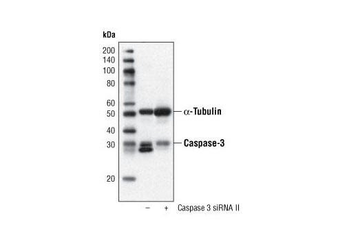
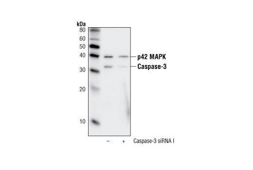
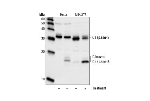
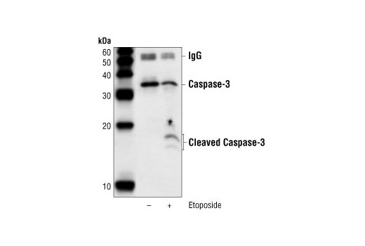
| Caspase-3 (8G10) Rabbit mAb | 9665 | 40 µl | 17, 19, 35 kDa | Rabbit IgG |
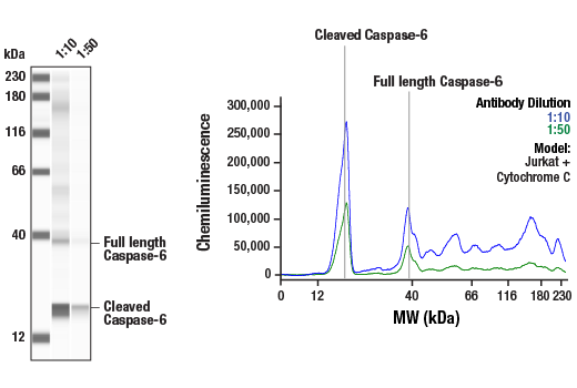
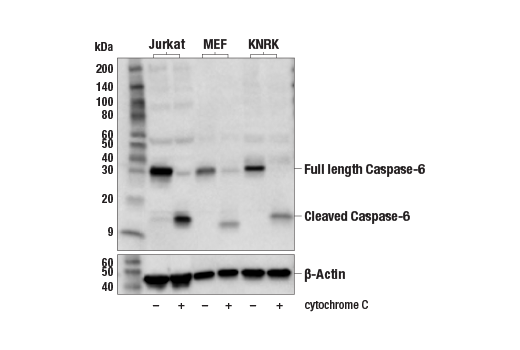
| Caspase-6 Antibody | 9762 | 40 µl | 15, 35 kDa | Rabbit |
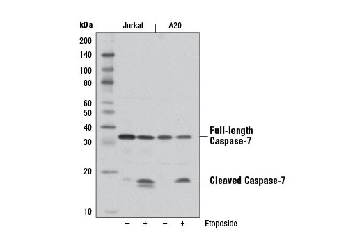
| Caspase-7 (D2Q3L) Rabbit mAb | 12827 | 40 µl | 20, 35 kDa | Rabbit IgG |
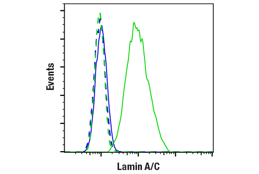
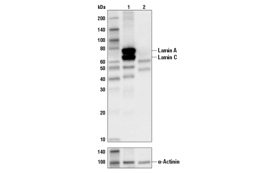
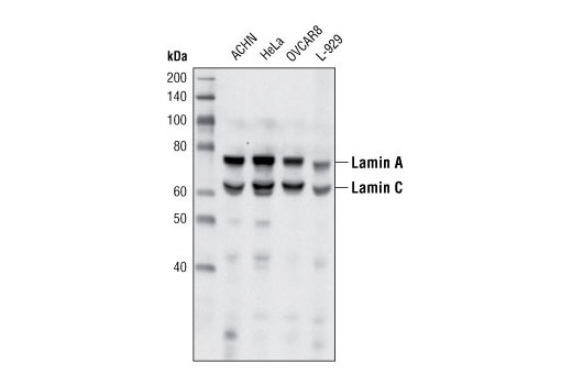
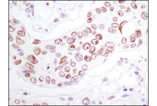
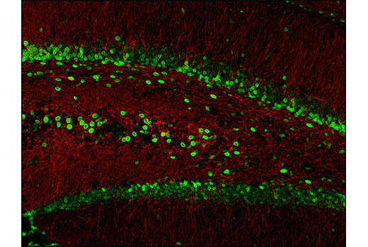
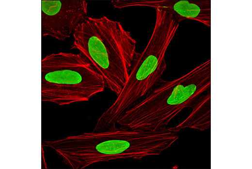
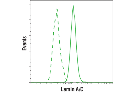
| Lamin A/C (4C11) Mouse mAb | 4777 | 40 µl | 74 (Lamin A), 63 (Lamin C) kDa | Mouse IgG2a |
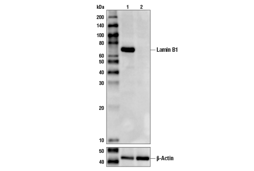
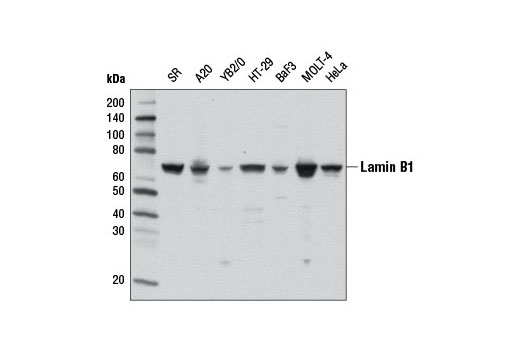
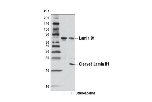
| Lamin B1 (D4Q4Z) Rabbit mAb | 12586 | 40 µl | 68, 25 kDa | Rabbit IgG |
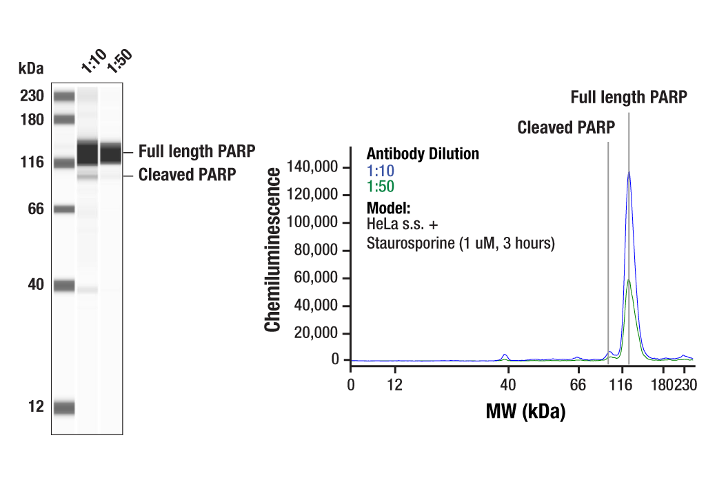
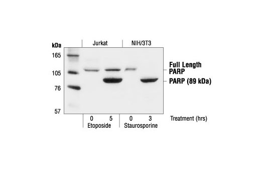
| PARP Antibody | 9542 | 40 µl | 89, 116 kDa | Rabbit |
| Anti-rabbit IgG, HRP-linked Antibody | 7074 | 100 µl | Goat | |
| Anti-mouse IgG, HRP-linked Antibody | 7076 | 100 µl | Horse |
Please visit cellsignal.com for individual component applications, species cross-reactivity, dilutions, protocols, and additional product information.
Description
The Effector Caspases and Substrates Antibody Sampler Kit provides an economical means to evaluate the activation of effector (executioner) caspases. The kit contains enough primary antibody to perform at least four western blots per primary antibody.
Storage
Background
Apoptosis is a regulated physiological process leading to cell death. Caspases, a family of cysteine acid proteases, are central regulators of apoptosis. Caspase-3 (CPP-32, Apoptain, Yama, SCA-1), Caspase-6 (Mch2), and Caspase-7 (CMH-1, Mch3, ICE-LAP3) are effector caspases functioning in cellular apoptotic processes (1-6). Upon apoptotic stimulation, initiator caspases such as caspase-9 (ICE-LAP6, Mch6) are cleaved and activated (7). The activated upstream caspases further process downstream executioner caspases by cleaving them into activated large and small subunits, thereby initiating a caspase cascade leading to apoptosis (4,6,8-10).
PARP, a 116 kDa nuclear poly (ADP-ribose) polymerase, appears to be involved in DNA repair in response to environmental stress (11). This protein can be cleaved by many ICE-like caspases in vitro (1,12) and is one of the main cleavage targets of caspase-3 in vivo (10,13). In human PARP, cleavage occurs between Asp214 and Gly215, which separates the PARP amino-terminal DNA binding domain (24 kDa) from the carboxy-terminal catalytic domain (89 kDa) (10,12). PARP helps cells to maintain their viability; cleavage of PARP facilitates cellular disassembly and serves as a marker of cells undergoing apoptosis (14).
Lamins are nuclear membrane structural components that are important in maintaining normal cell functions, such as cell cycle control, DNA replication, and chromatin organization (15-17). Lamins have been subdivided into types A and B. Type-A lamins consist of lamin A and C, which arise from alternative splicing of the lamin A gene LMNA. Lamin A and C are cleaved by caspases into large (41-50 kDa) and small (28 kDa) fragments, which can be used as markers for apoptosis (18,19). Type-B lamins consist of lamin B1 and B2, encoded by separate genes (20-22). Lamin B1 is also cleaved by caspases during apoptosis (23).
- Cohen, G.M. (1997) Biochem J 326 ( Pt 1), 1-16.
- Fernandes-Alnemri, T. et al. (1994) J Biol Chem 269, 30761-4.
- Faleiro, L. et al. (1997) EMBO J 16, 2271-81.
- Fernandes-Alnemri, T. et al. (1995) Cancer Res 55, 6045-52.
- Duan, H. et al. (1996) J Biol Chem 271, 1621-5.
- Lippke, J.A. et al. (1996) J Biol Chem 271, 1825-8.
- Li, P. et al. (1997) Cell 91, 479-89.
- Slee, E.A. et al. (1999) J Cell Biol 144, 281-92.
- MacFarlane, M. et al. (1997) J Cell Biol 137, 469-79.
- Nicholson, D.W. et al. (1995) Nature 376, 37-43.
- Satoh, M.S. and Lindahl, T. (1992) Nature 356, 356-8.
- Lazebnik, Y.A. et al. (1994) Nature 371, 346-7.
- Tewari, M. et al. (1995) Cell 81, 801-9.
- Oliver, F.J. et al. (1998) J Biol Chem 273, 33533-9.
- Dunbar, J.C. and Lu, H. (2000) Brain Res Bull 52, 123-6.
- Goldberg, M. et al. (1999) Crit Rev Eukaryot Gene Expr 9, 285-93.
- Yabuki, M. et al. (1999) Physiol Chem Phys Med NMR 31, 77-84.
- Rao, L. et al. (1996) J Cell Biol 135, 1441-55.
- Orth, K. et al. (1996) J Biol Chem 271, 16443-6.
- Biamonti, G. et al. (1992) Mol Cell Biol 12, 3499-506.
- Lin, F. and Worman, H.J. (1995) Genomics 27, 230-6.
- Pollard, K.M. et al. (1990) Mol Cell Biol 10, 2164-75.
- Chandler, J.M. et al. (1997) Biochem J 322 ( Pt 1), 19-23.
Background References
Trademarks and Patents
使用に関する制限
法的な権限を与えられたCSTの担当者が署名した書面によって別途明示的に合意された場合を除き、 CST、その関連会社または代理店が提供する製品には以下の条件が適用されます。お客様が定める条件でここに定められた条件に含まれるものを超えるもの、 または、ここに定められた条件と異なるものは、法的な権限を与えられたCSTの担当者が別途書面にて受諾した場合を除き、拒絶され、 いかなる効力も効果も有しません。
研究専用 (For Research Use Only) またはこれに類似する表示がされた製品は、 いかなる目的についても FDA または外国もしくは国内のその他の規制機関により承認、認可または許可を受けていません。 お客様は製品を診断もしくは治療目的で使用してはならず、また、製品に表示された内容に違反する方法で使用してはなりません。 CST が販売または使用許諾する製品は、エンドユーザーであるお客様に対し、使途を研究および開発のみに限定して提供されるものです。 診断、予防もしくは治療目的で製品を使用することまたは製品を再販売 (単独であるか他の製品等の一部であるかを問いません) もしくはその他の商業的利用の目的で購入することについては、CST から別途許諾を得る必要があります。 お客様は以下の事項を遵守しなければなりません。(a) CST の製品 (単独であるか他の資材と一緒であるかを問いません) を販売、使用許諾、貸与、寄付もしくはその他の態様で第三者に譲渡したり使用させたりしてはなりません。また、商用の製品を製造するために CST の製品を使用してはなりません。(b) 複製、改変、リバースエンジニアリング、逆コンパイル、 分解または他の方法により製品の構造または技術を解明しようとしてはなりません。また、 CST の製品またはサービスと競合する製品またはサービスを開発する目的で CST の製品を使用してはなりません。(c) CST の製品の商標、商号、ロゴ、特許または著作権に関する通知または表示を除去したり改変したりしてはなりません。(d) CST の製品をCST 製品販売条件(CST’s Product Terms of Sale) および該当する書面のみに従って使用しなければなりません。(e) CST の製品に関連してお客様が使用する第三者の製品またはサービスに関する使用許諾条件、 サービス提供条件またはこれに類する合意事項を遵守しなければなりません。