| Product Includes | Product # | Quantity | Mol. Wt | Isotype/Source |
|---|---|---|---|---|
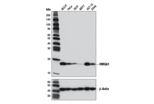
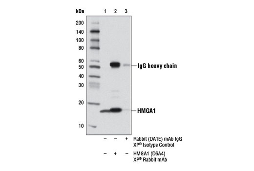
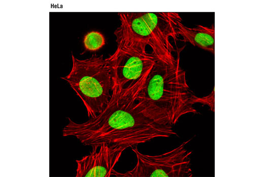
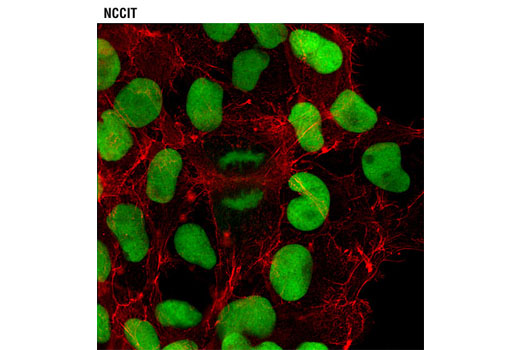
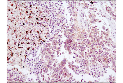
| HMGA1 (D6A4) XP® Rabbit mAb | 7777 | 40 µl | 18 kDa | Rabbit IgG |
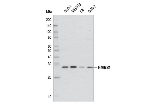
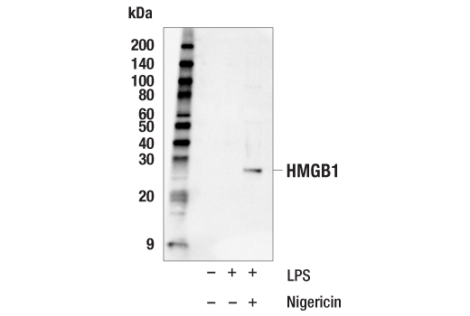
| HMGB1 (D3E5) Rabbit mAb | 6893 | 40 µl | 29 kDa | Rabbit IgG |
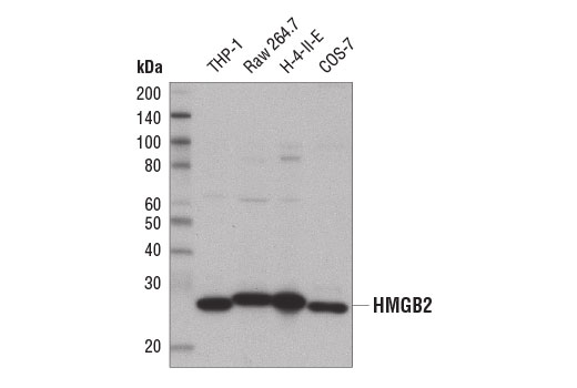
| HMGB2 (D1P9V) Rabbit mAb | 14163 | 40 µl | 28 kDa | Rabbit IgG |
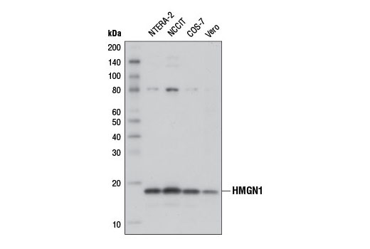
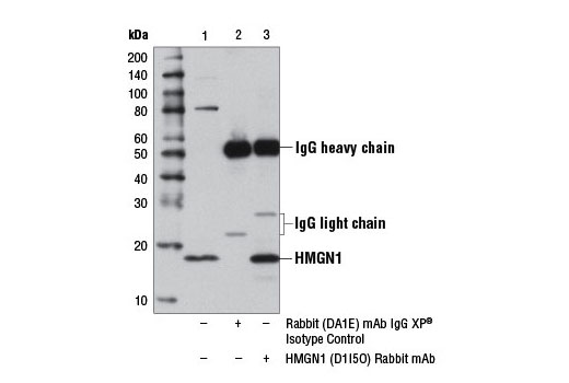
| HMGN1 (D1I5O) Rabbit mAb | 12734 | 40 µl | 18 kDa | Rabbit IgG |
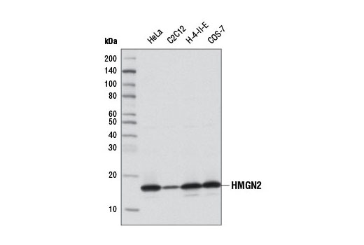
| HMGN2 (D9B9) XP® Rabbit mAb | 9437 | 40 µl | 17 kDa | Rabbit IgG |
| Anti-rabbit IgG, HRP-linked Antibody | 7074 | 100 µl | Goat |
Please visit cellsignal.com for individual component applications, species cross-reactivity, dilutions, protocols, and additional product information.
Description
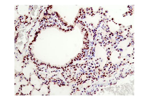
The High Mobility Group (HMG) Proteins Antibody Sampler Kit provides an economical means of detecting total protein from the HMG family members including HMGA1, HMGB1, HMGB2, HMGN1 and HMGN2. The kit contains enough primary antibody to perform four western blots per primary antibody.
Storage
Background
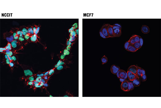
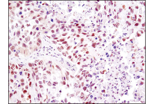
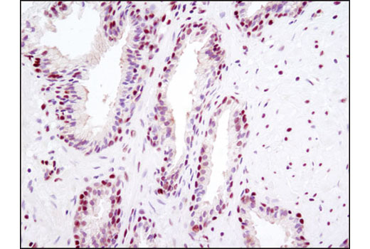
High mobility group (HMG) proteins are a superfamily of abundant and ubiquitous nuclear proteins that bind DNA without sequence specificity and induce structural changes to the chromatin fiber to regulate access to the underlying DNA (1). HMGA1, formerly known as HMG-I/Y, belongs to a family of high mobility group proteins known as HMGA. HMGA proteins are considered architectural transcription factors; they do not have direct transcriptional activation capacity, but instead regulate gene expression by changing DNA conformation through binding to AT-rich regions in the DNA and/or direct interaction with other transcription factors (2). HMGA1 is highly expressed during embryogenesis and in embryonic stem cells, but not in fully differentiated adult tissues (3,4). High mobility group protein B1 (HMGB1) and high mobility group protein B2 (HMGB2) belong to a family of highly conserved proteins that contain HMG box domains (5). HMGB1 is a widely expressed and highly abundant protein (6). HMGB2 is widely expressed during embryonic development, but it is restricted to lymphoid organs and testis in adult animals (7). While expression varies, the biochemical properties of the different family members may be indistinguishable. HMGB proteins are recruited by and help facilitate the assembly of site-specific DNA binding proteins to their cognate binding sites in chromatin. For example, HMGB1 and HMGB2 facilitate the binding of Hox proteins, Oct proteins, p53, Rel proteins, and steroid hormone receptor proteins to their target gene promoters (5,6). In addition to their functions in the nucleus, HMGB proteins play a significant role in extracellular signaling associated with inflammation. HMGB1 is massively released into the extracellular environment during cell necrosis, but not apoptosis. Extracellular HMGB1 "alarms" the innate immune system by acting as a chemoattractant for inflammatory cells triggering activation of T cells and dendritic cells. In addition, activated monocytes, macrophages, and dendritic cells also secrete HMGB1 (6). HMGB2 is secreted by myeloid cells and promotes proliferation and migration of endothelial cells by binding to the receptor for advanced glycation end products (RAGE) (8). The HMGN family of proteins, which includes five members (HMGN1-5) (1) function in transcriptional regulation and are recruited to gene promoters by transcription factors, such as estrogen receptor α (ERα), serum responsive factor (SRF), and PITX2, where they can facilitate either gene activation or repression (9-11). The expression of HMGN1 (also known as HMG14) and HMGN2 (also known as HMG17) is tightly linked to cellular differentiation. HMGN1 and HMNG2 are ubiquitous and highly expressed in all embryonic tissues. During mouse embryogenesis, expression is down-regulated throughout the embryo, except in committed but continuously renewing cell types undergoing active differentiation, such as the basal layer of the epithelium and kidney cells undergoing mesenchyme to epithelium transition (12,13).
- Hock, R. et al. (2007) Trends Cell Biol 17, 72-9.
- Cleynen, I. and Van de Ven, W.J. (2008) Int J Oncol 32, 289-305.
- Chiappetta, G. et al. (1996) Oncogene 13, 2439-46.
- Ben-Porath, I. et al. (2008) Nat Genet 40, 499-507.
- Thomas, J.O. and Travers, A.A. (2001) Trends Biochem Sci 26, 167-74.
- Müller, S. et al. (2004) J Intern Med 255, 332-43.
- Ronfani, L. et al. (2001) Development 128, 1265-73.
- Pusterla, T. et al. (2009) Autoimmunity 42, 308-10.
- Zhu, N. and Hansen, U. (2007) Mol Cell Biol 27, 8859-73.
- Amen, M. et al. (2008) Nucleic Acids Res 36, 462-76.
- Belova, G.I. et al. (2008) J Biol Chem 283, 8080-8.
- Furusawa, T. et al. (2006) Mol Cell Biol 26, 592-604.
- Lehtonen, S. and Lehtonen, E. (2001) Differentiation 67, 154-63.
Background References
Trademarks and Patents
使用に関する制限
法的な権限を与えられたCSTの担当者が署名した書面によって別途明示的に合意された場合を除き、 CST、その関連会社または代理店が提供する製品には以下の条件が適用されます。お客様が定める条件でここに定められた条件に含まれるものを超えるもの、 または、ここに定められた条件と異なるものは、法的な権限を与えられたCSTの担当者が別途書面にて受諾した場合を除き、拒絶され、 いかなる効力も効果も有しません。
研究専用 (For Research Use Only) またはこれに類似する表示がされた製品は、 いかなる目的についても FDA または外国もしくは国内のその他の規制機関により承認、認可または許可を受けていません。 お客様は製品を診断もしくは治療目的で使用してはならず、また、製品に表示された内容に違反する方法で使用してはなりません。 CST が販売または使用許諾する製品は、エンドユーザーであるお客様に対し、使途を研究および開発のみに限定して提供されるものです。 診断、予防もしくは治療目的で製品を使用することまたは製品を再販売 (単独であるか他の製品等の一部であるかを問いません) もしくはその他の商業的利用の目的で購入することについては、CST から別途許諾を得る必要があります。 お客様は以下の事項を遵守しなければなりません。(a) CST の製品 (単独であるか他の資材と一緒であるかを問いません) を販売、使用許諾、貸与、寄付もしくはその他の態様で第三者に譲渡したり使用させたりしてはなりません。また、商用の製品を製造するために CST の製品を使用してはなりません。(b) 複製、改変、リバースエンジニアリング、逆コンパイル、 分解または他の方法により製品の構造または技術を解明しようとしてはなりません。また、 CST の製品またはサービスと競合する製品またはサービスを開発する目的で CST の製品を使用してはなりません。(c) CST の製品の商標、商号、ロゴ、特許または著作権に関する通知または表示を除去したり改変したりしてはなりません。(d) CST の製品をCST 製品販売条件(CST’s Product Terms of Sale) および該当する書面のみに従って使用しなければなりません。(e) CST の製品に関連してお客様が使用する第三者の製品またはサービスに関する使用許諾条件、 サービス提供条件またはこれに類する合意事項を遵守しなければなりません。