| Product Includes | Product # | Quantity | Mol. Wt | Isotype/Source |
|---|---|---|---|---|
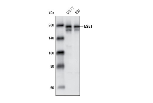
| ESET (C1C12) Rabbit mAb | 2196 | 40 µl | 180 kDa | Rabbit IgG |
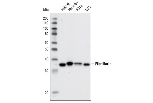
| Fibrillarin (C13C3) Rabbit mAb | 2639 | 40 µl | 37 kDa | Rabbit IgG |
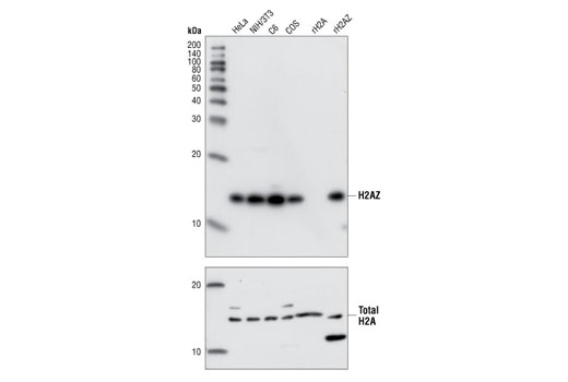
| Histone H2A.Z Antibody | 2718 | 40 µl | 14 kDa | Rabbit |
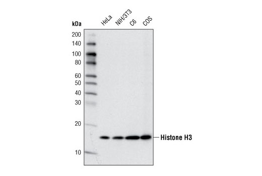
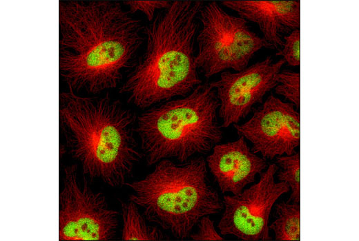
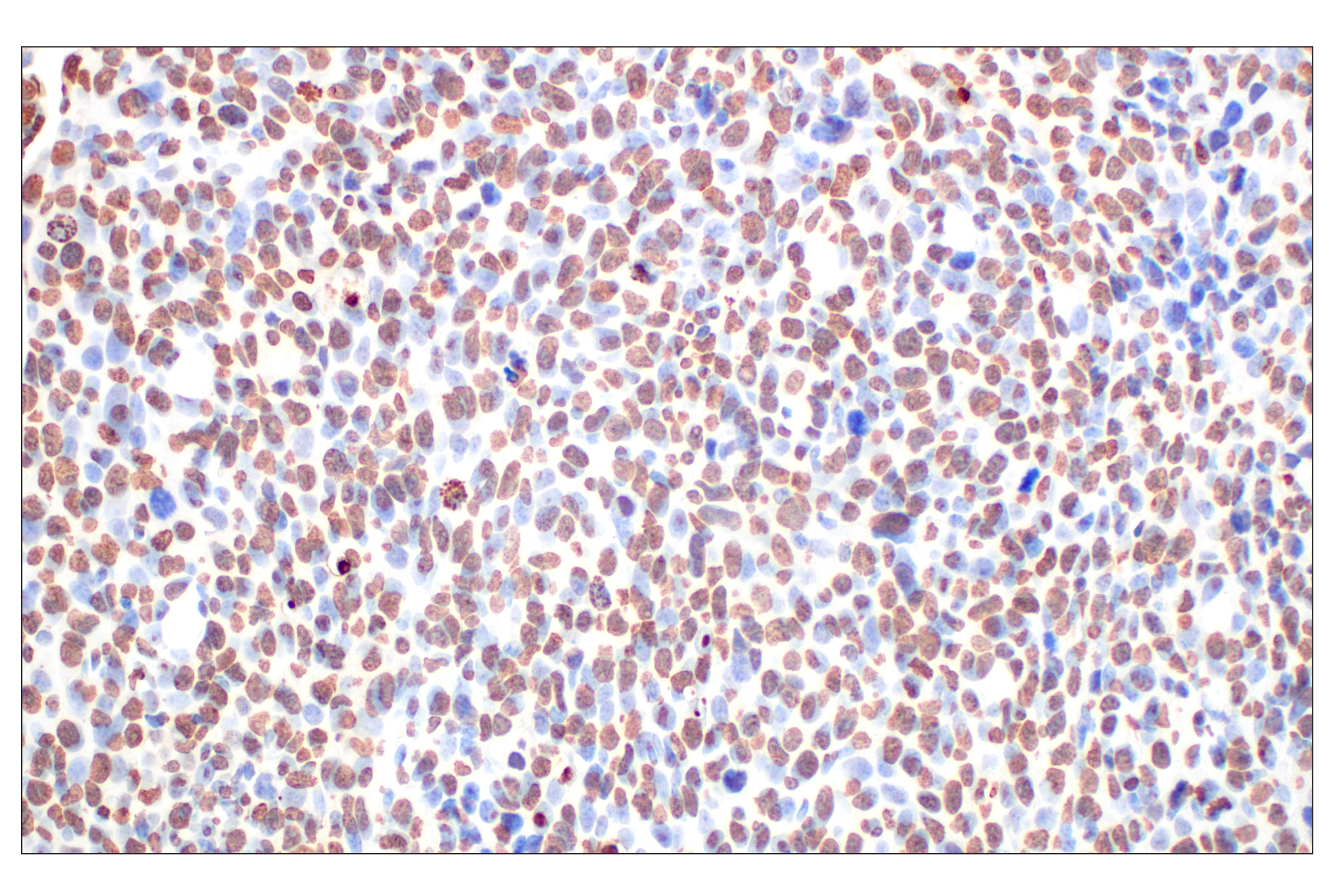
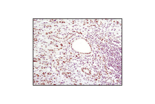
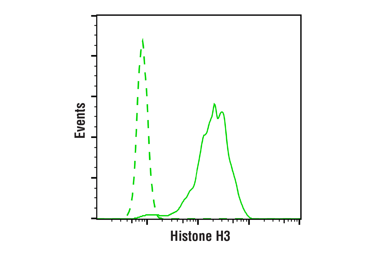
| Histone H3 (D1H2) XP® Rabbit mAb | 4499 | 40 µl | 17 kDa | Rabbit IgG |
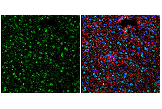
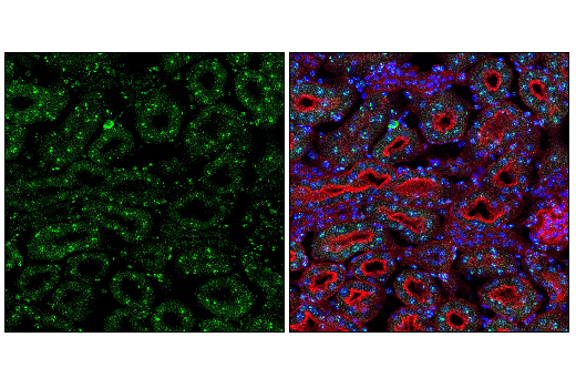
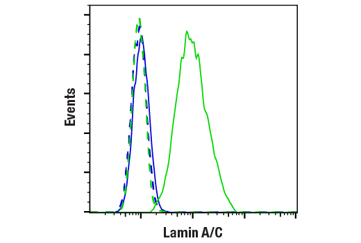
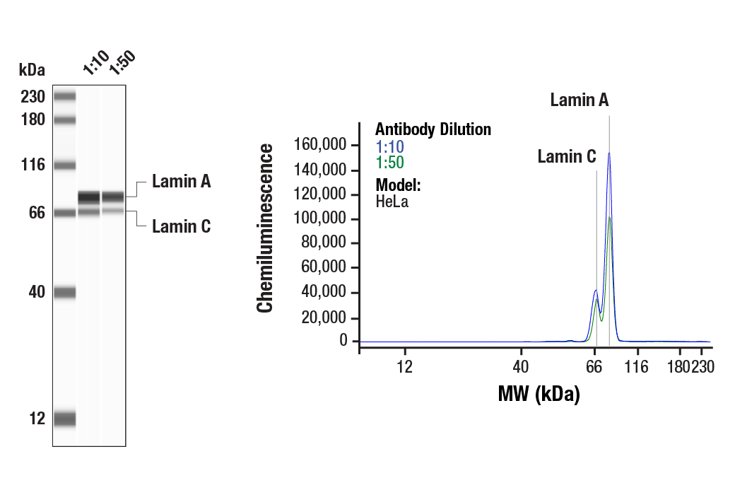
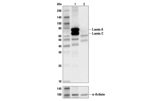
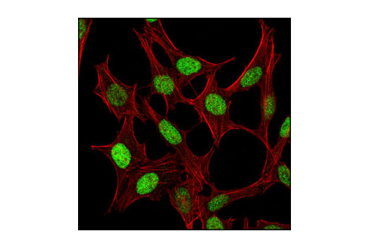
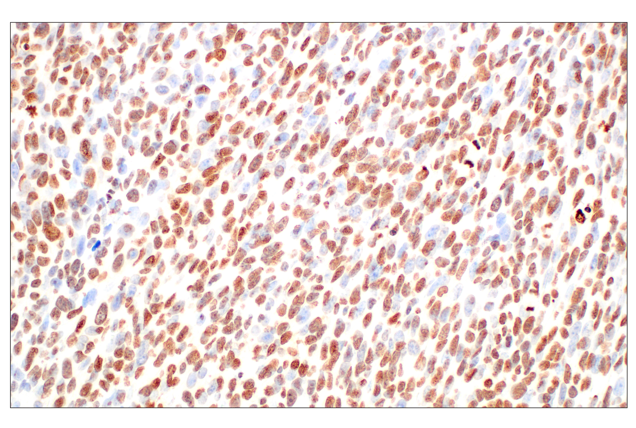
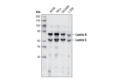
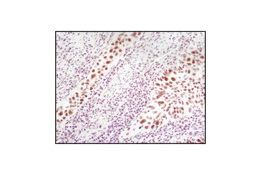
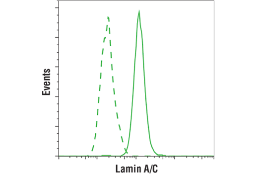
| Lamin A/C (4C11) Mouse mAb | 4777 | 40 µl | 74 (Lamin A), 63 (Lamin C) kDa | Mouse IgG2a |
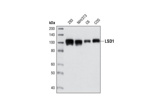
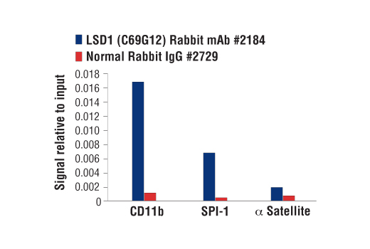
| LSD1 (C69G12) Rabbit mAb | 2184 | 40 µl | 110 kDa | Rabbit IgG |
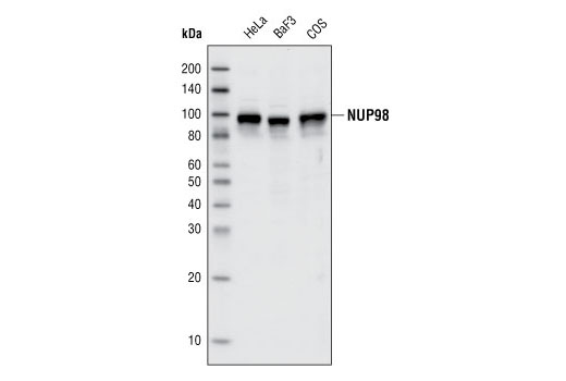
| NUP98 (C39A3) Rabbit mAb | 2598 | 40 µl | 98 kDa | Rabbit IgG |
| Anti-rabbit IgG, HRP-linked Antibody | 7074 | 100 µl | Goat | |
| Anti-mouse IgG, HRP-linked Antibody | 7076 | 100 µl | Horse |
Please visit cellsignal.com for individual component applications, species cross-reactivity, dilutions, protocols, and additional product information.
Description
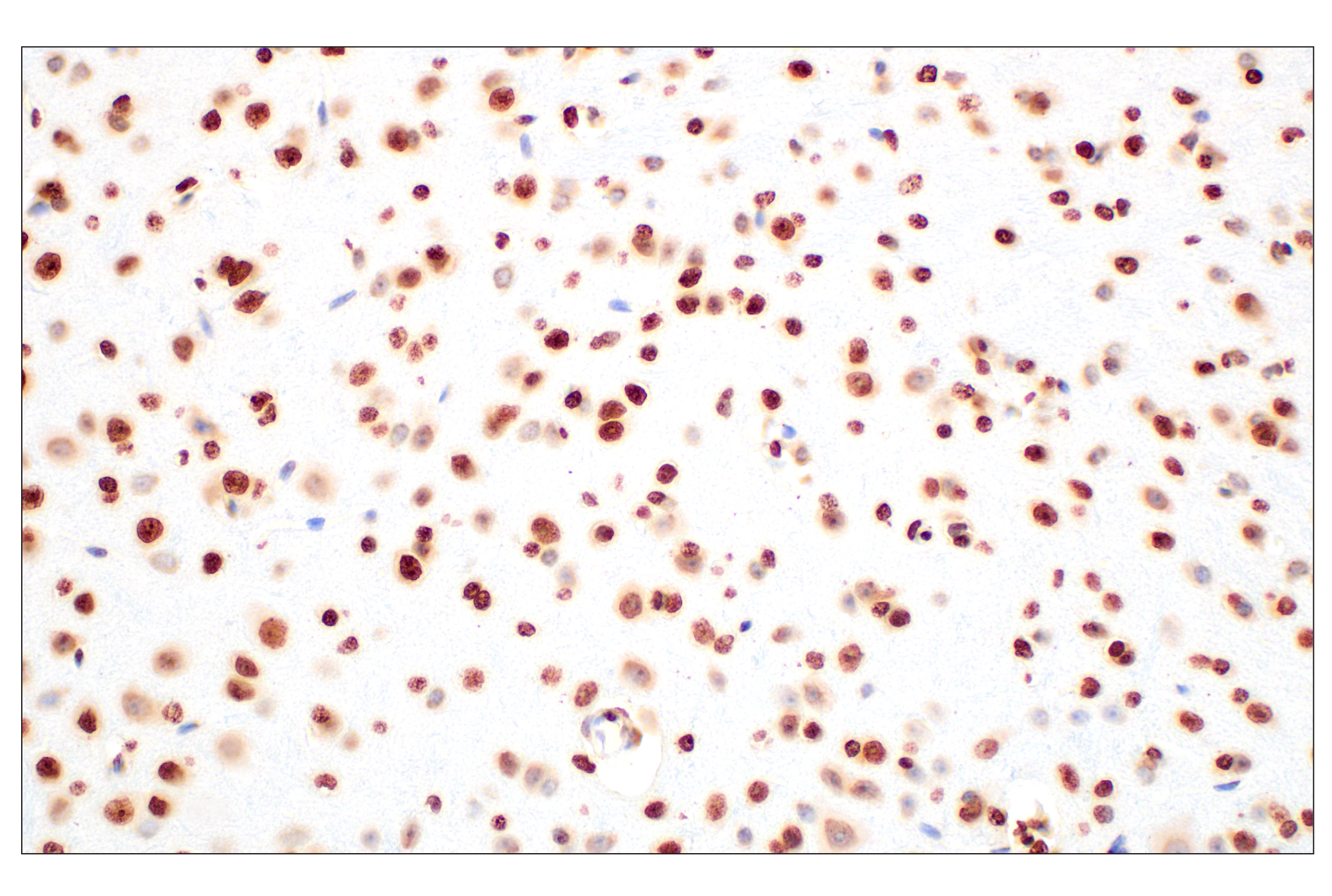
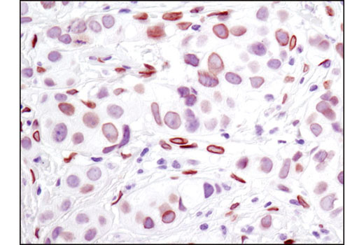
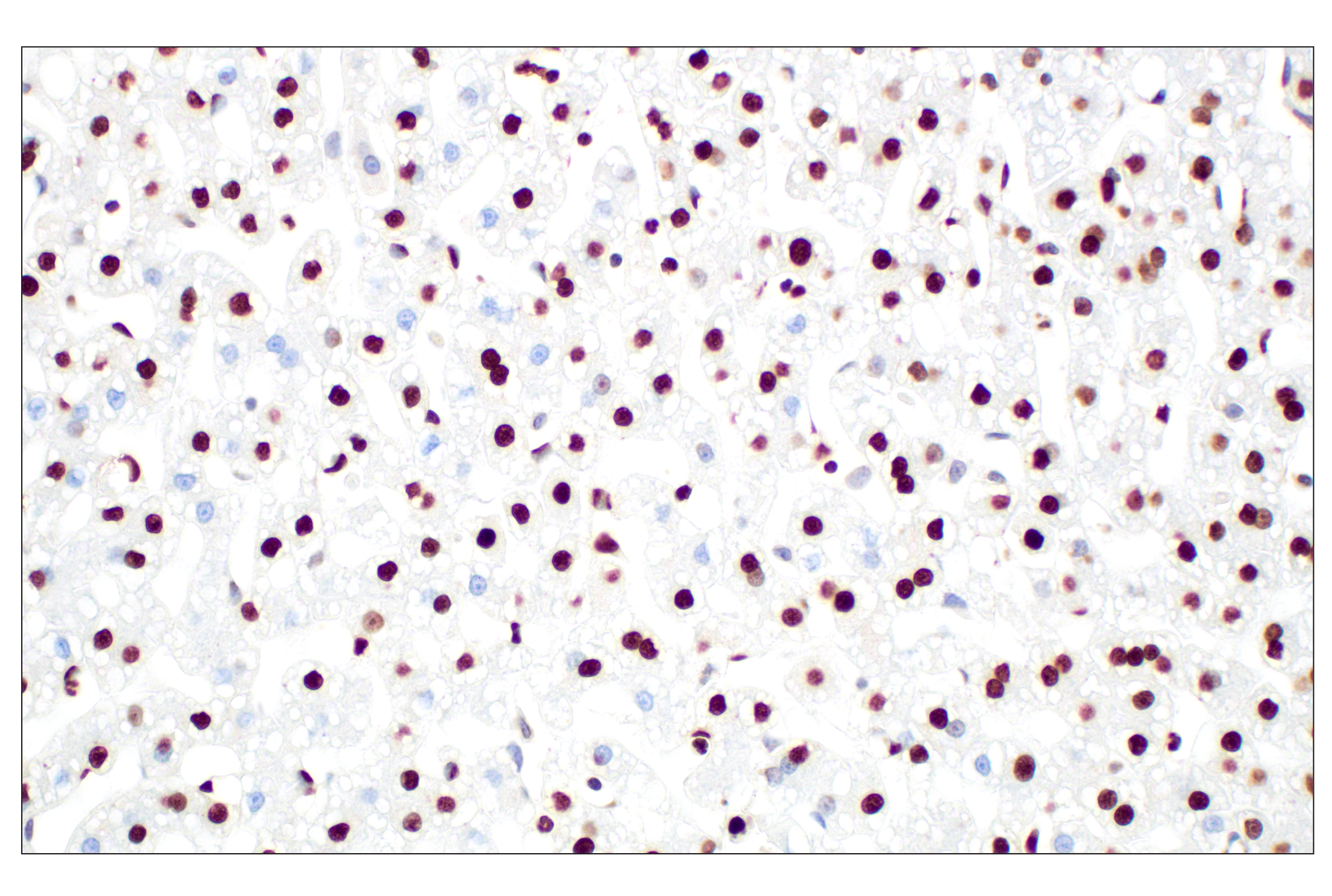
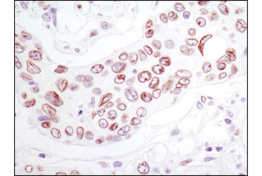
The Nucleus and Nuclear Envelope-Associated Marker Proteins Antibody Sampler Kit provides an economical means to evaluate relevant nuclear proteins. This kit contains enough primary antibody to perform at least four western blots per primary antibody.
Storage
Background
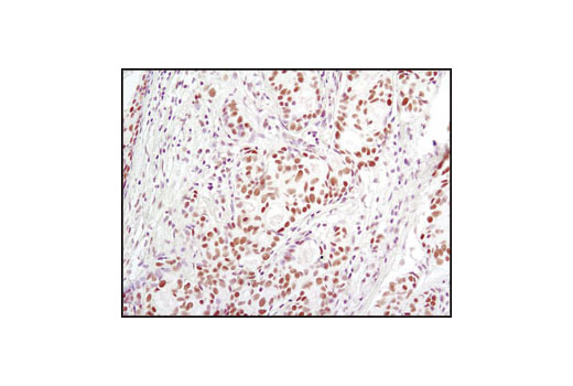
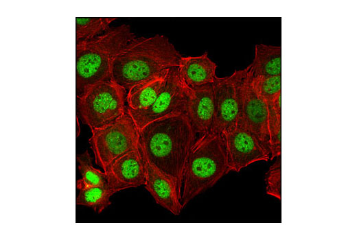
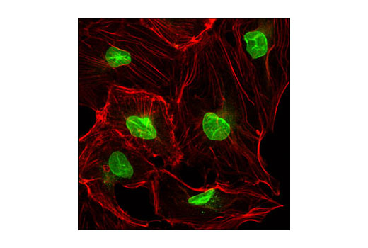
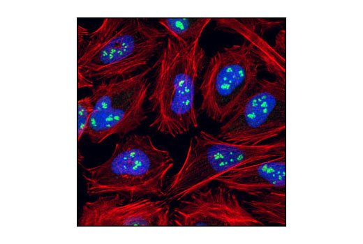
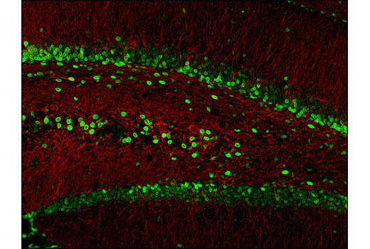
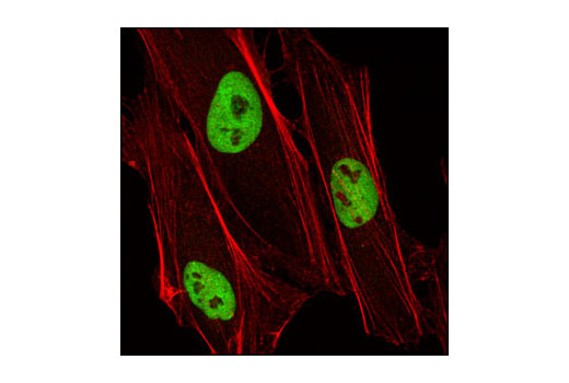
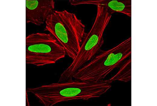
The Nucleus and Nuclear Envelope-Associated Marker Proteins Antibody Sampler Kit contains a variety of antibodies directed against established nuclear proteins (1). Histone H3 and histone H2A.Z are histone family members and components of nucleosomes, the primary building block of chromatin made up of DNA wound around eight core histone proteins. The amino-terminal tails of core histones undergo various post-translational modifications and have a direct effect on the accessibility of chromatin to transcription factors and, therefore, gene expression (2). ESET histone methyltransferase (3) and LSD1 histone demethylase (4) are both regulators of histone methylation and are chromatin-associated. Both NUP98 (5) and lamins (6) are located within the nuclear envelope (also known as the nuclear membrane). NUP98 is a component of the nuclear pore complex. Lamin A and lamin C are fibrous proteins contributing to nuclear structural and transcriptional regulation. Finally, fibrillarin (7) is located in fibrillar regions and Cajal bodies of nucleoli, where it functions to regulate RNA transcription and pre-rRNA processing.
- Workman, J.L. and Kingston, R.E. (1998) Annu Rev Biochem 67, 545-79.
- Jin, J. et al. (2005) Trends Biochem Sci 30, 680-7.
- Yang, L. et al. (2002) Oncogene 21, 148-52.
- Shi, Y. et al. (2004) Cell 119, 941-53.
- Fontoura, B.M. et al. (1999) J Cell Biol 144, 1097-112.
- Gruenbaum, Y. et al. (2000) J Struct Biol 129, 313-23.
- Tollervey, D. et al. (1993) Cell 72, 443-57.
Background References
Trademarks and Patents
使用に関する制限
法的な権限を与えられたCSTの担当者が署名した書面によって別途明示的に合意された場合を除き、 CST、その関連会社または代理店が提供する製品には以下の条件が適用されます。お客様が定める条件でここに定められた条件に含まれるものを超えるもの、 または、ここに定められた条件と異なるものは、法的な権限を与えられたCSTの担当者が別途書面にて受諾した場合を除き、拒絶され、 いかなる効力も効果も有しません。
研究専用 (For Research Use Only) またはこれに類似する表示がされた製品は、 いかなる目的についても FDA または外国もしくは国内のその他の規制機関により承認、認可または許可を受けていません。 お客様は製品を診断もしくは治療目的で使用してはならず、また、製品に表示された内容に違反する方法で使用してはなりません。 CST が販売または使用許諾する製品は、エンドユーザーであるお客様に対し、使途を研究および開発のみに限定して提供されるものです。 診断、予防もしくは治療目的で製品を使用することまたは製品を再販売 (単独であるか他の製品等の一部であるかを問いません) もしくはその他の商業的利用の目的で購入することについては、CST から別途許諾を得る必要があります。 お客様は以下の事項を遵守しなければなりません。(a) CST の製品 (単独であるか他の資材と一緒であるかを問いません) を販売、使用許諾、貸与、寄付もしくはその他の態様で第三者に譲渡したり使用させたりしてはなりません。また、商用の製品を製造するために CST の製品を使用してはなりません。(b) 複製、改変、リバースエンジニアリング、逆コンパイル、 分解または他の方法により製品の構造または技術を解明しようとしてはなりません。また、 CST の製品またはサービスと競合する製品またはサービスを開発する目的で CST の製品を使用してはなりません。(c) CST の製品の商標、商号、ロゴ、特許または著作権に関する通知または表示を除去したり改変したりしてはなりません。(d) CST の製品をCST 製品販売条件(CST’s Product Terms of Sale) および該当する書面のみに従って使用しなければなりません。(e) CST の製品に関連してお客様が使用する第三者の製品またはサービスに関する使用許諾条件、 サービス提供条件またはこれに類する合意事項を遵守しなければなりません。