| Product Includes | Product # | Quantity | Mol. Wt | Isotype/Source |
|---|---|---|---|---|
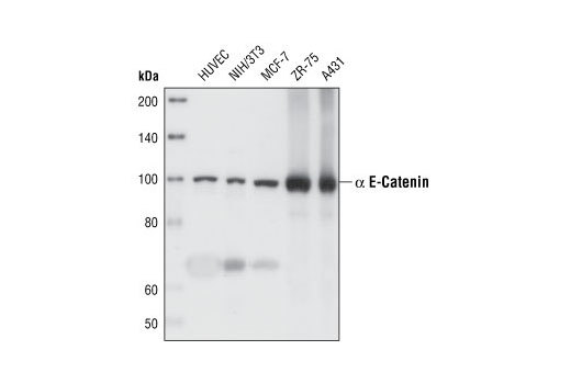
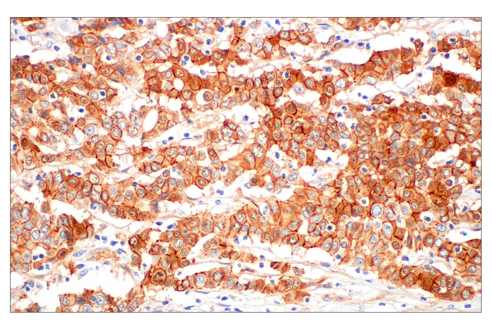
| α-E-Catenin (23B2) Rabbit mAb | 3240 | 40 µl | 100 kDa | Rabbit IgG |
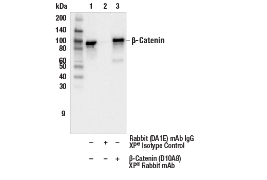
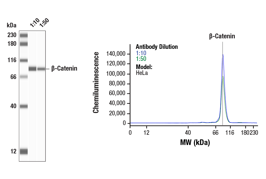
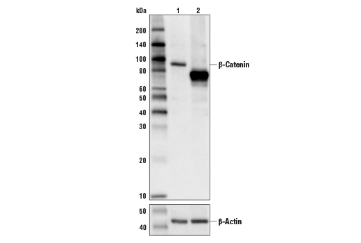
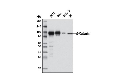
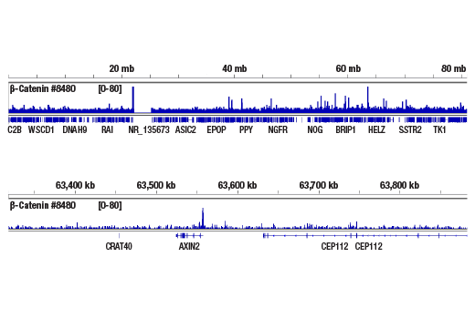
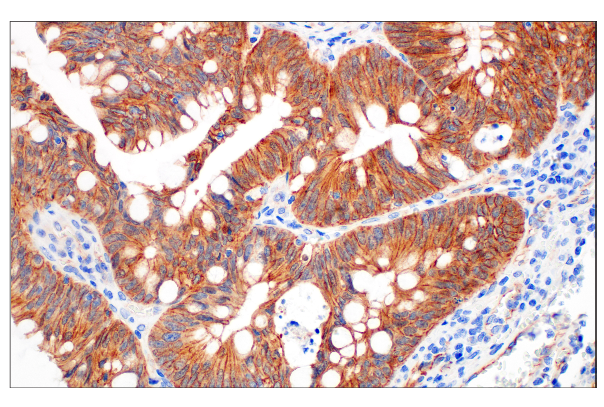
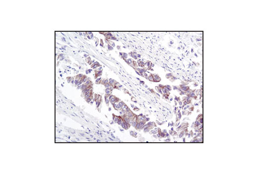
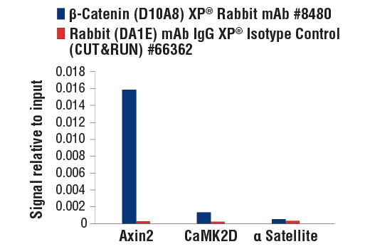
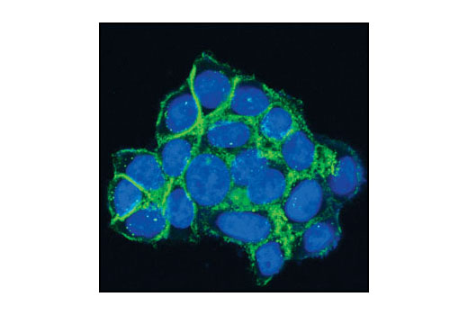
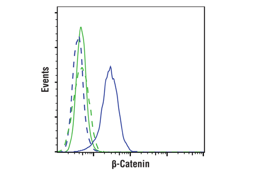
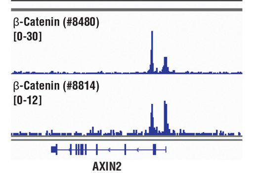
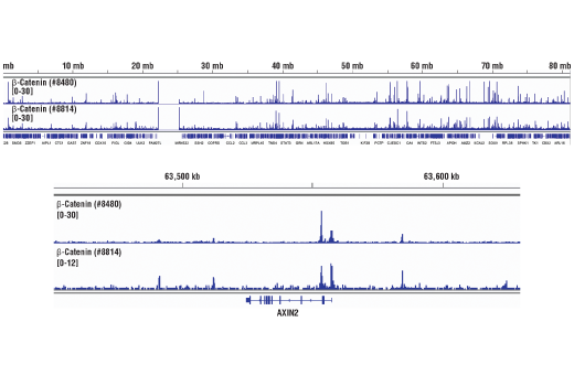
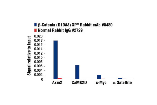
| β-Catenin (D10A8) XP® Rabbit mAb | 8480 | 40 µl | 92 kDa | Rabbit IgG |
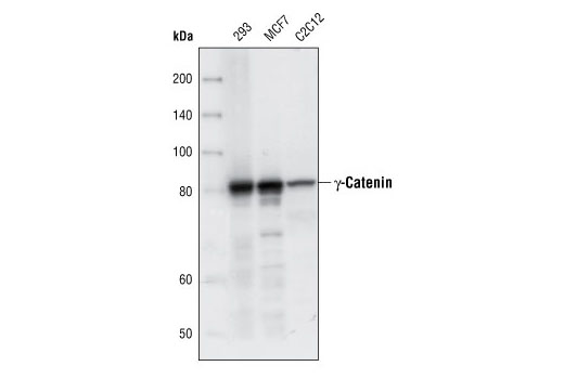
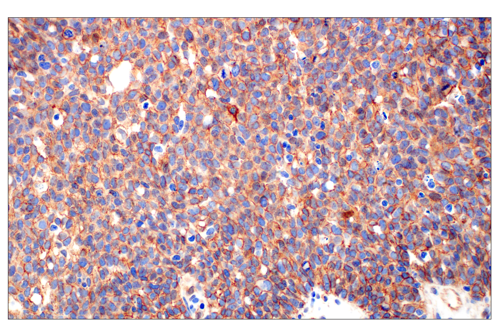
| γ-Catenin Antibody | 2309 | 40 µl | 83 kDa | Rabbit |
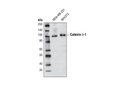
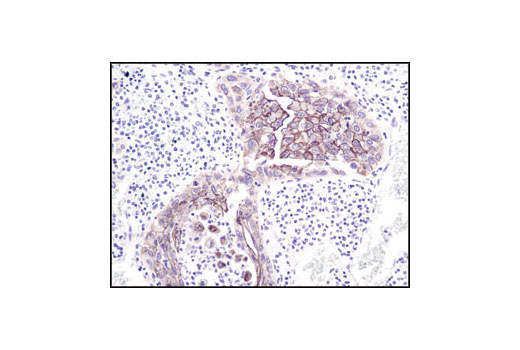
| Catenin δ-1 Antibody | 4989 | 40 µl | 100 kDa | Rabbit |
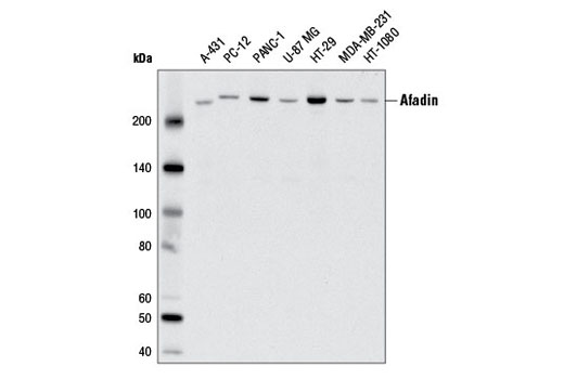
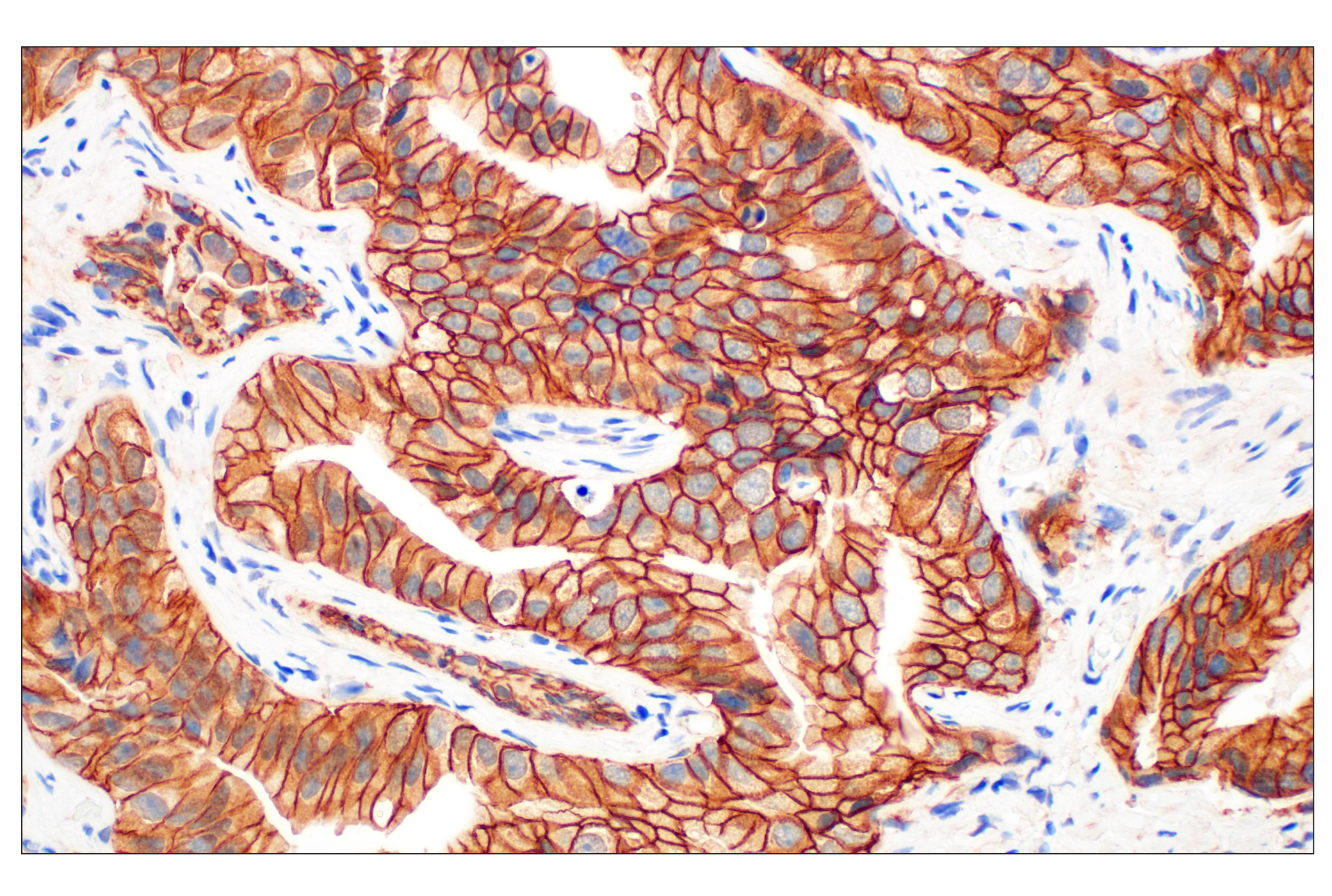
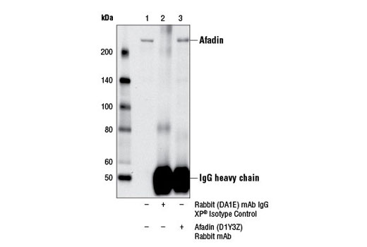
| Afadin (D1Y3Z) Rabbit mAb | 13531 | 40 µl | 205 kDa | Rabbit IgG |
| Anti-rabbit IgG, HRP-linked Antibody | 7074 | 100 µl | Goat |
Please visit cellsignal.com for individual component applications, species cross-reactivity, dilutions, protocols, and additional product information.
Description
The Adherens Junction Antibody Sampler Kit provides an economical means of detecting the protein components of adherens junctions. The kit includes enough antibody to perform four western blot experiments per primary antibody.
Storage
Background
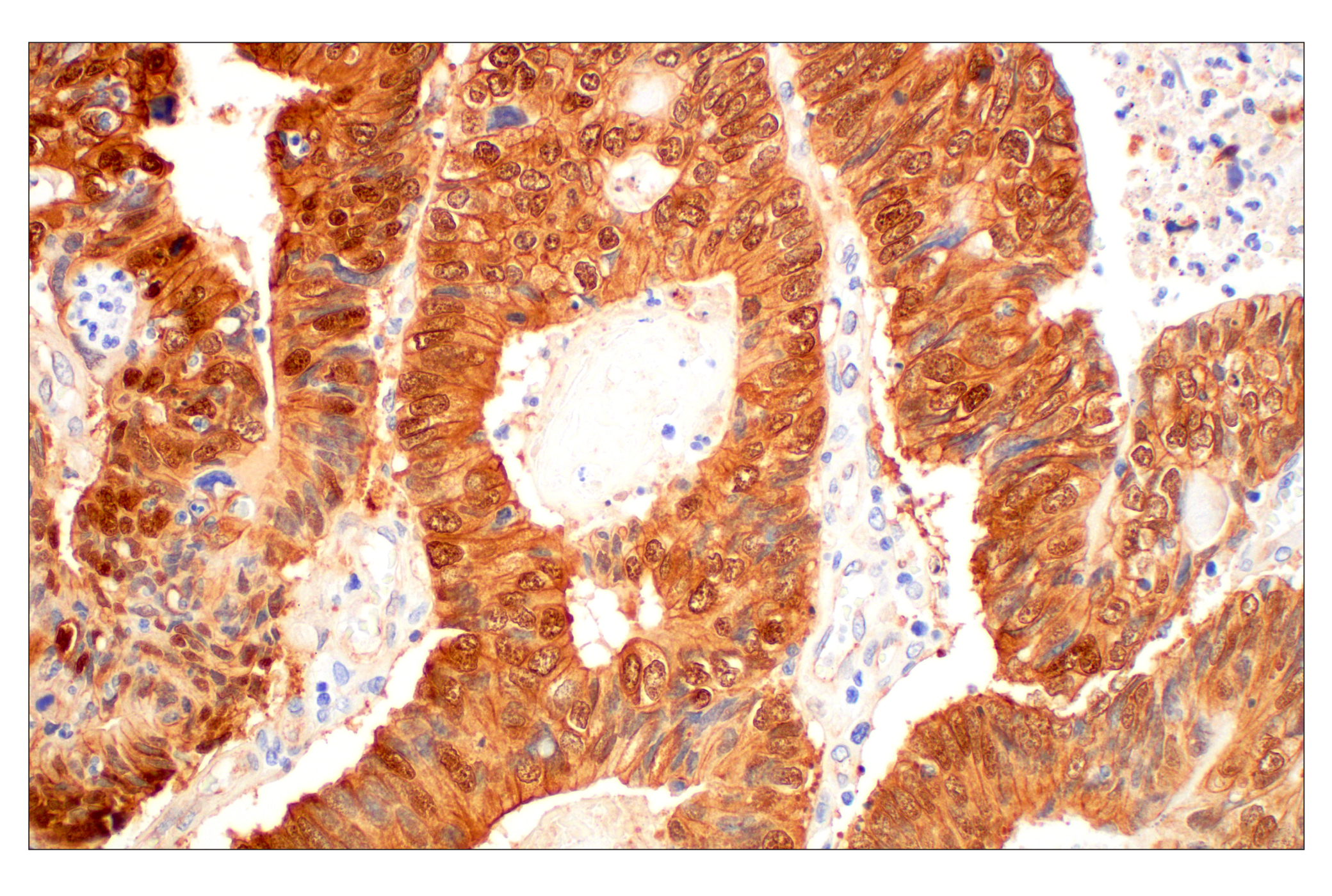
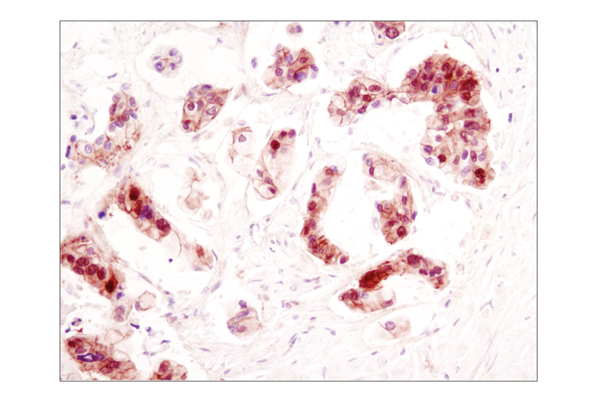
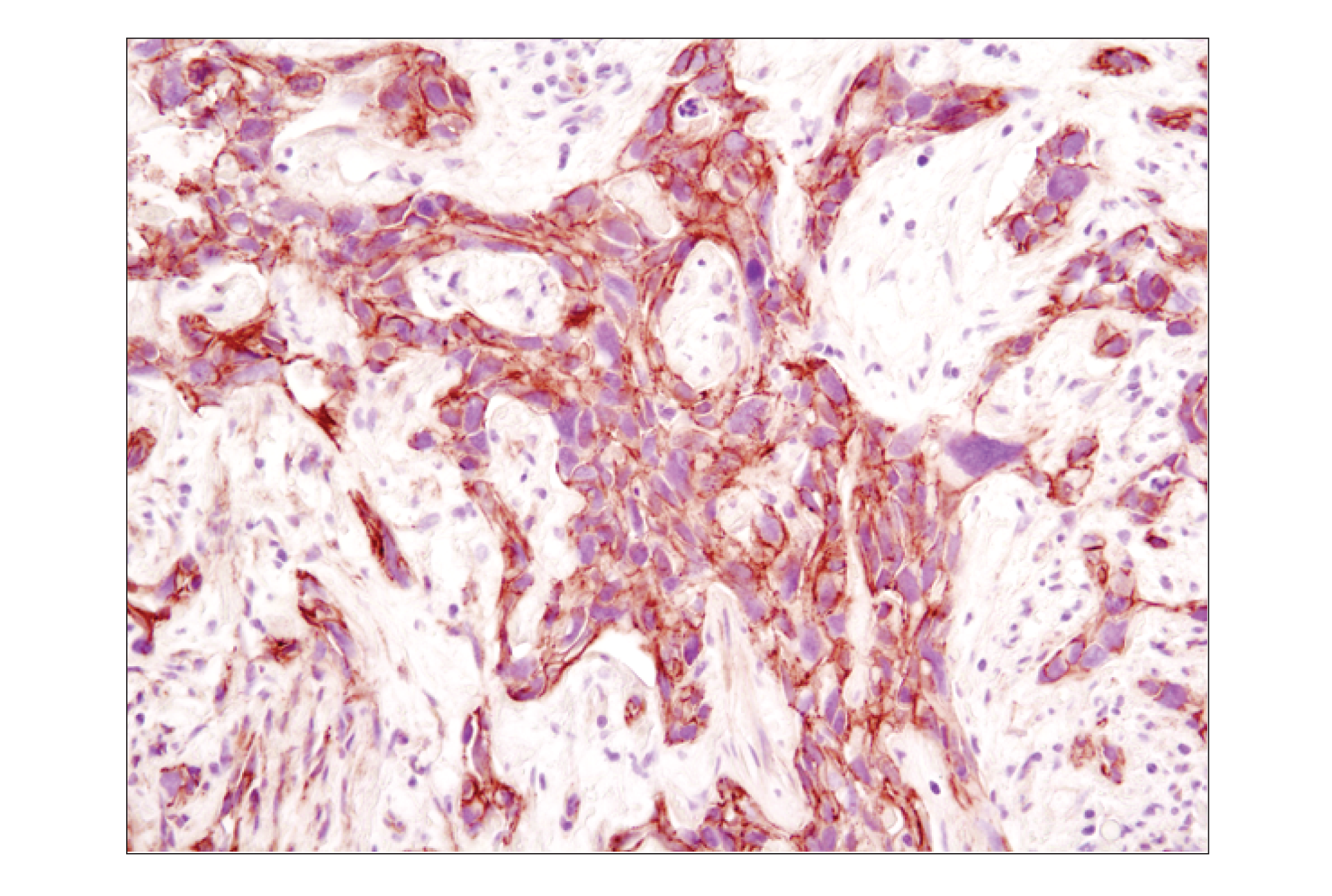
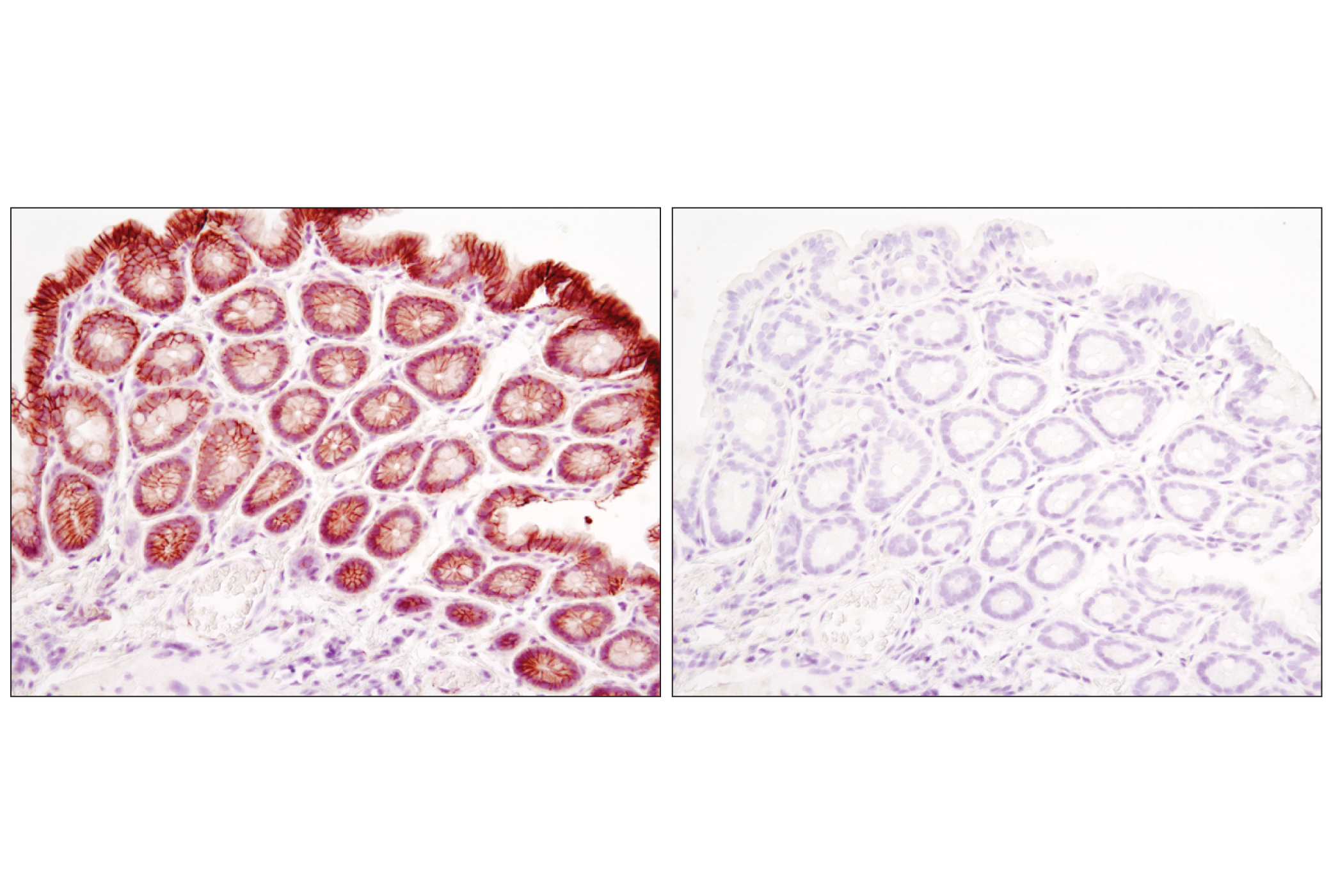
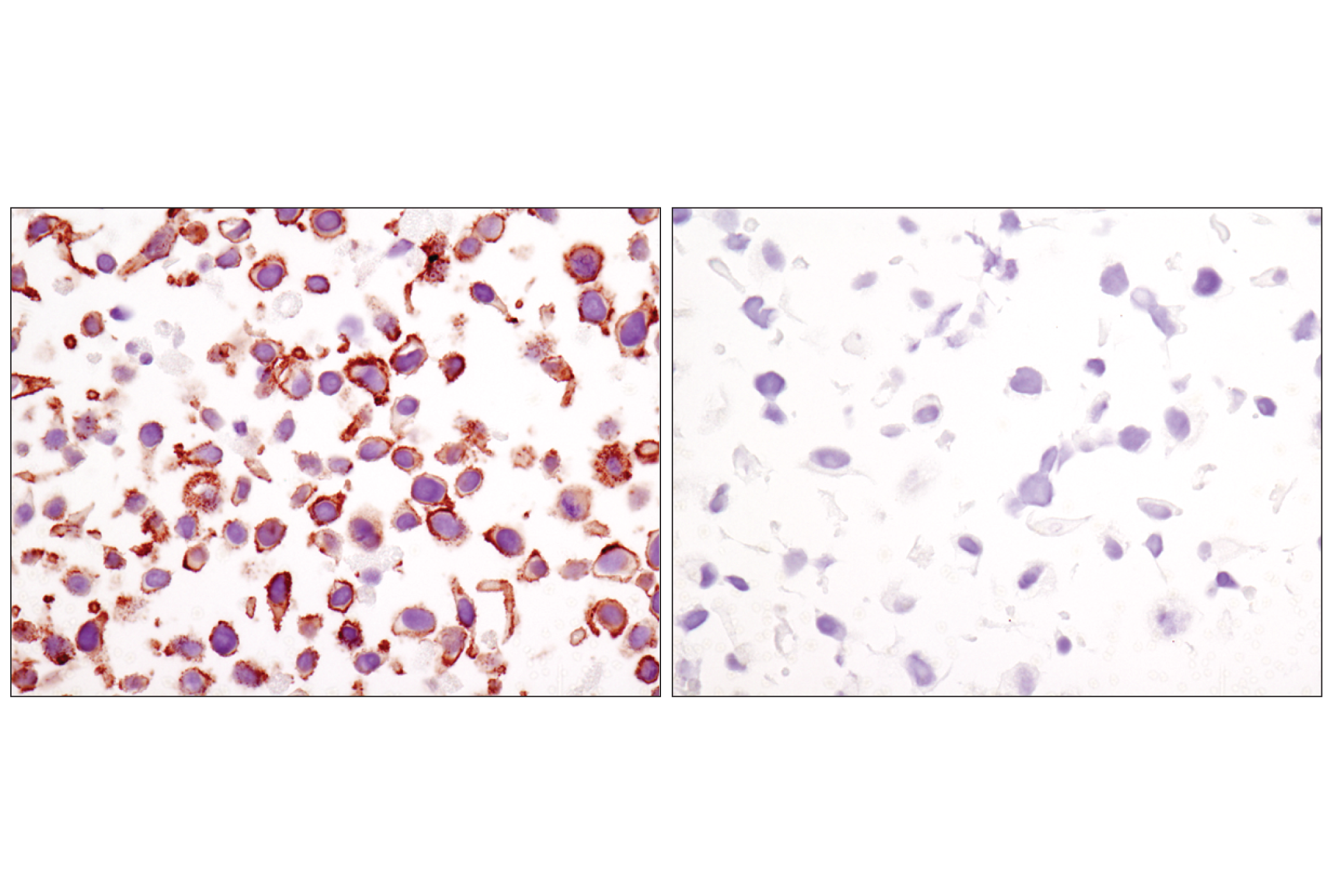
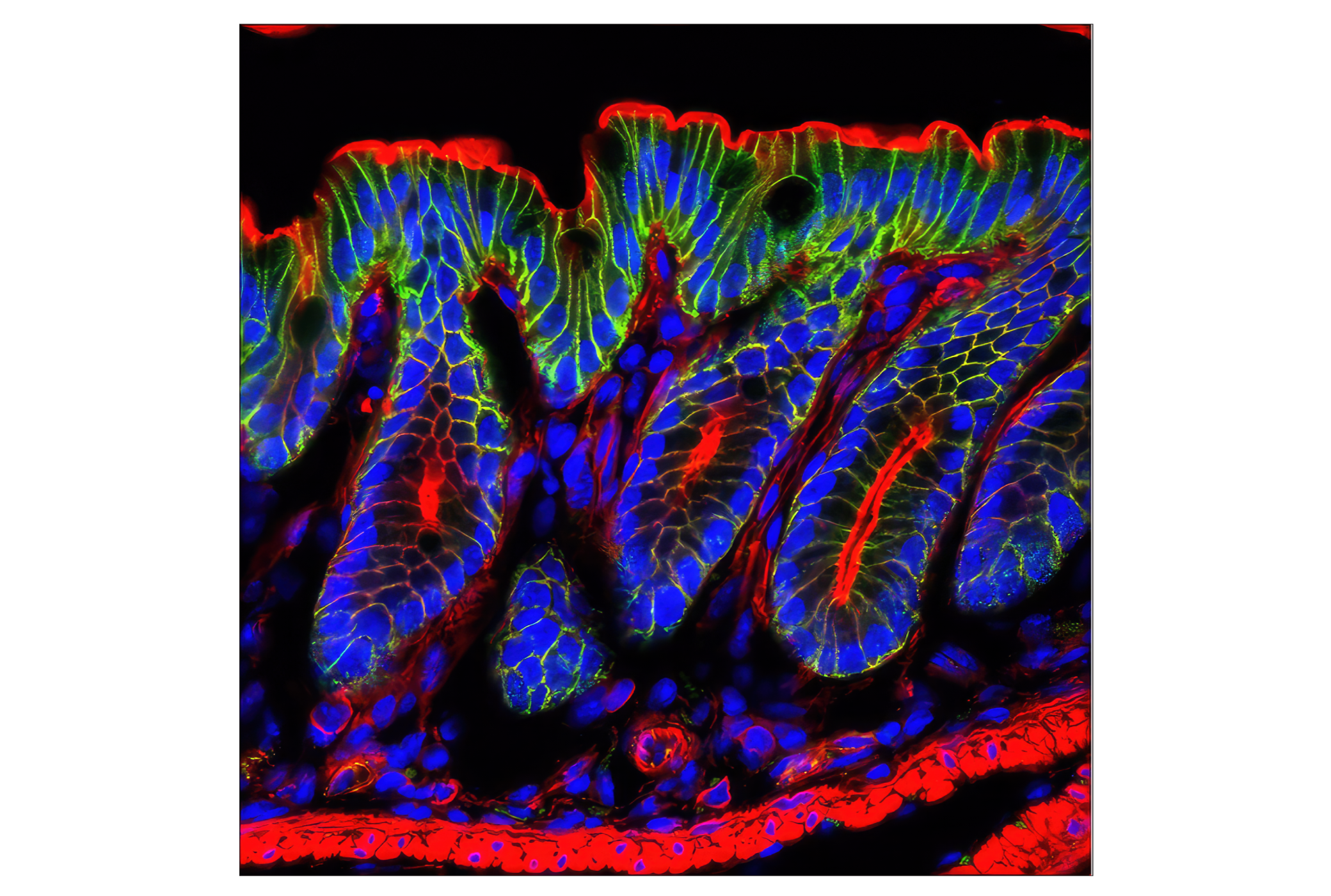
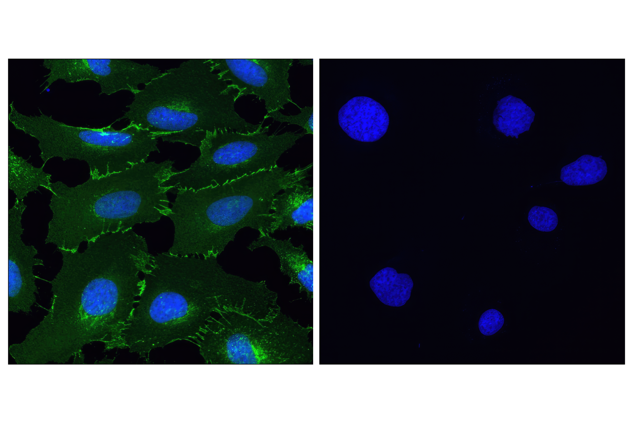
Adherens junctions are dynamic structures that form cell-cell contacts and are important in development, differentiation, tissue integrity, morphology and cell polarity. They are composed of the transmembrane proteins, cadherins, which bind cadherins on adjacent cells in a calcium-dependent manner. On the cytoplasmic side of adherens junctions, the classic model states that cadherins are linked to the cytoskeleton through β- and α-catenin. α-E-catenin is ubiquitously expressed, α-N-catenin is expressed in neuronal tissue, and α-T-catenin is primarily expressed in heart tissue. Research studies have demonstrated that loss of E-cadherin and α-E-catenin occurs during the progression of several human cancers, indicating that the breakdown of adherens junctions is important in cancer progression (reviewed in 1).
Research studies also suggest that, rather than acting as a static link between cadherins and actin, α-catenin regulates actin dynamics directly, possibly by competing with the actin nucleating arp2/3 complex (2,3). α-catenin also plays a role in regulating β-catenin-dependent transcriptional activity, affecting differentiation and response to Wnt signaling. α-catenin binds to β-catenin in the nucleus, preventing it from regulating transcription, and levels of both proteins appear to be regulated via proteasome-dependent degradation (4).
Afadin has two splice variants: l-afadin, which is ubiquitously expressed, and s-afadin, which is expressed predominantly in neural tissue. s-afadin is a shorter form lacking one of the three proline-rich regions found in l-afadin, as well as the carboxyl-terminal F-actin binding region (5). Human s-afadin is identical to AF-6, the ALL-1 fusion partner involved in acute myeloid leukemias (6). Recent research has also shown that afadin is involved in controlling the directionality of cell movement when it is localized at the leading edge of moving cells (7,8).
- Kobielak, A. and Fuchs, E. (2004) Nat Rev Mol Cell Biol 5, 614-25.
- Yamada, S. et al. (2005) Cell 123, 889-901.
- Drees, F. et al. (2005) Cell 123, 903-15.
- Hwang, S.G. et al. (2005) J Biol Chem 280, 12758-65.
- Mandai, K. et al. (1997) J Cell Biol 139, 517-28.
- Prasad, R. et al. (1993) Cancer Res 53, 5624-8.
- Miyata, M. et al. (2009) J Cell Sci 122, 4319-29.
- Miyata, M. et al. (2009) J Biol Chem 284, 24595-609.
Background References
Trademarks and Patents
使用に関する制限
法的な権限を与えられたCSTの担当者が署名した書面によって別途明示的に合意された場合を除き、 CST、その関連会社または代理店が提供する製品には以下の条件が適用されます。お客様が定める条件でここに定められた条件に含まれるものを超えるもの、 または、ここに定められた条件と異なるものは、法的な権限を与えられたCSTの担当者が別途書面にて受諾した場合を除き、拒絶され、 いかなる効力も効果も有しません。
研究専用 (For Research Use Only) またはこれに類似する表示がされた製品は、 いかなる目的についても FDA または外国もしくは国内のその他の規制機関により承認、認可または許可を受けていません。 お客様は製品を診断もしくは治療目的で使用してはならず、また、製品に表示された内容に違反する方法で使用してはなりません。 CST が販売または使用許諾する製品は、エンドユーザーであるお客様に対し、使途を研究および開発のみに限定して提供されるものです。 診断、予防もしくは治療目的で製品を使用することまたは製品を再販売 (単独であるか他の製品等の一部であるかを問いません) もしくはその他の商業的利用の目的で購入することについては、CST から別途許諾を得る必要があります。 お客様は以下の事項を遵守しなければなりません。(a) CST の製品 (単独であるか他の資材と一緒であるかを問いません) を販売、使用許諾、貸与、寄付もしくはその他の態様で第三者に譲渡したり使用させたりしてはなりません。また、商用の製品を製造するために CST の製品を使用してはなりません。(b) 複製、改変、リバースエンジニアリング、逆コンパイル、 分解または他の方法により製品の構造または技術を解明しようとしてはなりません。また、 CST の製品またはサービスと競合する製品またはサービスを開発する目的で CST の製品を使用してはなりません。(c) CST の製品の商標、商号、ロゴ、特許または著作権に関する通知または表示を除去したり改変したりしてはなりません。(d) CST の製品をCST 製品販売条件(CST’s Product Terms of Sale) および該当する書面のみに従って使用しなければなりません。(e) CST の製品に関連してお客様が使用する第三者の製品またはサービスに関する使用許諾条件、 サービス提供条件またはこれに類する合意事項を遵守しなければなりません。