IF-IC, DB
All
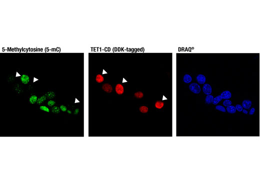
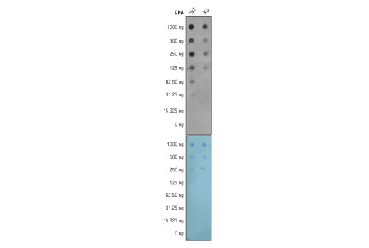
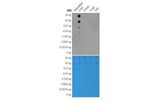
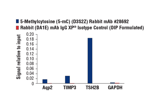
Endogenous
Rabbit IgG
Product Information
Product Usage Information
| Application | Dilution |
|---|---|
| Immunofluorescence (Immunocytochemistry) | 1:1600 |
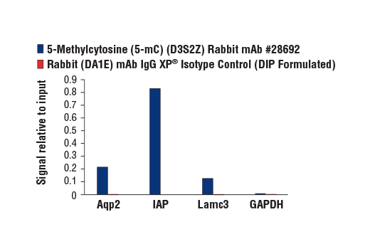
| DNA Dot Blot | 1:1000 |
Storage
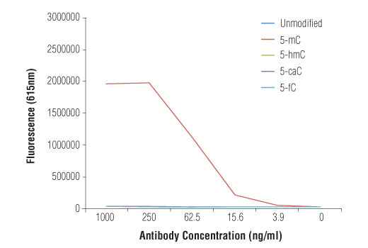
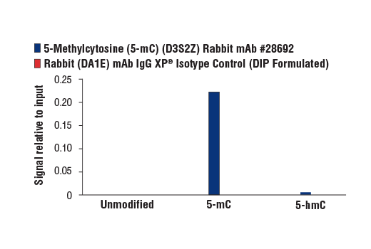
Specificity / Sensitivity
Species Reactivity:
All Species Expected
Source / Purification
Monoclonal antibody is produced by immunizing animals with 5-methylcytidine.
Background
Methylation of DNA at cytosine residues is a heritable, epigenetic modification that is critical for proper regulation of gene expression, genomic imprinting, and mammalian development (1,2). 5-methylcytosine is a repressive epigenetic mark established de novo by two enzymes, DNMT3a and DNMT3b, and is maintained by DNMT1 (3, 4). 5-methylcytosine was originally thought to be passively depleted during DNA replication. However, subsequent studies have shown that Ten-Eleven Translocation (TET) proteins TET1, TET2, and TET3 can catalyze the oxidation of methylated cytosine to 5-hydroxymethylcytosine (5-hmC) (5). Additionally, TET proteins can further oxidize 5-hmC to form 5-formylcytosine (5-fC) and 5-carboxylcytosine (5-caC), both of which are excised by thymine-DNA glycosylase (TDG), effectively linking cytosine oxidation to the base excision repair pathway and supporting active cytosine demethylation (6,7).
Normally DNA methylation occurs in a bimodal fashion, such that CpG dinucleotides are largely methylated across the genome, except in short stretches of CpG-rich sequences associated with gene promoters, known as CpG-islands, where methylation is virtually absent (8). Cancer cell genomes often undergo global hypomethylation, while CpG-islands become hypermethylated, causing their associated promoters to become repressed (9). There is evidence that a number of aberrantly hypermethylated CpG-islands found in carcinomas occur at tumor suppressor genes such as RB1, MLH1, and BRCA1 (10).
- Hermann, A. et al. (2004) Cell Mol Life Sci 61, 2571-87.
- Turek-Plewa, J. and Jagodziński, P.P. (2005) Cell Mol Biol Lett 10, 631-47.
- Okano, M. et al. (1999) Cell 99, 247-57.
- Li, E. et al. (1992) Cell 69, 915-26.
- Tahiliani, M. et al. (2009) Science 324, 930-5.
- He, Y.F. et al. (2011) Science 333, 1303-7.
- Ito, S. et al. (2011) Science 333, 1300-3.
- Suzuki, M.M. and Bird, A. (2008) Nat Rev Genet 9, 465-76.
- Berman, B.P. et al. (2012) Nat Genet 44, 40-6.
- Sproul, D. and Meehan, R.R. (2013) Brief Funct Genomics 12, 174-90.
Species Reactivity
Species reactivity is determined by testing in at least one approved application (e.g., western blot).
Applications Key
IF-IC: Immunofluorescence (Immunocytochemistry) DB: DNA Dot Blot
Cross-Reactivity Key
H: human M: mouse R: rat Hm: hamster Mk: monkey Vir: virus Mi: mink C: chicken Dm: D. melanogaster X: Xenopus Z: zebrafish B: bovine Dg: dog Pg: pig Sc: S. cerevisiae Ce: C. elegans Hr: horse GP: Guinea Pig Rab: rabbit All: all species expected
Trademarks and Patents
使用に関する制限
法的な権限を与えられたCSTの担当者が署名した書面によって別途明示的に合意された場合を除き、 CST、その関連会社または代理店が提供する製品には以下の条件が適用されます。お客様が定める条件でここに定められた条件に含まれるものを超えるもの、 または、ここに定められた条件と異なるものは、法的な権限を与えられたCSTの担当者が別途書面にて受諾した場合を除き、拒絶され、 いかなる効力も効果も有しません。
研究専用 (For Research Use Only) またはこれに類似する表示がされた製品は、 いかなる目的についても FDA または外国もしくは国内のその他の規制機関により承認、認可または許可を受けていません。 お客様は製品を診断もしくは治療目的で使用してはならず、また、製品に表示された内容に違反する方法で使用してはなりません。 CST が販売または使用許諾する製品は、エンドユーザーであるお客様に対し、使途を研究および開発のみに限定して提供されるものです。 診断、予防もしくは治療目的で製品を使用することまたは製品を再販売 (単独であるか他の製品等の一部であるかを問いません) もしくはその他の商業的利用の目的で購入することについては、CST から別途許諾を得る必要があります。 お客様は以下の事項を遵守しなければなりません。(a) CST の製品 (単独であるか他の資材と一緒であるかを問いません) を販売、使用許諾、貸与、寄付もしくはその他の態様で第三者に譲渡したり使用させたりしてはなりません。また、商用の製品を製造するために CST の製品を使用してはなりません。(b) 複製、改変、リバースエンジニアリング、逆コンパイル、 分解または他の方法により製品の構造または技術を解明しようとしてはなりません。また、 CST の製品またはサービスと競合する製品またはサービスを開発する目的で CST の製品を使用してはなりません。(c) CST の製品の商標、商号、ロゴ、特許または著作権に関する通知または表示を除去したり改変したりしてはなりません。(d) CST の製品をCST 製品販売条件(CST’s Product Terms of Sale) および該当する書面のみに従って使用しなければなりません。(e) CST の製品に関連してお客様が使用する第三者の製品またはサービスに関する使用許諾条件、 サービス提供条件またはこれに類する合意事項を遵守しなければなりません。