| Products Included | No. | Volume | Applicaton | Dilution | Reactivity |
|---|---|---|---|---|---|
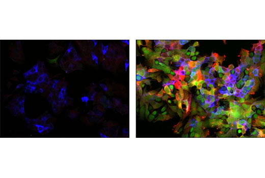
| Primary Cocktail | 8998 | 100 µl | Immunofluorescence (Immunocytochemistry) | 1:100 | Human Mouse Rat Monkey |
| Detection Cocktail | 8997 | 100 µl | Immunofluorescence (Immunocytochemistry) | 1:100 | N/A |
| Kit Analytes | Detection Dye | Ex(max) (nm) | Em(max) (nm) |
|---|---|---|---|
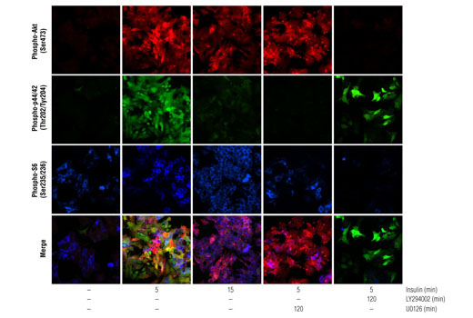
| Phospho-Akt (Ser473) | Alexa Fluor® 555 | 555 | 565 |
| Phospho-p44/42 Erk1/2 (Thr202/Tyr204) | Alexa Fluor® 488 | 495 | 519 |
| Phospho-S6 Ribosomal Protein (Ser235/236) | Alexa Fluor® 647 | 650 | 665 |
IF-IC
#P27361, #P31751, #Q9Y243, #P62753, #P28482, #P31749
5595, 208, 10000, 6194, 5594, 207
Product Information
Storage
Specificity / Sensitivity
Species Reactivity:
Human, Mouse, Rat, Monkey
Source / Purification
Monoclonal antibodies are produced by immunizing animals with synthetic phosphopeptides corresponding to residues surrounding Ser473 of human Akt, Thr202/Tyr204 of human p44 MAP kinase, and Ser235/Ser236 of human ribosomal protein S6.
Product Description
| MW (kDa) | n/a |
Background
Akt, also referred to as PKB or Rac, plays a critical role in controlling the balance between survival and apoptosis (1-3). This protein kinase is a downstream effector of phosphoinositide-3 kinase (PI3K), and is activated by phospholipid binding and activation loop phosphorylation at Thr308 by PDK1 (4), as well as by phosphorylation within the carboxy terminus at Ser473 by the mTOR-rictor complex (TORC2) (5). This pathway is down-regulated following dephosphorylation of phosphatidyl-inositol 3,4,5 triphosphate by PTEN, as well as by deactivation of PI3K with targeted small molecule inhibitors such as wortmannin and LY294002 (2,3,6,7).
p70 S6 kinase, a mitogen activated Ser/Thr protein kinase downstream of PI3K and the mTOR-raptor complex (mTORC1), phosphorylates the S6 protein of the 40S ribosomal subunit leading to an increase in translation of mRNA transcripts that contain an oligopyrimidine tract in their 5’ untranslated region (8). These particular mRNA transcripts (5’TOP) encode proteins involved in cell cycle progression, as well as ribosomal proteins and elongation factors necessary for translation (8,9). Important S6 ribosomal protein phosphorylation sites include several residues (Ser235, Ser236, Ser240 and Ser244) located within a small, carboxy-terminal region of the S6 protein (10,11).
Both p44 and p42 mitogen-activated protein (MAP) kinases (Erk1 and Erk2, respectively) play a critical role in the regulation of cell growth and differentiation (12-15). MAP kinases are activated by a wide variety of extracellular signals including growth and neurotrophic factors, cytokines, hormones, and neurotransmitters. Activation of MAP kinases occur through phosphorylation of Thr202/Tyr204 on human Erk1 and Thr185/Tyr187 on human Erk2 at the sequence T*EY* by a pair of upstream MAP kinase kinases (MEK1/2) (16,17). Erk proteins are negatively regulated by a family of dual specificity (Thr/Tyr) MAPK phosphatases, known as DUSPs or MKPs (18), along with MEK inhibitors such as U0126 and PD98059. Erk dependent phosphorylation of TSC2 at Ser663 leads to the functional inactivation of the TSC1/TSC2 inhibitory complex, and subsequent downstream activation of S6 ribosomal protein through the mTORC1/p70 S6K signaling cascade (19).
- Franke, T.F. et al. (1997) Cell 88, 435-7.
- Burgering, B.M. and Coffer, P.J. (1995) Nature 376, 599-602.
- Franke, T.F. et al. (1995) Cell 81, 727-36.
- Alessi, D.R. et al. (1996) EMBO J 15, 6541-51.
- Sarbassov, D.D. et al. (2005) Science 307, 1098-101.
- Myers, M.P. et al. (1998) Proc Natl Acad Sci U S A 95, 13513-8.
- Vlahos, C.J. et al. (1994) J Biol Chem 269, 5241-8.
- Peterson, R.T. and Schreiber, S.L. (1998) Curr Biol 8, R248-50.
- Jefferies, H.B. et al. (1997) EMBO J 16, 3693-704.
- Ferrari, S. et al. (1991) J Biol Chem 266, 22770-5.
- Flotow, H. and Thomas, G. (1992) J Biol Chem 267, 3074-8.
- Marshall, C.J. (1995) Cell 80, 179-85.
- Hunter, T. (1995) Cell 80, 225-36.
- Hill, C.S. and Treisman, R. (1995) Cell 80, 199-211.
- Cowley, S. et al. (1994) Cell 77, 841-52.
- Sturgill, T.W. et al. (1988) Nature 334, 715-8.
- Payne, D.M. et al. (1991) EMBO J 10, 885-92.
- Owens, D.M. and Keyse, S.M. (2007) Oncogene 26, 3203-13.
- Ma, L. et al. (2005) Cell 121, 179-93.
Species Reactivity
Species reactivity is determined by testing in at least one approved application (e.g., western blot).
Applications Key
IF-IC: Immunofluorescence (Immunocytochemistry)
Cross-Reactivity Key
H: human M: mouse R: rat Hm: hamster Mk: monkey Vir: virus Mi: mink C: chicken Dm: D. melanogaster X: Xenopus Z: zebrafish B: bovine Dg: dog Pg: pig Sc: S. cerevisiae Ce: C. elegans Hr: horse GP: Guinea Pig Rab: rabbit All: all species expected
Trademarks and Patents
使用に関する制限
法的な権限を与えられたCSTの担当者が署名した書面によって別途明示的に合意された場合を除き、 CST、その関連会社または代理店が提供する製品には以下の条件が適用されます。お客様が定める条件でここに定められた条件に含まれるものを超えるもの、 または、ここに定められた条件と異なるものは、法的な権限を与えられたCSTの担当者が別途書面にて受諾した場合を除き、拒絶され、 いかなる効力も効果も有しません。
研究専用 (For Research Use Only) またはこれに類似する表示がされた製品は、 いかなる目的についても FDA または外国もしくは国内のその他の規制機関により承認、認可または許可を受けていません。 お客様は製品を診断もしくは治療目的で使用してはならず、また、製品に表示された内容に違反する方法で使用してはなりません。 CST が販売または使用許諾する製品は、エンドユーザーであるお客様に対し、使途を研究および開発のみに限定して提供されるものです。 診断、予防もしくは治療目的で製品を使用することまたは製品を再販売 (単独であるか他の製品等の一部であるかを問いません) もしくはその他の商業的利用の目的で購入することについては、CST から別途許諾を得る必要があります。 お客様は以下の事項を遵守しなければなりません。(a) CST の製品 (単独であるか他の資材と一緒であるかを問いません) を販売、使用許諾、貸与、寄付もしくはその他の態様で第三者に譲渡したり使用させたりしてはなりません。また、商用の製品を製造するために CST の製品を使用してはなりません。(b) 複製、改変、リバースエンジニアリング、逆コンパイル、 分解または他の方法により製品の構造または技術を解明しようとしてはなりません。また、 CST の製品またはサービスと競合する製品またはサービスを開発する目的で CST の製品を使用してはなりません。(c) CST の製品の商標、商号、ロゴ、特許または著作権に関する通知または表示を除去したり改変したりしてはなりません。(d) CST の製品をCST 製品販売条件(CST’s Product Terms of Sale) および該当する書面のみに従って使用しなければなりません。(e) CST の製品に関連してお客様が使用する第三者の製品またはサービスに関する使用許諾条件、 サービス提供条件またはこれに類する合意事項を遵守しなければなりません。