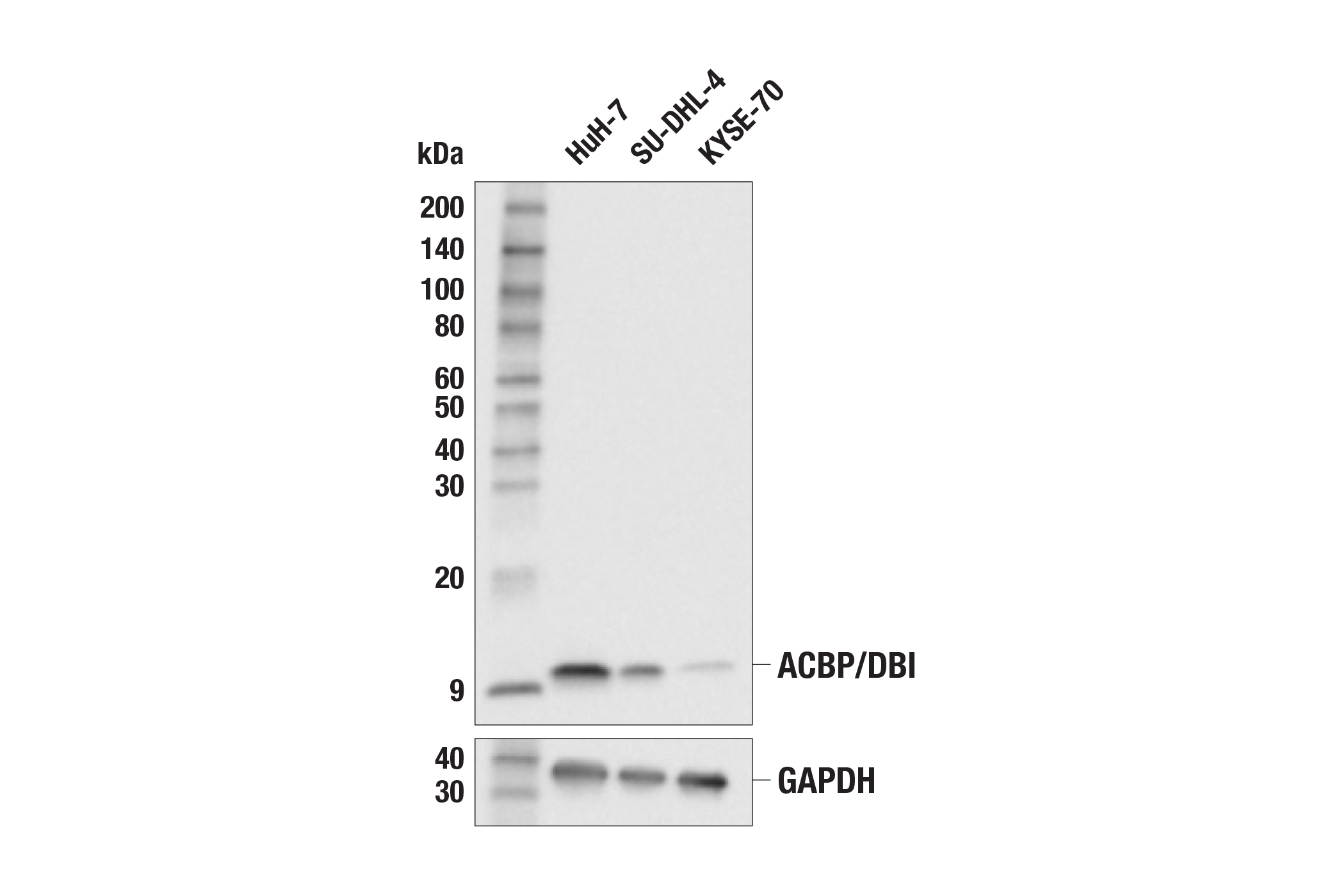
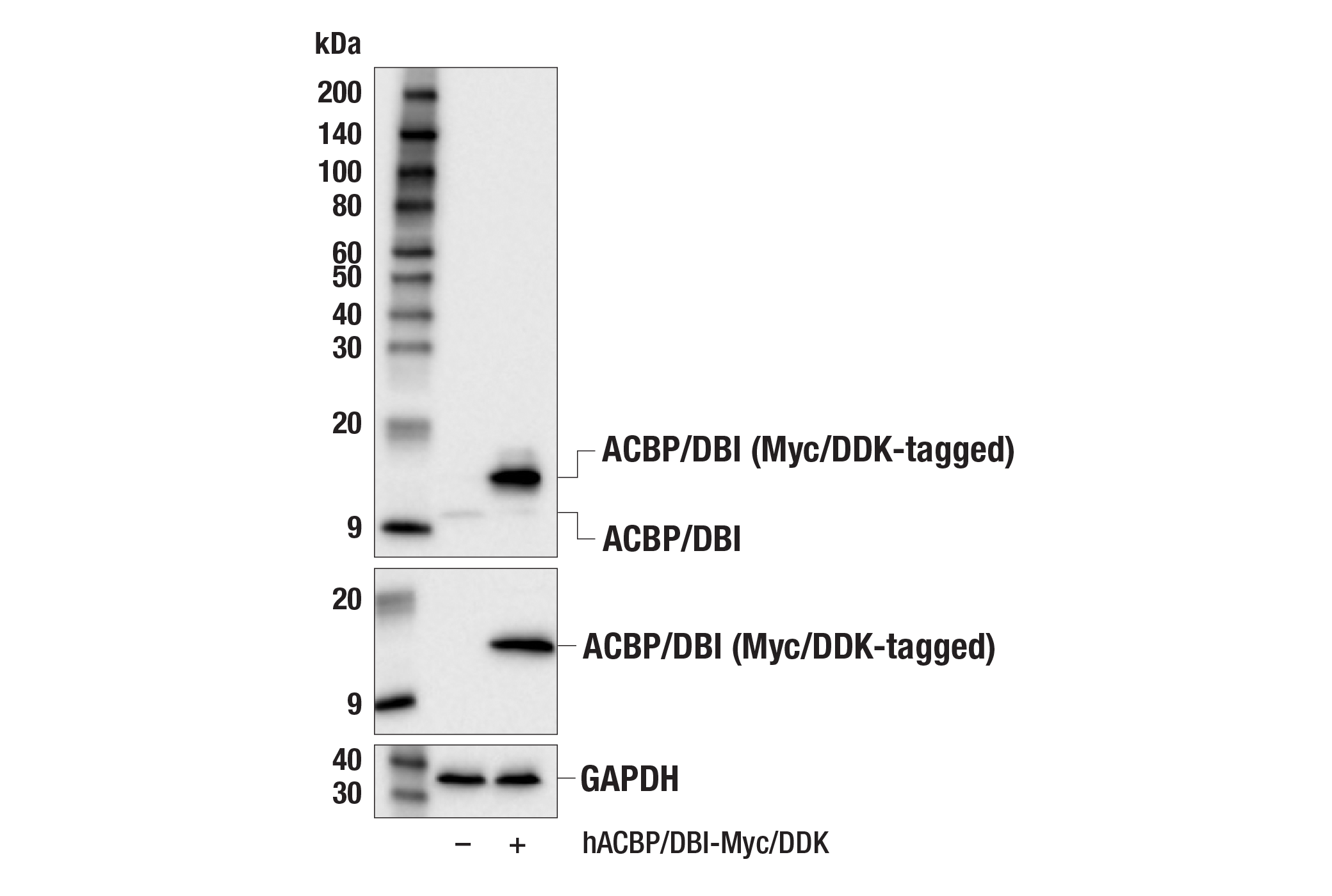
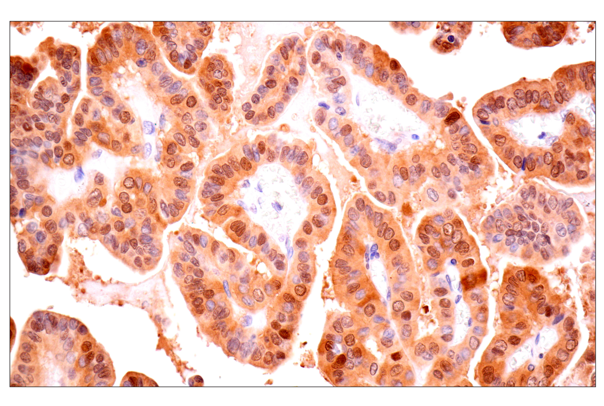
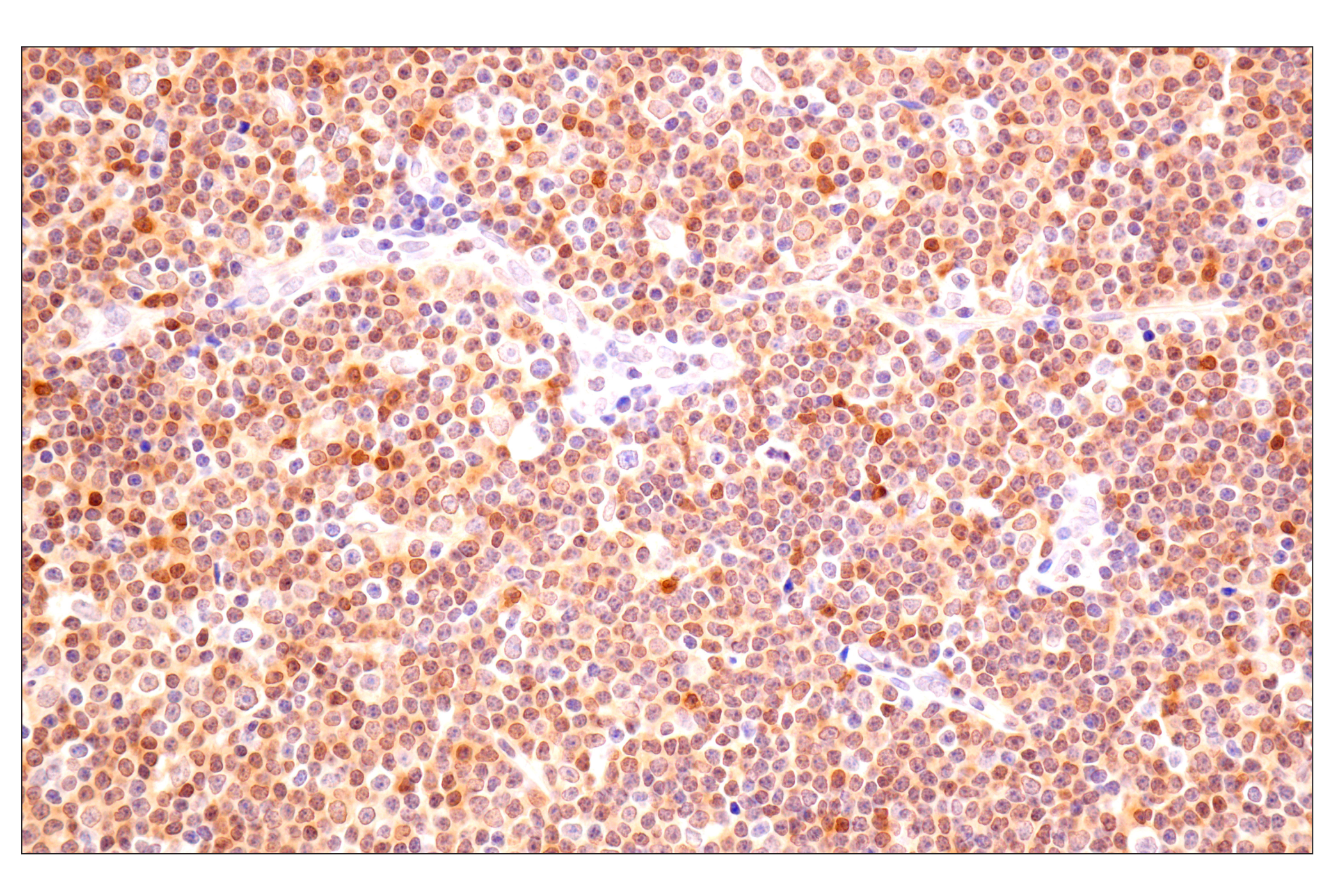
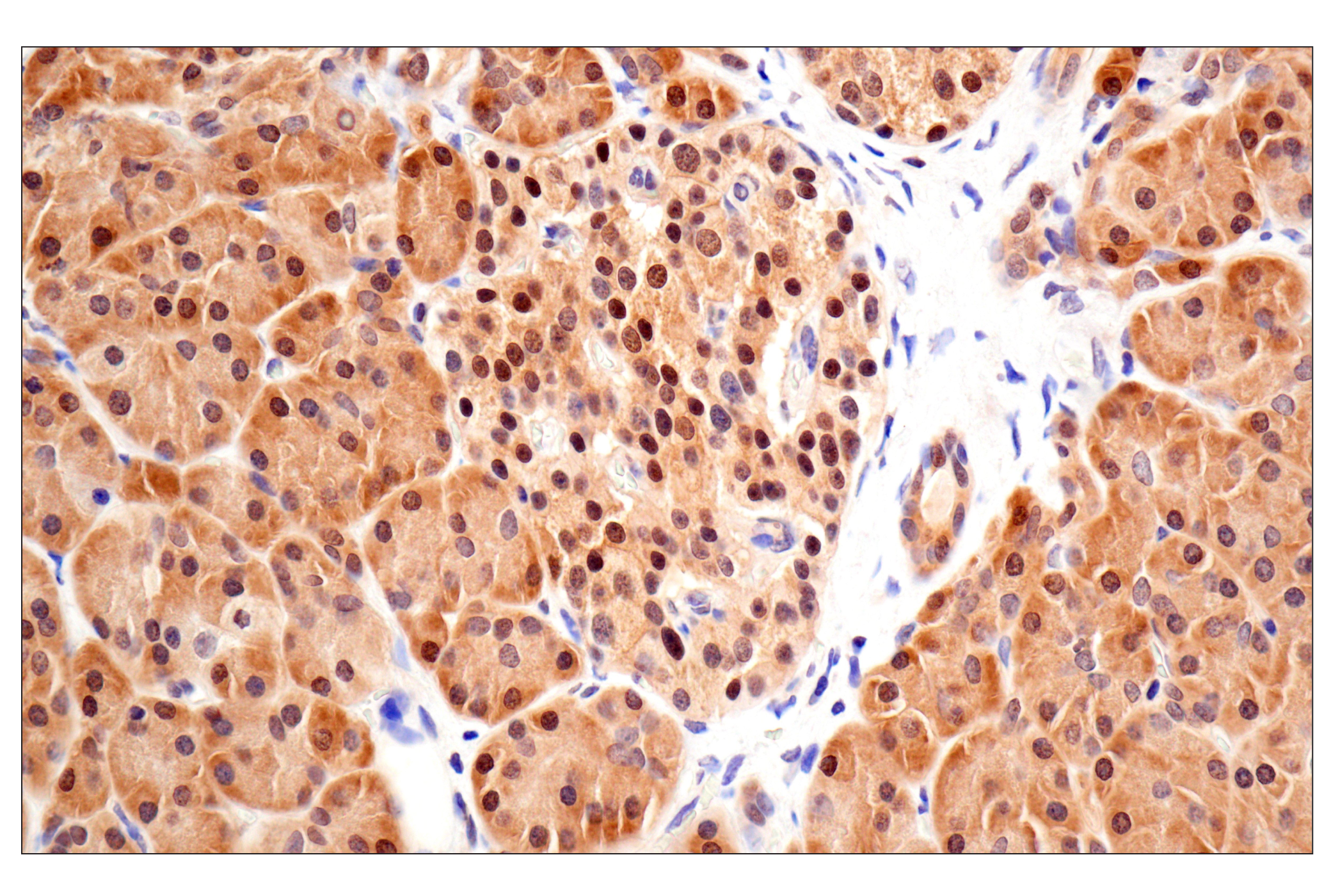
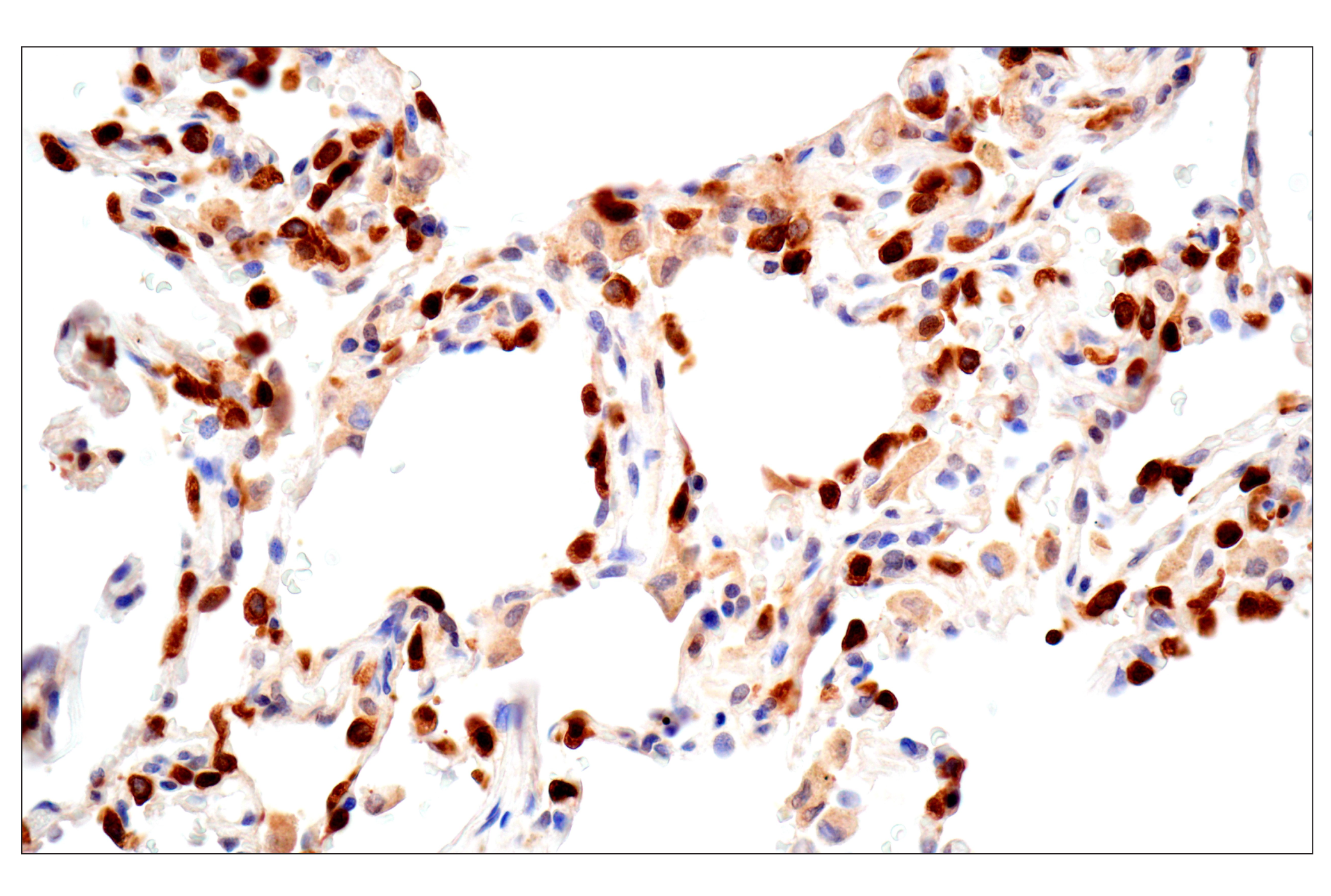
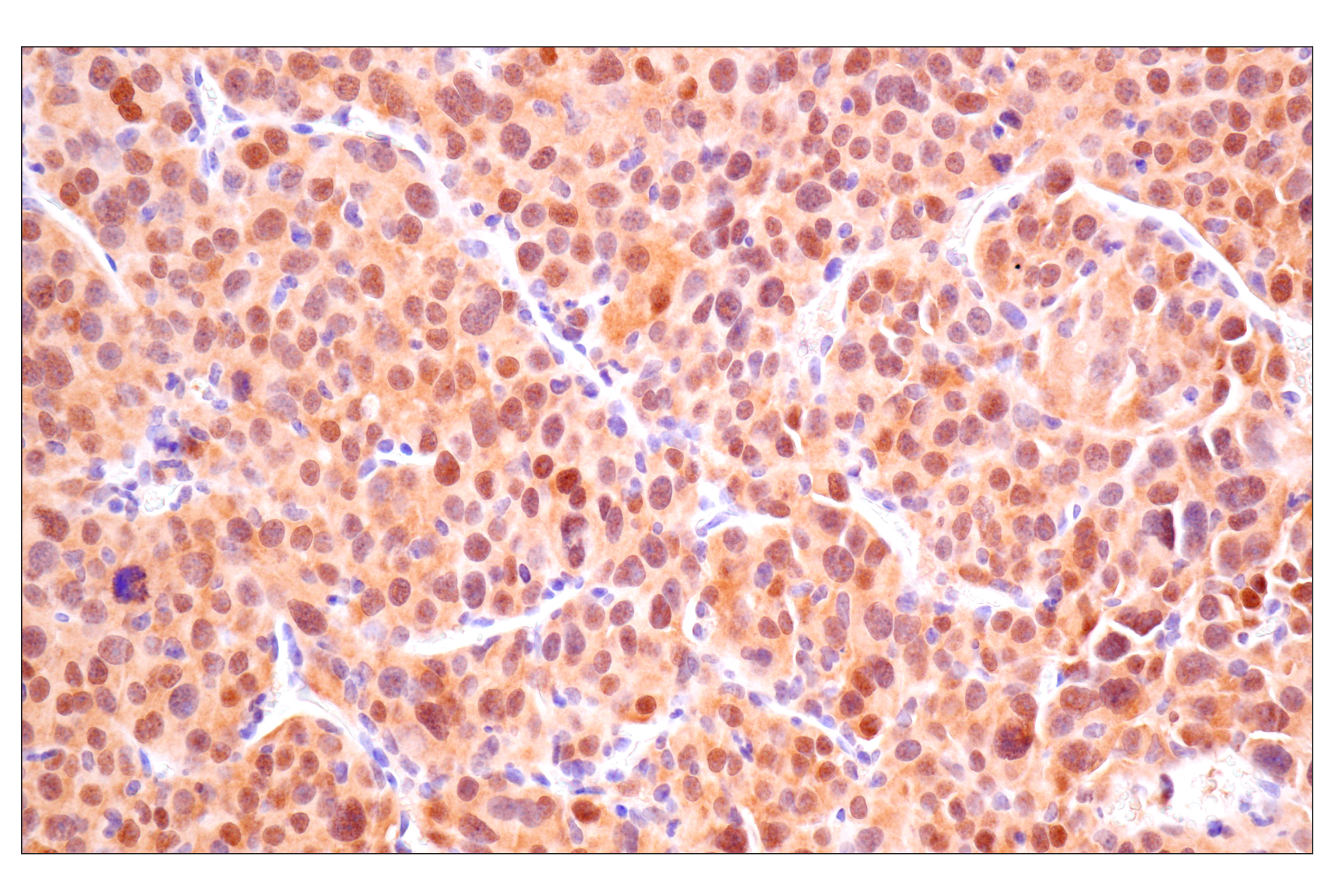
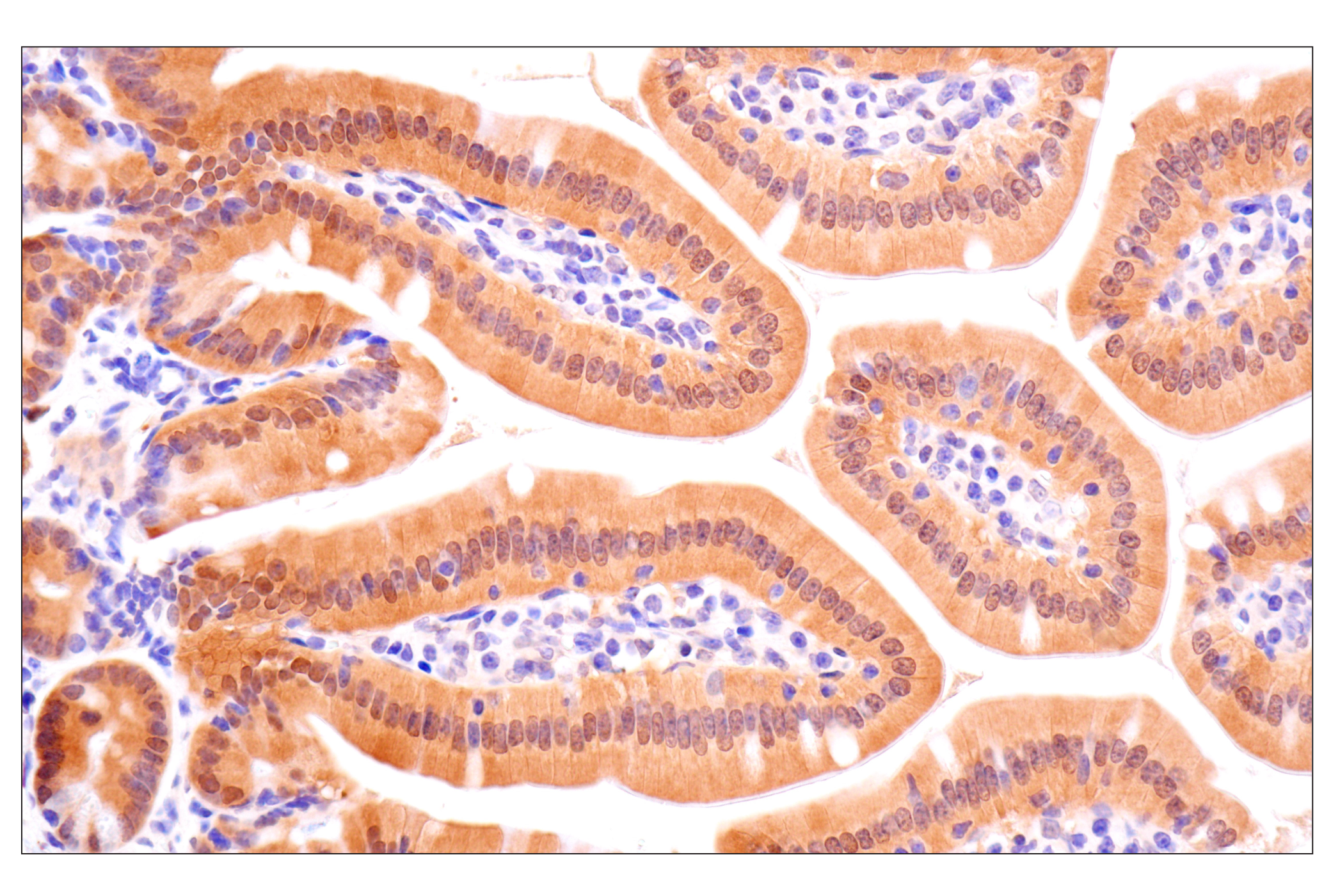
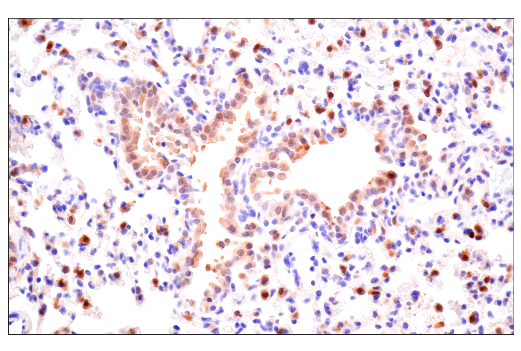
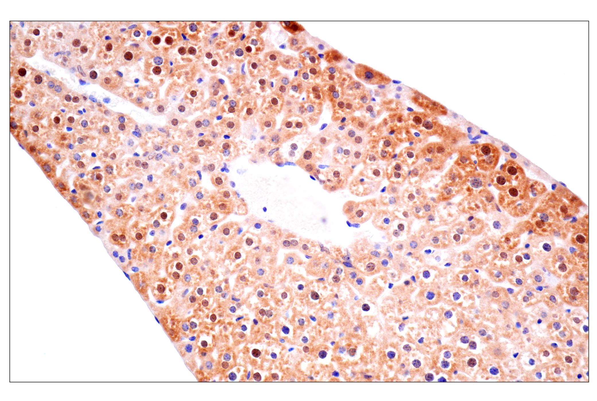
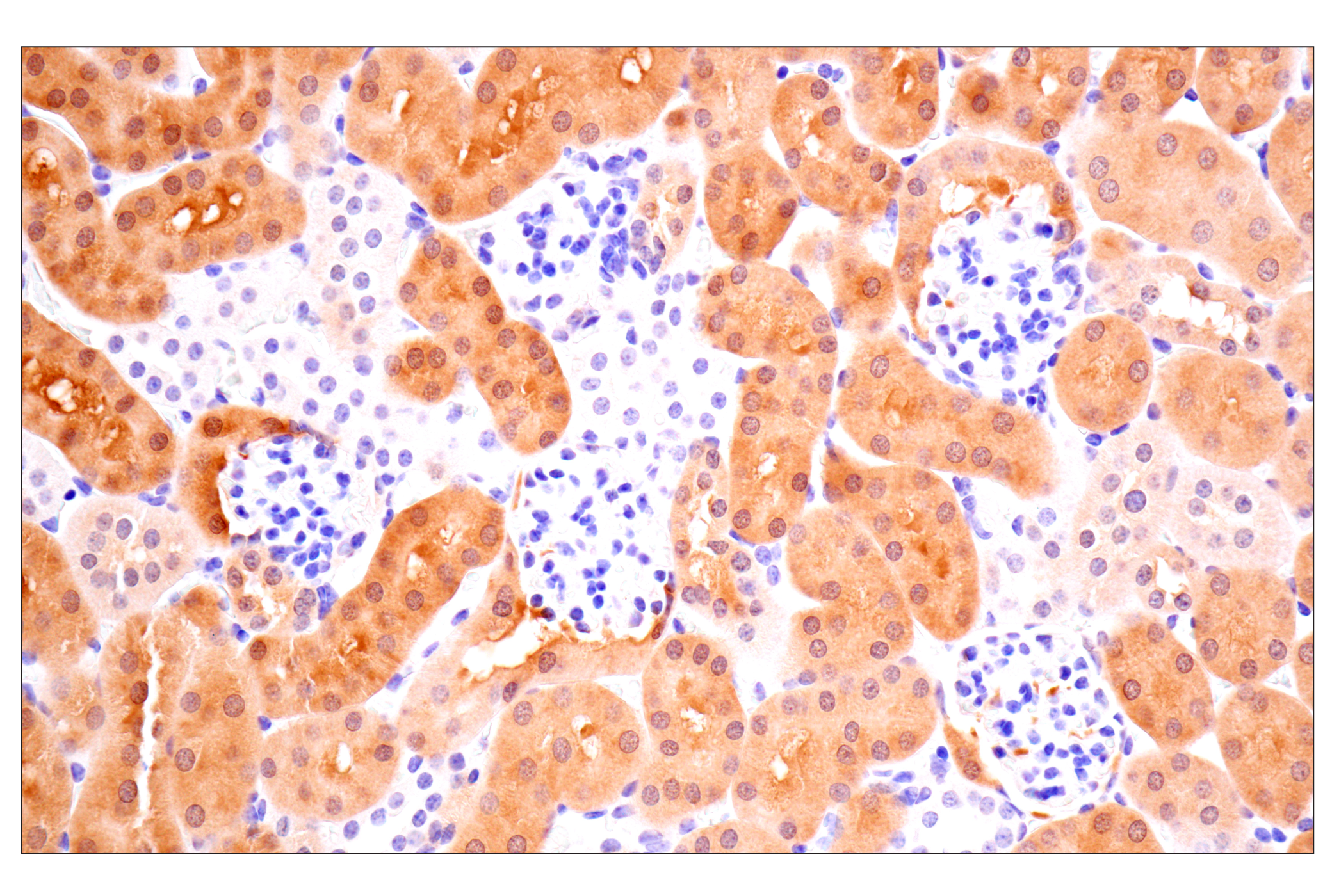
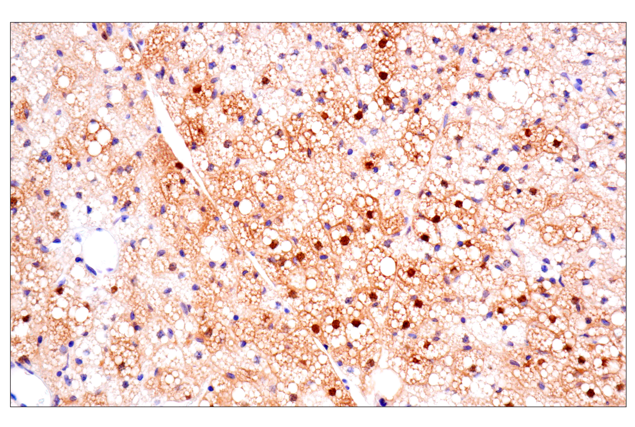
Revision 1

#40487Store at -20C

Cell Signaling Technology

Orders: 877-616-CELL (2355) [email protected]

Support: 877-678-TECH (8324)

Web: [email protected] cellsignal.com

3 Trask LaneDanversMassachusetts01923USA
For Research Use Only. Not for Use in Diagnostic Procedures.
Applications:

WB, IHC-P

REACTIVITY:

H M R

SENSITIVITY:

Endogenous

MW (kDa):

10

Source/Isotype:

Rabbit IgG

UniProt ID:

#P07108

Entrez-Gene Id:

1622

Product Information

Product Usage Information

Application Dilution
Western Blotting 1:1000
Immunohistochemistry (Paraffin) 1:500 - 1:2000

Storage

Supplied in 10 mM sodium HEPES (pH 7.5), 150 mM NaCl, 100 µg/mL BSA, 50% glycerol, and less than 0.02% sodium azide. Store at –20°C. Do not aliquot the antibody.

Specificity / Sensitivity

ACBP/DBI (E4V8V) XP® Rabbit mAb recognizes endogenous levels of total ACBP/DBI protein.

Species Reactivity:

Human, Mouse, Rat

Source / Purification

Monoclonal antibody is produced by immunizing animals with a recombinant human ACBP/DBI protein.
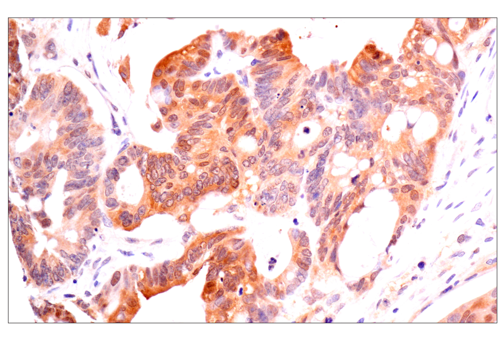
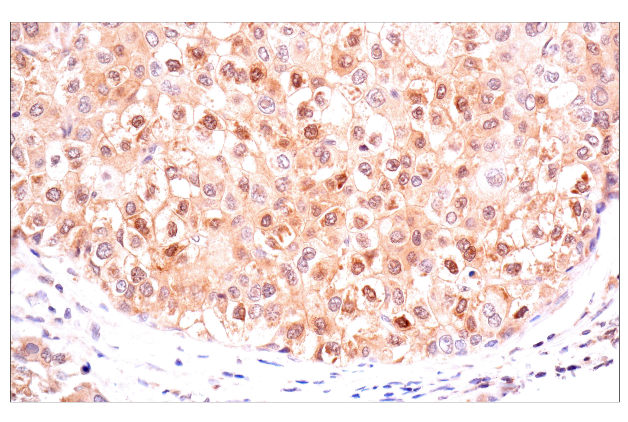
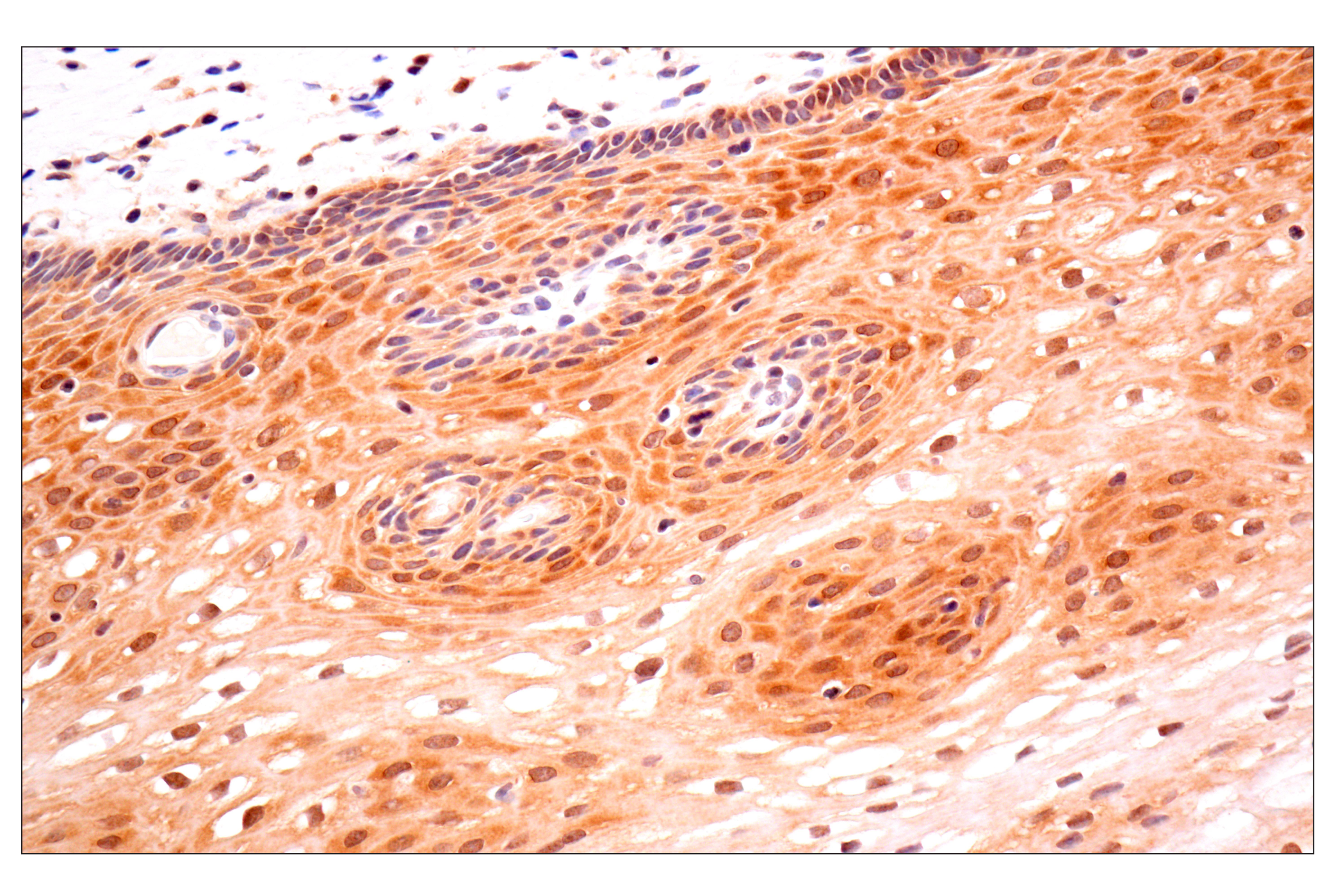
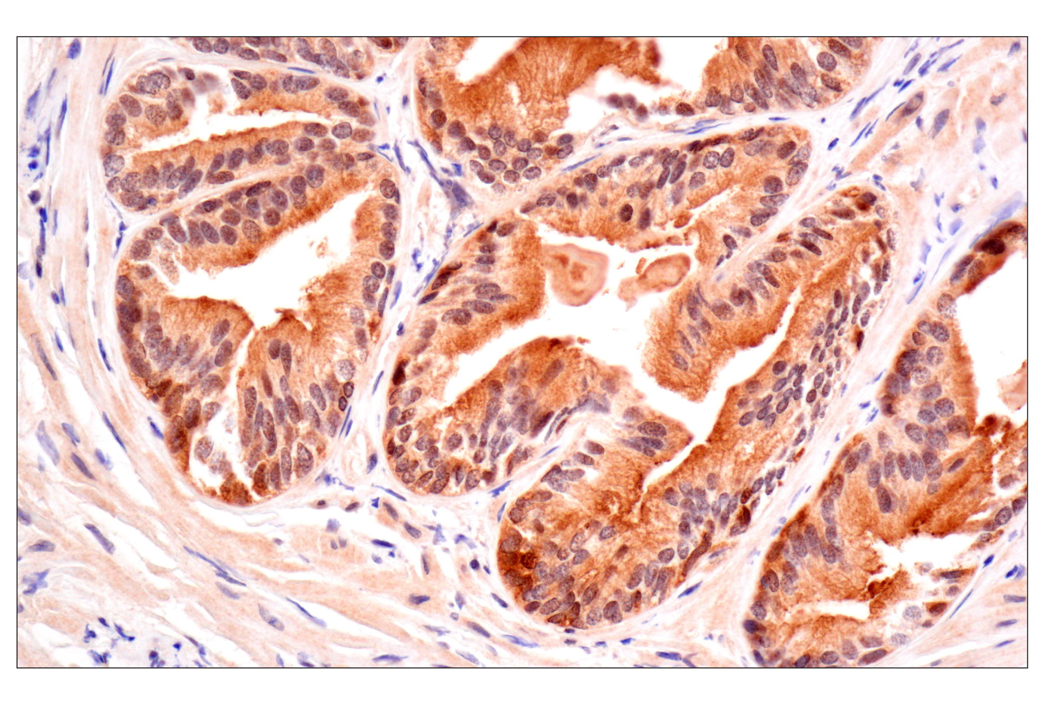
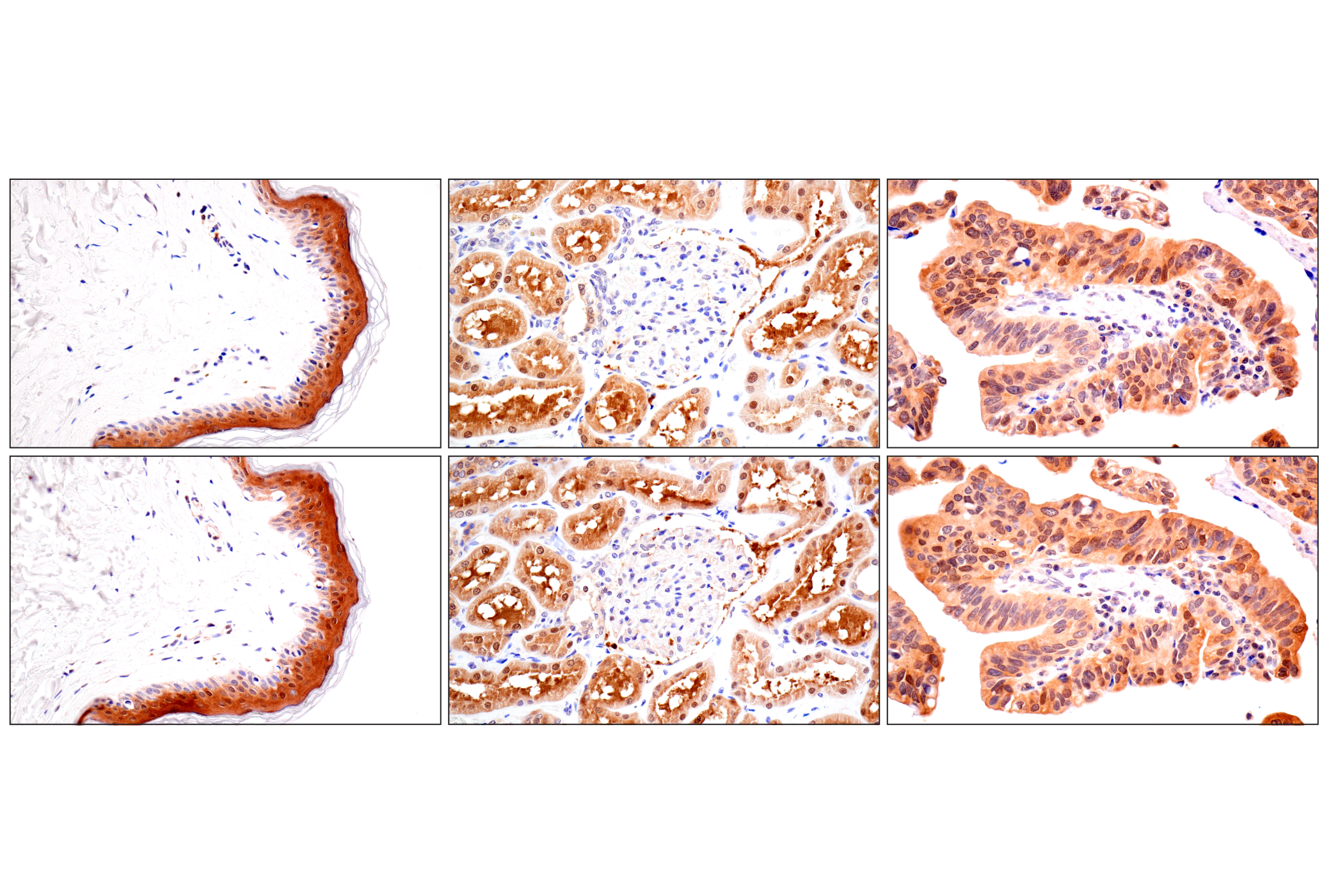
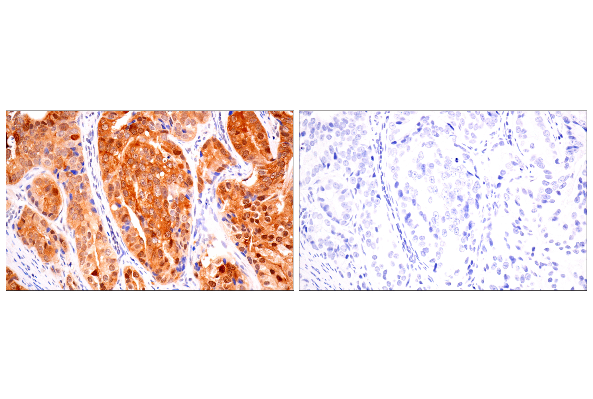
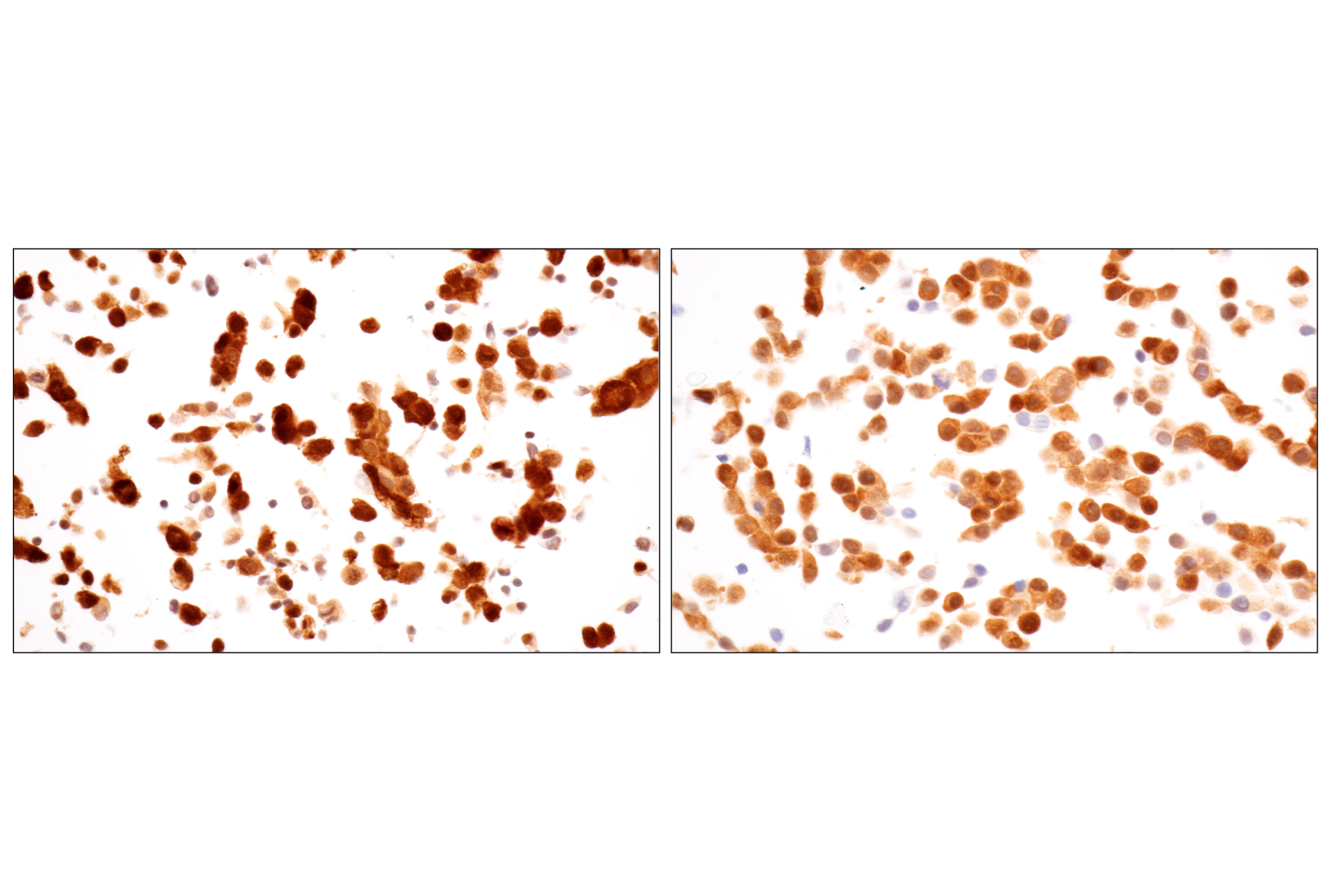
Background

Acyl-CoA-binding protein (ACBP) binds to long-chain acyl-CoA molecules and regulates lipid metabolism. This protein is also known as diazepam-binding inhibitor (DBI) and binds to the benzodiazepine-binding site of the γ-aminobutyric acid (GABA) type A receptor (GABAAR), regulating its activity (1). ACBP/DBI is highly expressed in glioblastoma and promotes glioblastoma growth by providing long-chain acyl-CoA molecules to mitochondria for fatty acid oxidation. Reduced ACBP/DBI expression leads to slower proliferation of glioblastoma (2,3). In addition, ACBP/DBI is a regulator of neural progenitor and stem cell proliferation in adult mouse neurogenic niches (4,5). Studies show that ACBP/DBI modulates embryonic neurogenesis of excitatory and inhibitory neurons through GABA signaling (6).

  1. Bravo-San Pedro, J.M. et al. (2019) Cell Metab 30, 754-767.e9.
  2. Duman, C. et al. (2019) Cell Metab 30, 274-289.e5.
  3. Bi, J. and Mischel, P.S. (2019) Cell Metab 30, 229-230.
  4. Alfonso, J. et al. (2012) Cell Stem Cell 10, 76-87.
  5. Dumitru, I. et al. (2017) Neuron 94, 125-137.e5.
  6. Everlien, I. et al. (2022) Neuron 110, 3139-3153.e6.

Species Reactivity

Species reactivity is determined by testing in at least one approved application (e.g., western blot).

Western Blot Buffer

IMPORTANT: For western blots, incubate membrane with diluted primary antibody in 5% w/v BSA, 1X TBS, 0.1% Tween® 20 at 4°C with gentle shaking, overnight.

Applications Key

WB: Western Blotting IHC-P: Immunohistochemistry (Paraffin)

Cross-Reactivity Key

H: human M: mouse R: rat Hm: hamster Mk: monkey Vir: virus Mi: mink C: chicken Dm: D. melanogaster X: Xenopus Z: zebrafish B: bovine Dg: dog Pg: pig Sc: S. cerevisiae Ce: C. elegans Hr: horse GP: Guinea Pig Rab: rabbit All: all species expected

Trademarks and Patents

Cell Signaling Technology is a trademark of Cell Signaling Technology, Inc.
XP is a registered trademark of Cell Signaling Technology, Inc.
All other trademarks are the property of their respective owners. Visit cellsignal.com/trademarks for more information.

使用に関する制限

法的な権限を与えられたCSTの担当者が署名した書面によって別途明示的に合意された場合を除き、 CST、その関連会社または代理店が提供する製品には以下の条件が適用されます。お客様が定める条件でここに定められた条件に含まれるものを超えるもの、 または、ここに定められた条件と異なるものは、法的な権限を与えられたCSTの担当者が別途書面にて受諾した場合を除き、拒絶され、 いかなる効力も効果も有しません。

研究専用 (For Research Use Only) またはこれに類似する表示がされた製品は、 いかなる目的についても FDA または外国もしくは国内のその他の規制機関により承認、認可または許可を受けていません。 お客様は製品を診断もしくは治療目的で使用してはならず、また、製品に表示された内容に違反する方法で使用してはなりません。 CST が販売または使用許諾する製品は、エンドユーザーであるお客様に対し、使途を研究および開発のみに限定して提供されるものです。 診断、予防もしくは治療目的で製品を使用することまたは製品を再販売 (単独であるか他の製品等の一部であるかを問いません) もしくはその他の商業的利用の目的で購入することについては、CST から別途許諾を得る必要があります。 お客様は以下の事項を遵守しなければなりません。(a) CST の製品 (単独であるか他の資材と一緒であるかを問いません) を販売、使用許諾、貸与、寄付もしくはその他の態様で第三者に譲渡したり使用させたりしてはなりません。また、商用の製品を製造するために CST の製品を使用してはなりません。(b) 複製、改変、リバースエンジニアリング、逆コンパイル、 分解または他の方法により製品の構造または技術を解明しようとしてはなりません。また、 CST の製品またはサービスと競合する製品またはサービスを開発する目的で CST の製品を使用してはなりません。(c) CST の製品の商標、商号、ロゴ、特許または著作権に関する通知または表示を除去したり改変したりしてはなりません。(d) CST の製品をCST 製品販売条件(CST’s Product Terms of Sale) および該当する書面のみに従って使用しなければなりません。(e) CST の製品に関連してお客様が使用する第三者の製品またはサービスに関する使用許諾条件、 サービス提供条件またはこれに類する合意事項を遵守しなければなりません。

Revision 1
#40487

ACBP/DBI (E4V8V) XP® Rabbit mAb

Western Blotting Image 1: ACBP/DBI (E4V8V) XP® Rabbit mAb Expand Image
Western blot analysis of extracts from HuH-7, SU-DHL-4, and KYSE-70 cells using ACBP/DBI (E4V8V) XP® Rabbit mAb (upper) or GAPDH (D16H11) XP® Rabbit mAb #5174 (lower). Lower expression of ACBP/DBI protein in KYSE-70 cells is consistent with the predicted expression pattern.
Western Blotting Image 2: ACBP/DBI (E4V8V) XP® Rabbit mAb Expand Image
Western blot analysis of extracts from 293T cells, mock transfected (-) or transfected with a construct expressing Myc/DDK-tagged human ACBP/DBI protein (hACBP/DBI-Myc/DDK; +), using ACBP/DBI (E4V8V) XP® Rabbit mAb (upper), Myc-Tag (71D10) Rabbit mAb #2278 (middle), or GAPDH (D16H11) XP® Rabbit mAb #5174 (lower).
Immunohistochemistry Image 1: ACBP/DBI (E4V8V) XP® Rabbit mAb Expand Image
Immunohistochemical analysis of paraffin-embedded human papillary thyroid carcinoma using ACBP/DBI (E4V8V) XP® Rabbit mAb.
Immunohistochemistry Image 2: ACBP/DBI (E4V8V) XP® Rabbit mAb Expand Image
Immunohistochemical analysis of paraffin-embedded human B-cell non-Hodgkin lymphoma using ACBP/DBI (E4V8V) XP® Rabbit mAb.
Immunohistochemistry Image 3: ACBP/DBI (E4V8V) XP® Rabbit mAb Expand Image

Immunohistochemical analysis of paraffin-embedded human colon carcinoma using ACBP/DBI (E4V8V) XP® Rabbit mAb.
Immunohistochemistry Image 4: ACBP/DBI (E4V8V) XP® Rabbit mAb Expand Image
Immunohistochemical analysis of paraffin-embedded human hepatocellular carcinoma using ACBP/DBI (E4V8V) XP® Rabbit mAb.
Immunohistochemistry Image 5: ACBP/DBI (E4V8V) XP® Rabbit mAb Expand Image
Immunohistochemical analysis of paraffin-embedded normal human esophagus using ACBP/DBI (E4V8V) XP® Rabbit mAb.
Immunohistochemistry Image 6: ACBP/DBI (E4V8V) XP® Rabbit mAb Expand Image

Immunohistochemical analysis of paraffin-embedded normal human prostate using ACBP/DBI (E4V8V) XP® Rabbit mAb.

Immunohistochemistry Image 7: ACBP/DBI (E4V8V) XP® Rabbit mAb Expand Image
Immunohistochemical analysis of paraffin-embedded normal human pancreas using ACBP/DBI (E4V8V) XP® Rabbit mAb.
Immunohistochemistry Image 8: ACBP/DBI (E4V8V) XP® Rabbit mAb Expand Image
Immunohistochemical analysis of paraffin-embedded normal human lung using ACBP/DBI (E4V8V) XP® Rabbit mAb.
Immunohistochemistry Image 9: ACBP/DBI (E4V8V) XP® Rabbit mAb Expand Image
Immunohistochemical analysis of paraffin-embedded Renca syngeneic tumor using ACBP/DBI (E4V8V) XP® Rabbit mAb.
Immunohistochemistry Image 10: ACBP/DBI (E4V8V) XP® Rabbit mAb Expand Image
Immunohistochemical analysis of paraffin-embedded mouse small intestine using ACBP/DBI (E4V8V) XP® Rabbit mAb.
Immunohistochemistry Image 11: ACBP/DBI (E4V8V) XP® Rabbit mAb Expand Image
Immunohistochemical analysis of paraffin-embedded mouse lung using ACBP/DBI (E4V8V) XP® Rabbit mAb.
Immunohistochemistry Image 12: ACBP/DBI (E4V8V) XP® Rabbit mAb Expand Image
Immunohistochemical analysis of paraffin-embedded mouse liver using ACBP/DBI (E4V8V) XP® Rabbit mAb.
Immunohistochemistry Image 13: ACBP/DBI (E4V8V) XP® Rabbit mAb Expand Image
Immunohistochemical analysis of paraffin-embedded mouse kidney using ACBP/DBI (E4V8V) XP® Rabbit mAb.
Immunohistochemistry Image 14: ACBP/DBI (E4V8V) XP® Rabbit mAb Expand Image
Immunohistochemical analysis of paraffin-embedded mouse brown fat using ACBP/DBI (E4V8V) XP® Rabbit mAb.
Immunohistochemistry Image 15: ACBP/DBI (E4V8V) XP® Rabbit mAb Expand Image
Immunohistochemical analysis of paraffin-embedded normal human skin (left), normal kidney (middle), or esophageal carcinoma (right) using ACBP/DBI (E4V8V) XP® Rabbit mAb (top) or ACBP/DBI (E6K8G) XP® Rabbit mAb #45365 (bottom). These two antibodies detect unique epitopes on human ACBP/DBI protein. The similar staining patterns obtained with both antibodies help to confirm the specificity of the staining.
Immunohistochemistry Image 16: ACBP/DBI (E4V8V) XP® Rabbit mAb Expand Image
Immunohistochemical analysis of paraffin-embedded human prostate adenocarcinoma using ACBP/DBI (E4V8V) XP® Rabbit mAb (left) compared to concentration-matched Rabbit (DA1E) mAb IgG XP® Isotype Control #3900 (right).
Immunohistochemistry Image 17: ACBP/DBI (E4V8V) XP® Rabbit mAb Expand Image
Immunohistochemical analysis of paraffin-embedded HuH-7 cell pellet (left, high-expressing) or KYSE-70 cell pellet (right, low-expressing) using ACBP/DBI (E4V8V) XP® Rabbit mAb.