| Product Includes | Product # | Quantity | Mol. Wt | Isotype/Source |
|---|---|---|---|---|
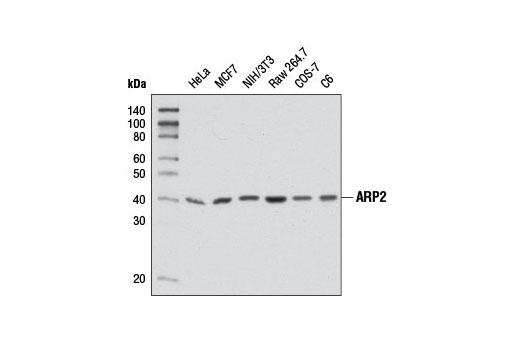
| ARP2 (D85D5) Rabbit mAb | 5614 | 40 µl | 44 kDa | Rabbit IgG |
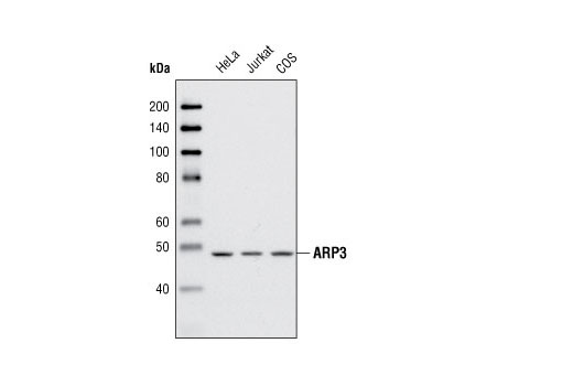
| ARP3 Antibody | 4738 | 40 µl | 47 kDa | Rabbit |
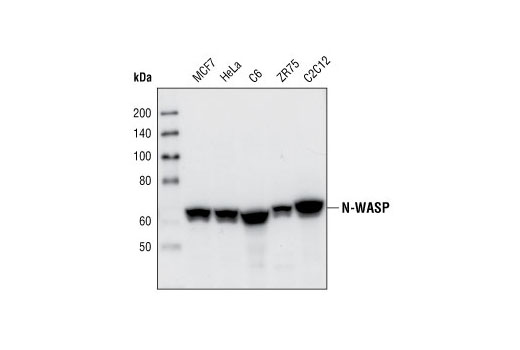
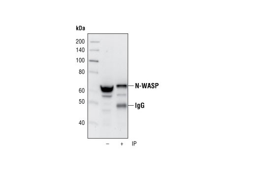
| N-WASP (30D10) Rabbit mAb | 4848 | 40 µl | 65 kDa | Rabbit IgG |
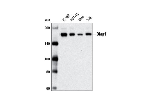
| Diap1 Antibody | 5486 | 40 µl | 150 kDa | Rabbit |
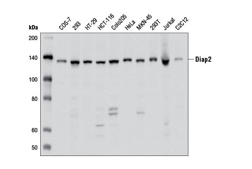
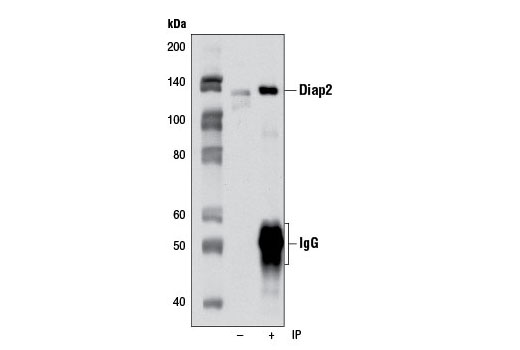
| Diap2 Antibody | 5474 | 40 µl | 130 kDa | Rabbit |
| Anti-rabbit IgG, HRP-linked Antibody | 7074 | 100 µl | Goat |
Please visit cellsignal.com for individual component applications, species cross-reactivity, dilutions, protocols, and additional product information.
Description
The Actin Nucleation Antibody Sampler Kit provides an economical means to evaluate the presence and status of actin nucleation. The kit contains enough primary and secondary antibodies to perform four western blot experiments per primary antibody.
Storage
Background
Actin nucleation, the formation of new actin filaments from existing filaments, affects actin filament structure during cell motility, division, and intracellular trafficking. An important actin nucleation protein complex is the highly conserved ARP2/3 complex, consisting of ARP2, ARP3, and ARPC1-5. The ARP2/3 complex promotes branching of an existing actin filament and formation of a daughter filament following activation by nucleation-promoting factors, such as WASP/WAVE or cortactin (1). The formation of podosomes, small cellular projections that degrade the extracellular matrix, is enhanced by ARP2/3 complex action. ARP2/3 competes with caldesmon, an actin binding protein shown to negatively affect podosome formation (2). Along with N-WASP, the ARP2/3 complex regulates nuclear actin filament nucleation and controls actin polymerization during transcription (3).
Formins are a family of large multidomain actin nucleation/polymerization proteins characterized by their catalytic FH2 domains. The mammalian diaphanous-related formin (mDia/diap) subfamily, including mDia1/diap1, mDia2/diap3 and mDia3/diap2, are effectors of Rho family small GTPases. In response to Rho, mDia/diap proteins are involved in the regulation of multiple cell functions including cytoskeletal dynamics, migration, adhesion, polarity, and cell shape (reviewed in 4,5).
mDia1/diap1 is activated by GTP-bound Rho, leading to Rho-associated kinase (ROCK)-dependent stress fiber formation (6,7). Rho activation of mDia1 has also been shown to regulate serum response factor (SRF)-dependent transcription (8), and has been implicated in human cancer phenotypes such as ras-mediated transformation, metastasis, and invasion (reviewed in 9).
mDia3/diap2, activated by the Rho family small GTPase cdc42, regulates the attachment of microtubules to the kinetochore during mitosis in mammalian cells (10).
Rho-dependent activation of mDia2/diap3 is important in assembly of the contractile ring during cytokinesis (11,12).
- Goley, E.D. and Welch, M.D. (2006) Nat. Rev. Mol. Cell Biol. 7, 713-726.
- Morita, T. et al. (2007) J. Biol. Chem. 282, 8454-8463.
- Yoo, Y. et al. (2007) J. Biol. Chem. 282, 7616-7623.
- Schönichen, A. and Geyer, M. (2010) Biochim Biophys Acta 1803, 152-63.
- Chesarone, M.A. et al. (2010) Nat Rev Mol Cell Biol 11, 62-74.
- Watanabe, N. et al. (1999) Nat Cell Biol 1, 136-43.
- Ishizaki, T. et al. (2001) Nat Cell Biol 3, 8-14.
- Copeland, J.W. and Treisman, R. (2002) Mol Biol Cell 13, 4088-99.
- Narumiya, S. et al. (2009) Cancer Metastasis Rev 28, 65-76.
- Yasuda, S. et al. (2004) Nature 428, 767-71.
- Watanabe, S. et al. (2010) Mol Biol Cell 21, 3193-204.
- Watanabe, S. et al. (2008) Mol Biol Cell 19, 2328-38.
Background References
Trademarks and Patents
使用に関する制限
法的な権限を与えられたCSTの担当者が署名した書面によって別途明示的に合意された場合を除き、 CST、その関連会社または代理店が提供する製品には以下の条件が適用されます。お客様が定める条件でここに定められた条件に含まれるものを超えるもの、 または、ここに定められた条件と異なるものは、法的な権限を与えられたCSTの担当者が別途書面にて受諾した場合を除き、拒絶され、 いかなる効力も効果も有しません。
研究専用 (For Research Use Only) またはこれに類似する表示がされた製品は、 いかなる目的についても FDA または外国もしくは国内のその他の規制機関により承認、認可または許可を受けていません。 お客様は製品を診断もしくは治療目的で使用してはならず、また、製品に表示された内容に違反する方法で使用してはなりません。 CST が販売または使用許諾する製品は、エンドユーザーであるお客様に対し、使途を研究および開発のみに限定して提供されるものです。 診断、予防もしくは治療目的で製品を使用することまたは製品を再販売 (単独であるか他の製品等の一部であるかを問いません) もしくはその他の商業的利用の目的で購入することについては、CST から別途許諾を得る必要があります。 お客様は以下の事項を遵守しなければなりません。(a) CST の製品 (単独であるか他の資材と一緒であるかを問いません) を販売、使用許諾、貸与、寄付もしくはその他の態様で第三者に譲渡したり使用させたりしてはなりません。また、商用の製品を製造するために CST の製品を使用してはなりません。(b) 複製、改変、リバースエンジニアリング、逆コンパイル、 分解または他の方法により製品の構造または技術を解明しようとしてはなりません。また、 CST の製品またはサービスと競合する製品またはサービスを開発する目的で CST の製品を使用してはなりません。(c) CST の製品の商標、商号、ロゴ、特許または著作権に関する通知または表示を除去したり改変したりしてはなりません。(d) CST の製品をCST 製品販売条件(CST’s Product Terms of Sale) および該当する書面のみに従って使用しなければなりません。(e) CST の製品に関連してお客様が使用する第三者の製品またはサービスに関する使用許諾条件、 サービス提供条件またはこれに類する合意事項を遵守しなければなりません。