| Product Includes | Product # | Quantity | Mol. Wt | Isotype/Source |
|---|---|---|---|---|
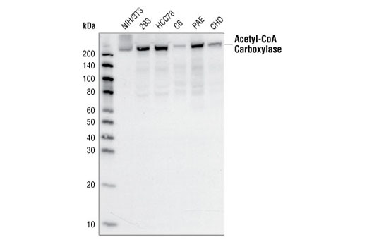
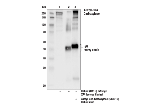
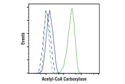
| Acetyl-CoA Carboxylase (C83B10) Rabbit mAb | 3676 | 20 µl | 280 kDa | Rabbit IgG |
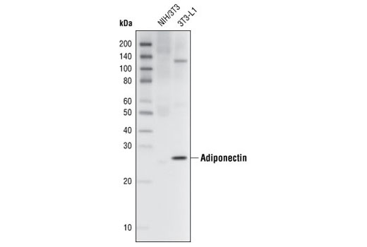
| Adiponectin (C45B10) Rabbit mAb | 2789 | 20 µl | 27 kDa | Rabbit IgG |
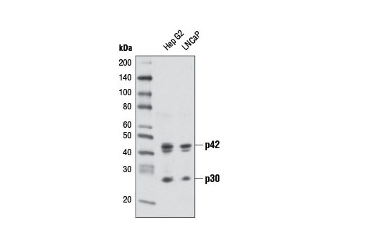
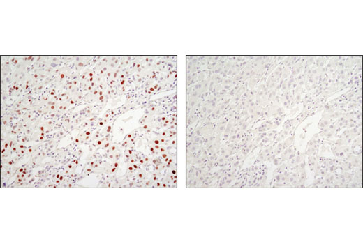
| C/EBPα (D56F10) XP® Rabbit mAb | 8178 | 20 µl | 42, 28 kDa | Rabbit IgG |
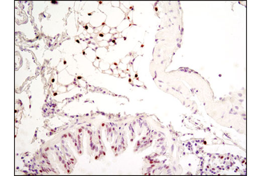
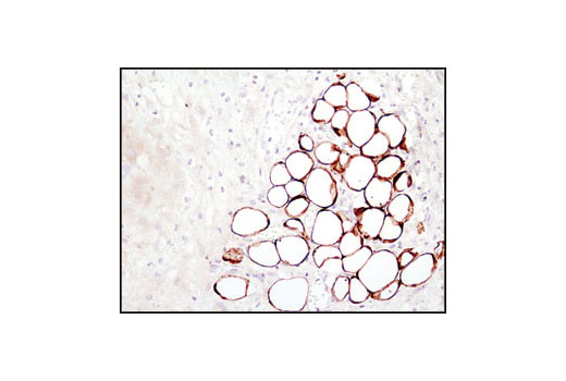
| FABP4 Antibody | 2120 | 20 µl | 15 kDa | Rabbit |
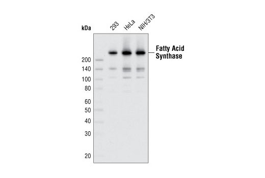
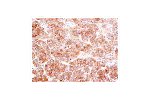
| Fatty Acid Synthase (C20G5) Rabbit mAb | 3180 | 20 µl | 273 kDa | Rabbit IgG |
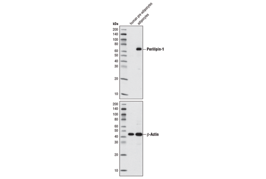
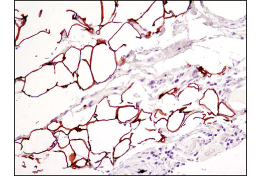
| Perilipin-1 (D1D8) XP® Rabbit mAb | 9349 | 20 µl | 62 kDa | Rabbit IgG |
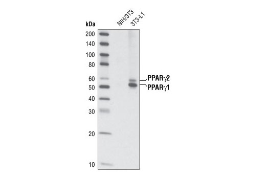
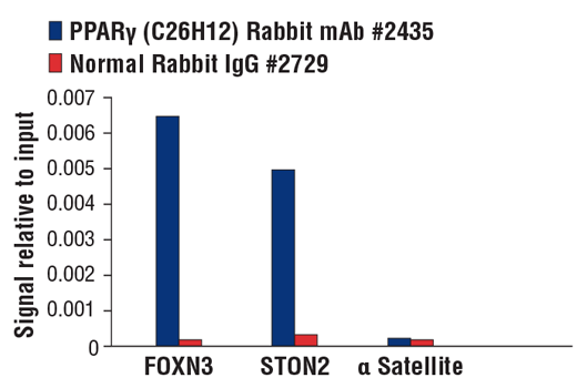
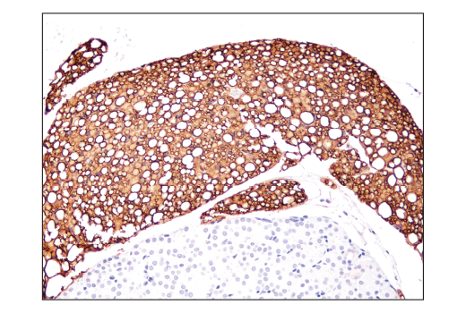
| PPARγ (C26H12) Rabbit mAb | 2435 | 20 µl | 53, 57 kDa | Rabbit IgG |
| Anti-rabbit IgG, HRP-linked Antibody | 7074 | 100 µl | Goat |
Please visit cellsignal.com for individual component applications, species cross-reactivity, dilutions, protocols, and additional product information.
Description
The Adipogenesis Marker Antibody Sampler Kit provides an economical means to evaluate proteins involved in the regulation of adipogenesis. The kit includes enough antibody to perform two western blot experiments with each primary antibody.
Storage
Background
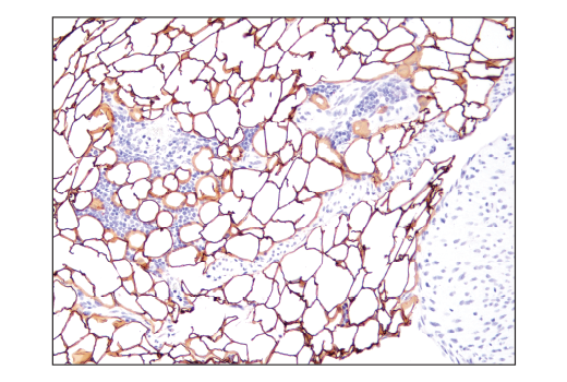
Adipocytes are the primary cellular component of adipose tissue and play a key role in the storage of triacylglycerol. Adipogenesis is the cellular process where preadipocytes differentiate into adipocytes.
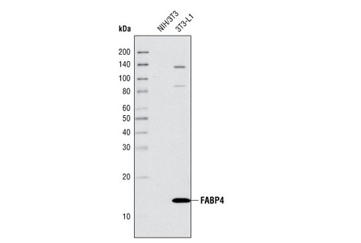
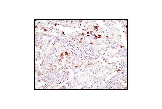
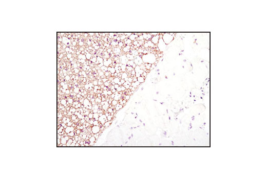
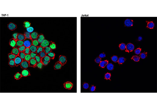
Fatty acid binding proteins (FABPs) act as cytoplasmic lipid chaperones by binding fatty acids and lipids for transport to various cellular pathways (1,2). The predominant fatty acid binding protein found in adipocytes is FABP4.
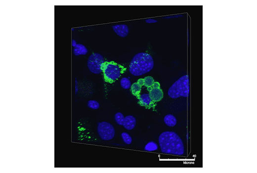
Adiponectin is an adipokine expressed exclusively in brown and white adipocytes and is secreted into the blood. It exists in three major forms: a low molecular weight trimer, a medium molecular weight hexamer and a high molecular weight multimer (3). Decreased adiponectin levels are seen in obese and insulin-resistant mice and humans (4), suggesting that this adipokine is critical for maintenance of insulin sensitivity.
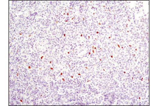
Peroxisome proliferator-activated receptor γ (PPARγ) is a transcriptional activator preferentially expressed in adipocytes, vascular smooth muscle cells, and macrophages (5,6).
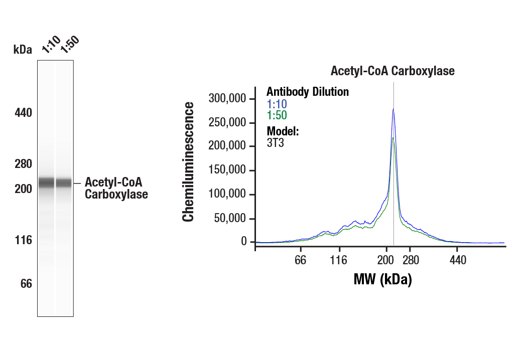
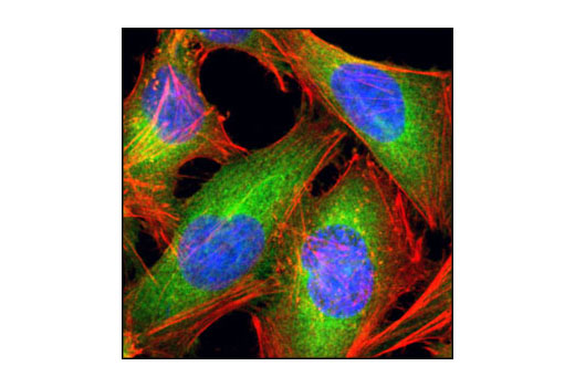
Acetyl-CoA carboxylase (ACC) is a key fatty acid biosynthesis and oxidation enzyme that is responsible for the carboxylation of acetyl-CoA to malonyl-CoA, (7). Phosphorylation of acetyl-CoA carboxylase by AMPK at Ser79 or by PKA at Ser1200 inhibits ACC enzymatic activity (8). ACC is a potential target of anti-obesity drugs (9,10).
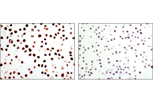
CCAAT/enhancer-binding proteins (C/EBPs) transcription factors are critical for cellular differentiation, terminal function, and the inflammatory response (11). Phosphorylation of C/EBPα at Thr222, Thr226, and Ser230 by GSK-3 may be required for adipogenesis (12).
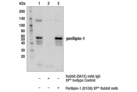
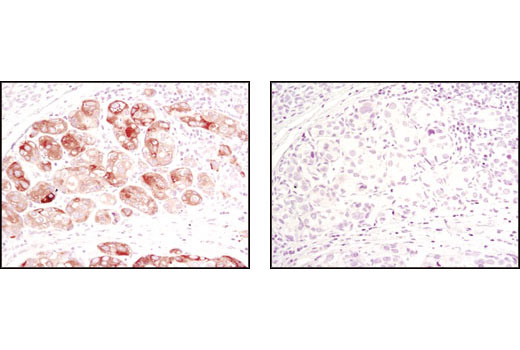
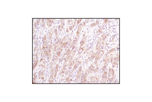
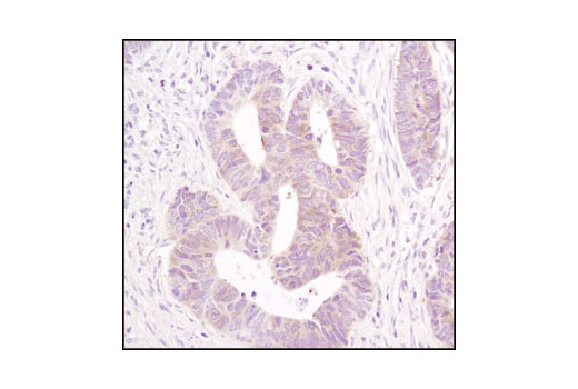
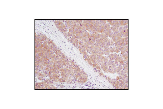
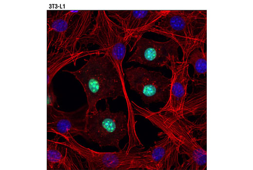
Perilipin localizes to the periphery of lipid droplets and serves as a protective coating against lipases. Evidence suggests that PKA regulates lipolysis by phosphorylating perilipin (13-17), resulting in a conformational change that exposes lipid droplets to endogenous, hormone-sensitive lipases (14). Hence, perilipin plays a pivotal role in lipid storage (14,17).
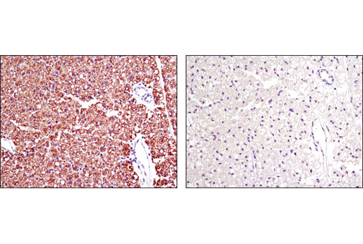
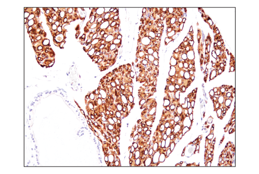
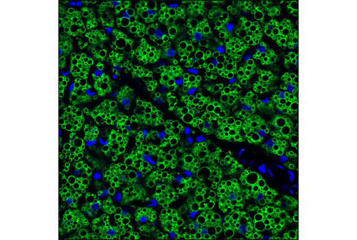
Fatty acid synthase (FASN) catalyzes the synthesis of long-chain fatty acids from acetyl-CoA and malonyl-CoA. FASN is active as a homodimer with seven different catalytic activities and produces lipids in the liver for export to metabolically active tissues or storage in adipose tissue. In most other human tissues, FASN is minimally expressed since they rely on circulating fatty acids for new structural lipid synthesis (18).
- Tuncman, G. et al. (2006) Proc Natl Acad Sci U S A 103, 6970-5.
- Haunerland, N.H. and Spener, F. (2004) Prog Lipid Res 43, 328-49.
- Kadowaki, T. et al. (2006) J Clin Invest 116, 1784-92.
- Hu, E. et al. (1996) J Biol Chem 271, 10697-703.
- Tontonoz, P. et al. (1995) Curr Opin Genet Dev 5, 571-6.
- Rosen, E.D. et al. (1999) Mol Cell 4, 611-7.
- Castle, J.C. et al. (2009) PLoS One 4, e4369.
- Ha, J. et al. (1994) J Biol Chem 269, 22162-8.
- Abu-Elheiga, L. et al. (2001) Science 291, 2613-6.
- Levert, K.L. et al. (2002) J Biol Chem 277, 16347-50.
- Lekstrom-Himes, J. and Xanthopoulos, K.G. (1998) J Biol Chem 273, 28545-8.
- Ross, S.E. et al. (1999) Mol Cell Biol 19, 8433-41.
- Greenberg, A.S. et al. (1991) J Biol Chem 266, 11341-6.
- Brasaemle, D.L. (2007) J Lipid Res 48, 2547-59.
- Ducharme, N.A. and Bickel, P.E. (2008) Endocrinology 149, 942-9.
- Egan, J.J. et al. (1990) J Biol Chem 265, 18769-75.
- Brasaemle, D.L. et al. (2009) Mol Cell Biochem 326, 15-21.
- Katsurada, A. et al. (1990) Eur J Biochem 190, 427-33.
Background References
Trademarks and Patents
使用に関する制限
法的な権限を与えられたCSTの担当者が署名した書面によって別途明示的に合意された場合を除き、 CST、その関連会社または代理店が提供する製品には以下の条件が適用されます。お客様が定める条件でここに定められた条件に含まれるものを超えるもの、 または、ここに定められた条件と異なるものは、法的な権限を与えられたCSTの担当者が別途書面にて受諾した場合を除き、拒絶され、 いかなる効力も効果も有しません。
研究専用 (For Research Use Only) またはこれに類似する表示がされた製品は、 いかなる目的についても FDA または外国もしくは国内のその他の規制機関により承認、認可または許可を受けていません。 お客様は製品を診断もしくは治療目的で使用してはならず、また、製品に表示された内容に違反する方法で使用してはなりません。 CST が販売または使用許諾する製品は、エンドユーザーであるお客様に対し、使途を研究および開発のみに限定して提供されるものです。 診断、予防もしくは治療目的で製品を使用することまたは製品を再販売 (単独であるか他の製品等の一部であるかを問いません) もしくはその他の商業的利用の目的で購入することについては、CST から別途許諾を得る必要があります。 お客様は以下の事項を遵守しなければなりません。(a) CST の製品 (単独であるか他の資材と一緒であるかを問いません) を販売、使用許諾、貸与、寄付もしくはその他の態様で第三者に譲渡したり使用させたりしてはなりません。また、商用の製品を製造するために CST の製品を使用してはなりません。(b) 複製、改変、リバースエンジニアリング、逆コンパイル、 分解または他の方法により製品の構造または技術を解明しようとしてはなりません。また、 CST の製品またはサービスと競合する製品またはサービスを開発する目的で CST の製品を使用してはなりません。(c) CST の製品の商標、商号、ロゴ、特許または著作権に関する通知または表示を除去したり改変したりしてはなりません。(d) CST の製品をCST 製品販売条件(CST’s Product Terms of Sale) および該当する書面のみに従って使用しなければなりません。(e) CST の製品に関連してお客様が使用する第三者の製品またはサービスに関する使用許諾条件、 サービス提供条件またはこれに類する合意事項を遵守しなければなりません。