Revision 7

#74417Store at -20C

Cell Signaling Technology

Orders: 877-616-CELL (2355) [email protected]

Support: 877-678-TECH (8324)

Web: [email protected] cellsignal.com

3 Trask LaneDanversMassachusetts01923USA
For Research Use Only. Not for Use in Diagnostic Procedures.
Applications:

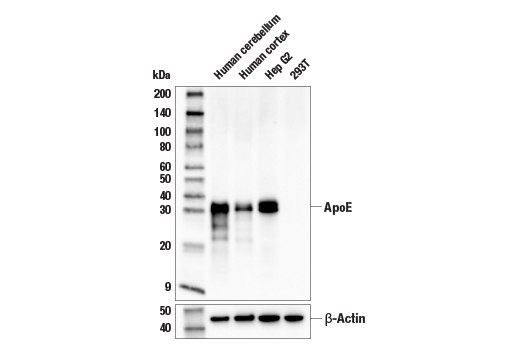
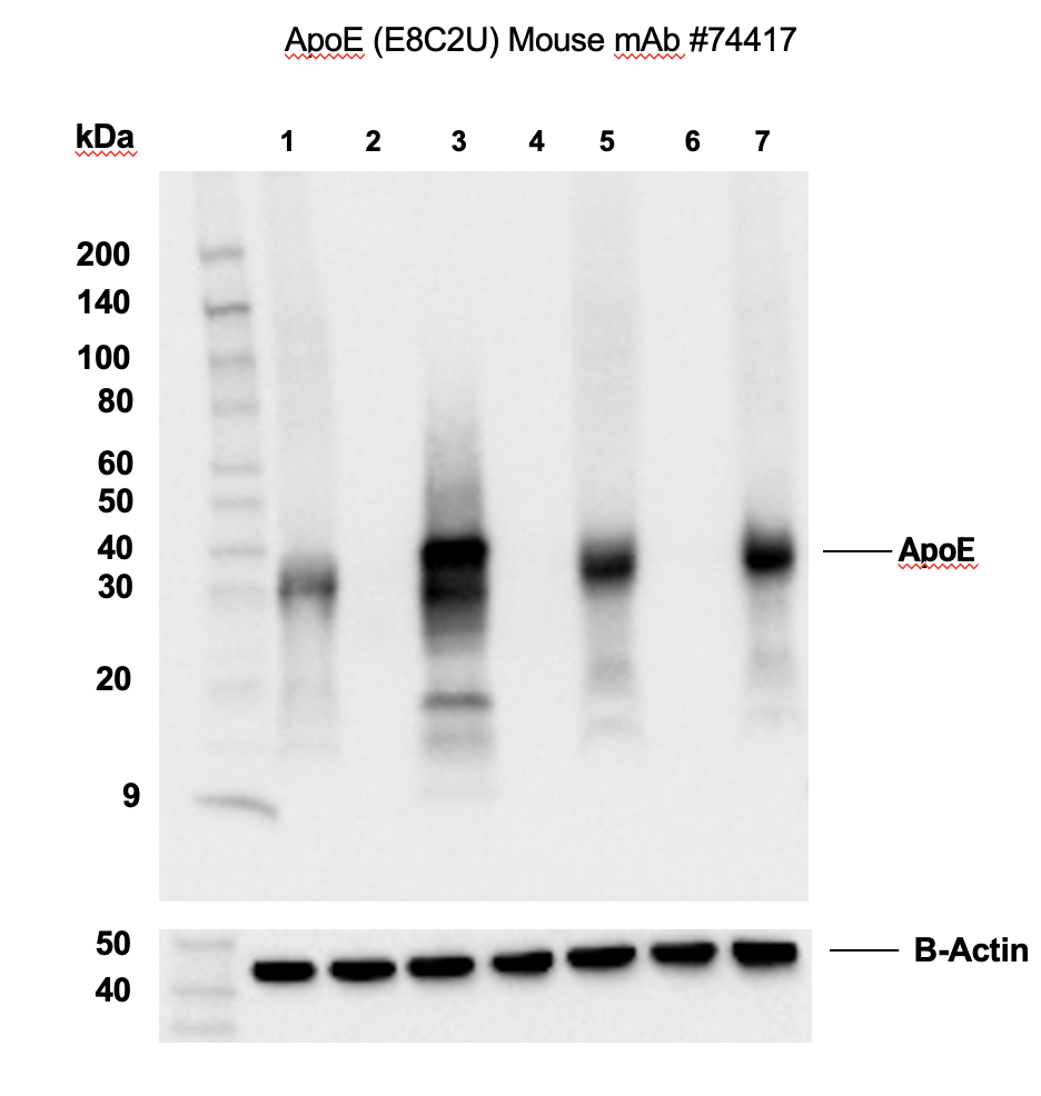
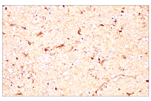
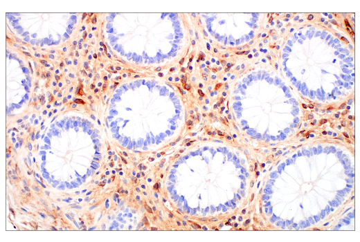
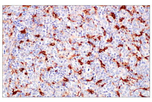
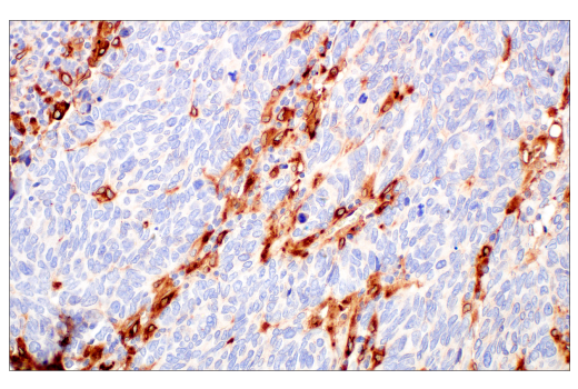
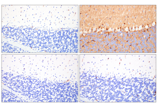
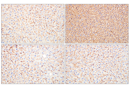
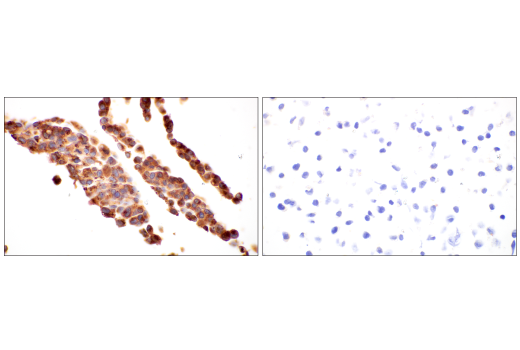
WB, IHC-P, IF-F, IF-IC

REACTIVITY:

H

SENSITIVITY:

Endogenous

MW (kDa):

35

Source/Isotype:

Mouse IgG2b

UniProt ID:

#P02649

Entrez-Gene Id:

348

Product Information

Product Usage Information

Application Dilution
Western Blotting 1:1000
Immunohistochemistry (Paraffin) 1:50 - 1:200
Immunofluorescence (Frozen) 1:50 - 1:200
Immunofluorescence (Immunocytochemistry) 1:50 - 1:200

Storage

Supplied in 10 mM sodium HEPES (pH 7.5), 150 mM NaCl, 100 µg/mL BSA, 50% glycerol, and less than 0.02% sodium azide. Store at –20°C. Do not aliquot the antibody.

For a carrier free (BSA and azide free) version of this product see product #31516.

Specificity / Sensitivity

ApoE (pan) (E8C2U) Mouse mAb recognizes endogenous levels of total ApoE protein.

Species Reactivity:

Human

Source / Purification

Monoclonal antibody is produced by immunizing animals with a synthetic peptide corresponding to residues near the carboxy terminus of human ApoE protein.

Background

Apolipoproteins are plasma lipoproteins that function as transporters of lipids and cholesterol in the circulatory system. Chylomicrons are a fundamental class of apolipoproteins containing very low-density lipoproteins (VLDL), intermediate-density lipoproteins (IDL), low-density lipoproteins (LDL), and high-density lipoproteins (HDL) (1,2).
Human ApoE has three isoforms: ApoE2, ApoE3, and ApoE4. These three isoforms differ in the combination of cysteine and arginine residues located at positions 130 and 176. The ApoE4 isoform contains arginine at both locations. Research studies have linked ApoE4 function to neuronal plasticity, synaptogenesis, and neurodegenerative diseases (3). ApoE4 is produced in the liver and brain, although it is widely expressed in other tissues, such as the lung, spleen, and ovary. Investigators have established the ApoE4 allele as a genetic risk factor for Alzheimer’s disease (AD), accounting for 50-60% of the genetic variation in the disease (4). Research studies indicate that patients expressing ApoE4 have a reduced capacity for synaptic plasticity, an earlier age of onset of AD, and an increase in amyloid-beta (Aβ) deposition. The increase in Aβ suggests a role for ApoE4 in the impairment of amyloid clearance (5).

  1. Kwiterovich, P.O. (2000) Am J Cardiol 86, 5L-10L.
  2. Hussain, M.M. (2000) Atherosclerosis 148, 1-15.
  3. Raber, J. et al. Neurobiol Aging 25, 641-50.
  4. Corder, E.H. et al. (1993) Science 261, 921-3.
  5. Holtzman, D.M. et al. (2000) Proc Natl Acad Sci U S A 97, 2892-7.

Species Reactivity

Species reactivity is determined by testing in at least one approved application (e.g., western blot).

Western Blot Buffer

IMPORTANT: For western blots, incubate membrane with diluted primary antibody in 5% w/v BSA, 1X TBS, 0.1% Tween® 20 at 4°C with gentle shaking, overnight.

Applications Key

WB: Western Blotting IHC-P: Immunohistochemistry (Paraffin) IF-F: Immunofluorescence (Frozen) IF-IC: Immunofluorescence (Immunocytochemistry)

Cross-Reactivity Key

H: human M: mouse R: rat Hm: hamster Mk: monkey Vir: virus Mi: mink C: chicken Dm: D. melanogaster X: Xenopus Z: zebrafish B: bovine Dg: dog Pg: pig Sc: S. cerevisiae Ce: C. elegans Hr: horse GP: Guinea Pig Rab: rabbit All: all species expected

Trademarks and Patents

Cell Signaling Technology is a trademark of Cell Signaling Technology, Inc.
SignalStain is a registered trademark of Cell Signaling Technology, Inc.
All other trademarks are the property of their respective owners. Visit cellsignal.com/trademarks for more information.

使用に関する制限

法的な権限を与えられたCSTの担当者が署名した書面によって別途明示的に合意された場合を除き、 CST、その関連会社または代理店が提供する製品には以下の条件が適用されます。お客様が定める条件でここに定められた条件に含まれるものを超えるもの、 または、ここに定められた条件と異なるものは、法的な権限を与えられたCSTの担当者が別途書面にて受諾した場合を除き、拒絶され、 いかなる効力も効果も有しません。

研究専用 (For Research Use Only) またはこれに類似する表示がされた製品は、 いかなる目的についても FDA または外国もしくは国内のその他の規制機関により承認、認可または許可を受けていません。 お客様は製品を診断もしくは治療目的で使用してはならず、また、製品に表示された内容に違反する方法で使用してはなりません。 CST が販売または使用許諾する製品は、エンドユーザーであるお客様に対し、使途を研究および開発のみに限定して提供されるものです。 診断、予防もしくは治療目的で製品を使用することまたは製品を再販売 (単独であるか他の製品等の一部であるかを問いません) もしくはその他の商業的利用の目的で購入することについては、CST から別途許諾を得る必要があります。 お客様は以下の事項を遵守しなければなりません。(a) CST の製品 (単独であるか他の資材と一緒であるかを問いません) を販売、使用許諾、貸与、寄付もしくはその他の態様で第三者に譲渡したり使用させたりしてはなりません。また、商用の製品を製造するために CST の製品を使用してはなりません。(b) 複製、改変、リバースエンジニアリング、逆コンパイル、 分解または他の方法により製品の構造または技術を解明しようとしてはなりません。また、 CST の製品またはサービスと競合する製品またはサービスを開発する目的で CST の製品を使用してはなりません。(c) CST の製品の商標、商号、ロゴ、特許または著作権に関する通知または表示を除去したり改変したりしてはなりません。(d) CST の製品をCST 製品販売条件(CST’s Product Terms of Sale) および該当する書面のみに従って使用しなければなりません。(e) CST の製品に関連してお客様が使用する第三者の製品またはサービスに関する使用許諾条件、 サービス提供条件またはこれに類する合意事項を遵守しなければなりません。

Revision 7
#74417

ApoE (pan) (E8C2U) Mouse mAb

Western Blotting Image 1: ApoE (pan) (E8C2U) Mouse mAb Expand Image
Western blot analysis of extracts from various tissues and cell lines using ApoE (pan) (E8C2U) Mouse mAb (upper) or β-Actin (D6A8) Rabbit mAb #8457 (lower).
Western Blotting Image 2: ApoE (pan) (E8C2U) Mouse mAb Expand Image
Western blot analysis of extracts from HepG2 cells (lane 1), 293T mock transfected (lane 2) or transiently transfected with a construct expressing ApoE4 (lane 3), whole liver extracts from wild type C57BL/6NTac model #B6-F mice (lane 4), or ApoE4 knock-in (model 1549-F) (lane 5), whole brain extracts from wild type C57BL/6NTac mice (lane 6), or ApoE4 knock-in (lane 7) using ApoE (E8C2U) Mouse mAb (upper) and β-Actin (D6A8) Rabbit mAb #8457 (lower). Mice from Taconic Biosciences, Inc.
Immunohistochemistry Image 1: ApoE (pan) (E8C2U) Mouse mAb Expand Image
Immunohistochemical analysis of paraffin-embedded normal human brain using ApoE (pan) (E8C2U) Mouse mAb.
Immunohistochemistry Image 2: ApoE (pan) (E8C2U) Mouse mAb Expand Image
Immunohistochemical analysis of paraffin-embedded normal human colon using ApoE (pan) (E8C2U) Mouse mAb.
Immunohistochemistry Image 3: ApoE (pan) (E8C2U) Mouse mAb Expand Image
Immunohistochemical analysis of paraffin-embedded normal human lymph node using ApoE (pan) (E8C2U) Mouse mAb.
Immunohistochemistry Image 4: ApoE (pan) (E8C2U) Mouse mAb Expand Image
Immunohistochemical analysis of paraffin-embedded normal human placenta using ApoE (pan) (E8C2U) Mouse mAb.
Immunohistochemistry Image 5: ApoE (pan) (E8C2U) Mouse mAb Expand Image
Immunohistochemical analysis of paraffin-embedded human hepatocellular carcinoma using ApoE (pan) (E8C2U) Mouse mAb.
Immunohistochemistry Image 6: ApoE (pan) (E8C2U) Mouse mAb Expand Image
Immunohistochemical analysis of paraffin-embedded human urothelial carcinoma using ApoE (pan) (E8C2U) Mouse mAb.
Immunohistochemistry Image 7: ApoE (pan) (E8C2U) Mouse mAb Expand Image
Immunohistochemical analysis of paraffin-embedded human B-cell non-Hodgkin lymphoma using ApoE (pan) (E8C2U) Mouse mAb.
Immunohistochemistry Image 8: ApoE (pan) (E8C2U) Mouse mAb Expand Image
Immunohistochemical analysis of paraffin-embedded human small cell carcinoma of the salivary gland using ApoE (pan) (E8C2U) Mouse mAb.
Immunohistochemistry Image 9: ApoE (pan) (E8C2U) Mouse mAb Expand Image
Immunohistochemical analysis of paraffin-embedded brain from C57BL/6NTac (wt/wt, model #B6-F, left) and APOE4 knock-in (hu/hu, model #1549-F, right) mice using ApoE (pan) (E8C2U) Mouse mAb (top) compared to concentration-matched Mouse (E7Q5L) mAb IgG2b Isotype Control #53484 (bottom). Mice from Taconic Biosciences, Inc. Note non-specific mouse on mouse signal in samples with enhanced specific signal in the APOE4 knock-in tissue.
Immunohistochemistry Image 10: ApoE (pan) (E8C2U) Mouse mAb Expand Image
Immunohistochemical analysis of paraffin-embedded liver from C57BL/6NTac (wt/wt, model #B6-F, left) and APOE4 knock-in (hu/hu, model #1549-F, right) mice using ApoE (pan) (E8C2U) Mouse mAb (left) compared to concentration-matched Mouse (E7Q5L) mAb IgG2b Isotype Control #53484 (right). Mice from Taconic Biosciences, Inc. Note non-specific mouse on mouse signal in samples with enhanced specific signal in the APOE4 knock-in tissue.
Immunohistochemistry Image 11: ApoE (pan) (E8C2U) Mouse mAb Expand Image
Immunohistochemical analysis of paraffin-embedded Hep G2 cell pellet (left, positive) or 293T cell pellet (right, negative) using ApoE (pan) (E8C2U) Mouse mAb.
Immunofluorescence Image 1: ApoE (pan) (E8C2U) Mouse mAb Expand Image
Confocal immunofluorescent analysis of Hep G2 cells (left, positive) and HCT 116 cells (right, negative) using ApoE (pan) (E8C2U) Mouse mAb (green), DyLight 554 Phalloidin #13054 (red), and DAPI #4083 (blue).