WB, IP, IHC-P, IF-F
H M R
Endogenous
28
Rabbit IgG
#P55087
361
Product Information
Product Usage Information
| Application | Dilution |
|---|---|
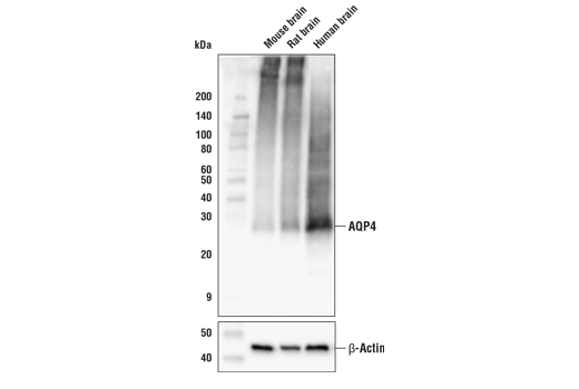
| Western Blotting | 1:1000 |
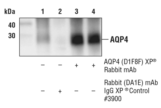
| Immunoprecipitation | 1:100 |
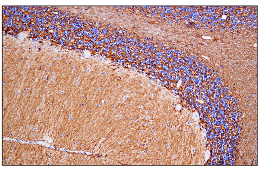
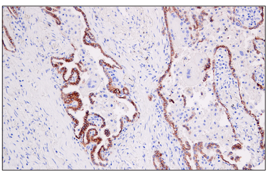
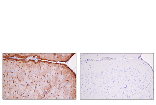
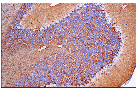
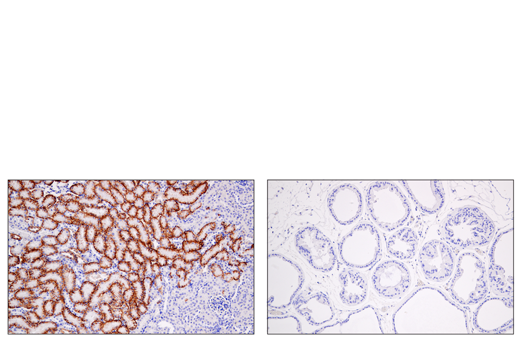
| Immunohistochemistry (Paraffin) | 1:50 - 1:200 |
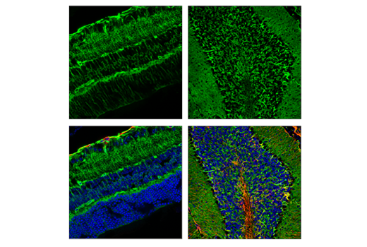
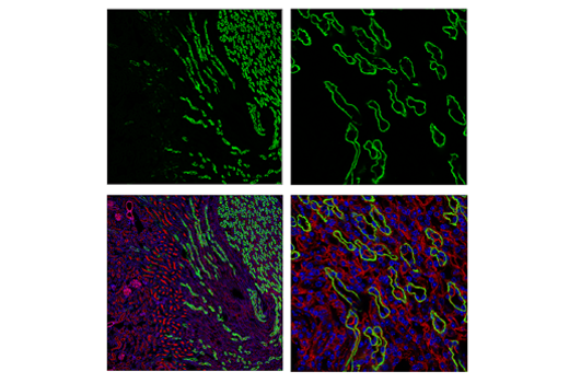
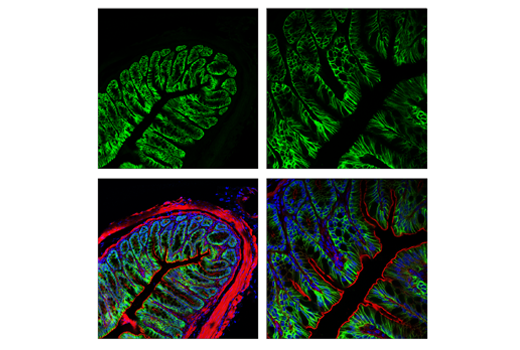
| Immunofluorescence (Frozen) | 1:800 - 1:3200 |
Storage
For a carrier free (BSA and azide free) version of this product see product #39374.
Specificity / Sensitivity
Species Reactivity:
Human, Mouse, Rat
Source / Purification
Monoclonal antibody is produced by immunizing animals with recombinant protein specific to the carboxy terminus of human AQP4 protein.
Background
Aquaporins (AQP) are integral membrane proteins that serve as channels in the transfer of water and small solutes across the membrane. There are 13 isoforms of AQP that express in different types of cells and tissues (1,2). AQP1 is found in blood vessels, kidney, eye, and ear. AQP2 is found in the kidney, and it has been shown that the lack of AQP2 results in diabetes (1,3). AQP4 is present in the brain, where it is enriched in astrocytes (1,2,4). AQP5 is found in the salivary and lacrimal gland, AQP6 in intracellular vesicles in the kidney, AQP7 in adipocytes, AQP8 in kidney, testis, and liver, AQP9 is present in liver and leukocytes, and AQP10-11 in the intestine (1,3,4). AQPs are essential for the function of cells and organs. It has been shown that AQP1 and AQP4 regulate the water homeostasis in astrocytes, preventing cerebral edema caused by solute imbalance (5). Several studies have shown the involvement of AQPs in the development of inflammatory processes, including cells of innate and adaptive immunity (6,7).
AQP4 is expressed in brain cells, including neurons, but enriched in astrocytes, as well as in the peripheral nervous system (4,7). AQP4 influences synaptic plasticity and the lack of this protein in the brain may cause memory and learning impairment via glutamate transporter 1 (GLT1) in Alzheimer's disease (8,9,10). It's suggested that AQP4 could play a role in the clearance of β-amyloid, and it may influence the transport of potassium and calcium in Alzheimer's disease (8,9). In Parkinson's disease, the lack of AQP4 results in an increased susceptibility of neurons from the substantia nigra and the ventral tegmental area for MPTP (11).
- Takata, K. et al. (2004) Prog Histochem Cytochem 39, 1-83.
- Echevarría, M. and Ilundáin, A.A. (1998) J Physiol Biochem 54, 107-18.
- Gomes, A. et al. (2018) Front Chem 6, 238.
- Xu, M. et al. (2017) Adv Exp Med Biol 969, 81-103.
- Kobayashi, H. et al. (2004) J Pharmacol Sci 96, 264-70.
- Meli, R. et al. (2018) Front Physiol 9, 101.
- Ishibashi, K. et al. (1998) Biochem Biophys Res Commun 244, 268-74.
- Hubbard, J.A. et al. (2018) Brain Res Bull 136, 118-129.
- Lan, Y.L. et al. (2016) Neural Plast 2016, 4626593.
- Lan, Y.L. et al. (2016) Mol Neurobiol 53, 5300-9.
- Zhang, J. et al. (2016) Neurosci Lett 614, 7-15.
Species Reactivity
Species reactivity is determined by testing in at least one approved application (e.g., western blot).
Western Blot Buffer
IMPORTANT: For western blots, incubate membrane with diluted primary antibody in 5% w/v BSA, 1X TBS, 0.1% Tween® 20 at 4°C with gentle shaking, overnight.
Applications Key
WB: Western Blotting IP: Immunoprecipitation IHC-P: Immunohistochemistry (Paraffin) IF-F: Immunofluorescence (Frozen)
Cross-Reactivity Key
H: human M: mouse R: rat Hm: hamster Mk: monkey Vir: virus Mi: mink C: chicken Dm: D. melanogaster X: Xenopus Z: zebrafish B: bovine Dg: dog Pg: pig Sc: S. cerevisiae Ce: C. elegans Hr: horse GP: Guinea Pig Rab: rabbit All: all species expected
Trademarks and Patents
使用に関する制限
法的な権限を与えられたCSTの担当者が署名した書面によって別途明示的に合意された場合を除き、 CST、その関連会社または代理店が提供する製品には以下の条件が適用されます。お客様が定める条件でここに定められた条件に含まれるものを超えるもの、 または、ここに定められた条件と異なるものは、法的な権限を与えられたCSTの担当者が別途書面にて受諾した場合を除き、拒絶され、 いかなる効力も効果も有しません。
研究専用 (For Research Use Only) またはこれに類似する表示がされた製品は、 いかなる目的についても FDA または外国もしくは国内のその他の規制機関により承認、認可または許可を受けていません。 お客様は製品を診断もしくは治療目的で使用してはならず、また、製品に表示された内容に違反する方法で使用してはなりません。 CST が販売または使用許諾する製品は、エンドユーザーであるお客様に対し、使途を研究および開発のみに限定して提供されるものです。 診断、予防もしくは治療目的で製品を使用することまたは製品を再販売 (単独であるか他の製品等の一部であるかを問いません) もしくはその他の商業的利用の目的で購入することについては、CST から別途許諾を得る必要があります。 お客様は以下の事項を遵守しなければなりません。(a) CST の製品 (単独であるか他の資材と一緒であるかを問いません) を販売、使用許諾、貸与、寄付もしくはその他の態様で第三者に譲渡したり使用させたりしてはなりません。また、商用の製品を製造するために CST の製品を使用してはなりません。(b) 複製、改変、リバースエンジニアリング、逆コンパイル、 分解または他の方法により製品の構造または技術を解明しようとしてはなりません。また、 CST の製品またはサービスと競合する製品またはサービスを開発する目的で CST の製品を使用してはなりません。(c) CST の製品の商標、商号、ロゴ、特許または著作権に関する通知または表示を除去したり改変したりしてはなりません。(d) CST の製品をCST 製品販売条件(CST’s Product Terms of Sale) および該当する書面のみに従って使用しなければなりません。(e) CST の製品に関連してお客様が使用する第三者の製品またはサービスに関する使用許諾条件、 サービス提供条件またはこれに類する合意事項を遵守しなければなりません。