| Product Includes | Product # | Quantity | Mol. Wt | Isotype/Source |
|---|---|---|---|---|
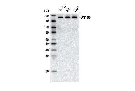
| AS160 (C69A7) Rabbit mAb | 2670 | 20 µl | 160 kDa | Rabbit |
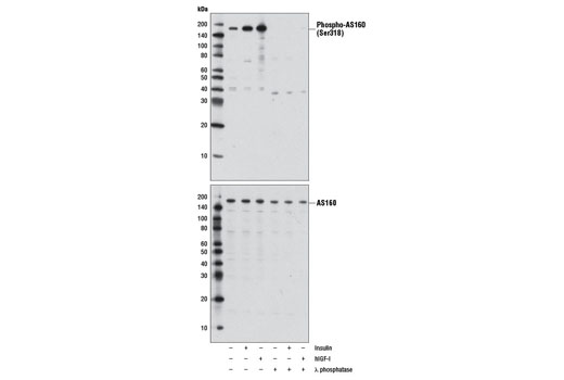
| Phospho-AS160 (Ser318) (D3D11) Rabbit mAb | 8619 | 20 µl | 160 kDa | Rabbit IgG |
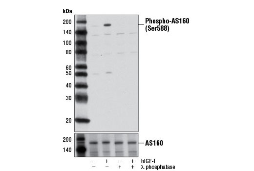
| Phospho-AS160 (Ser588) (D8E4) Rabbit mAb | 8730 | 20 µl | 160 kDa | Rabbit IgG |
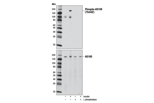
| Phospho-AS160 (Thr642) (D27E6) Rabbit mAb | 8881 | 20 µl | 160 kDa | Rabbit IgG |
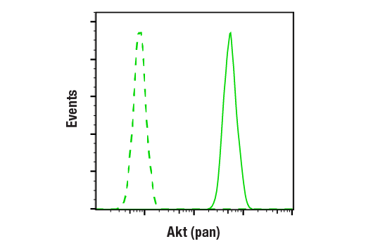
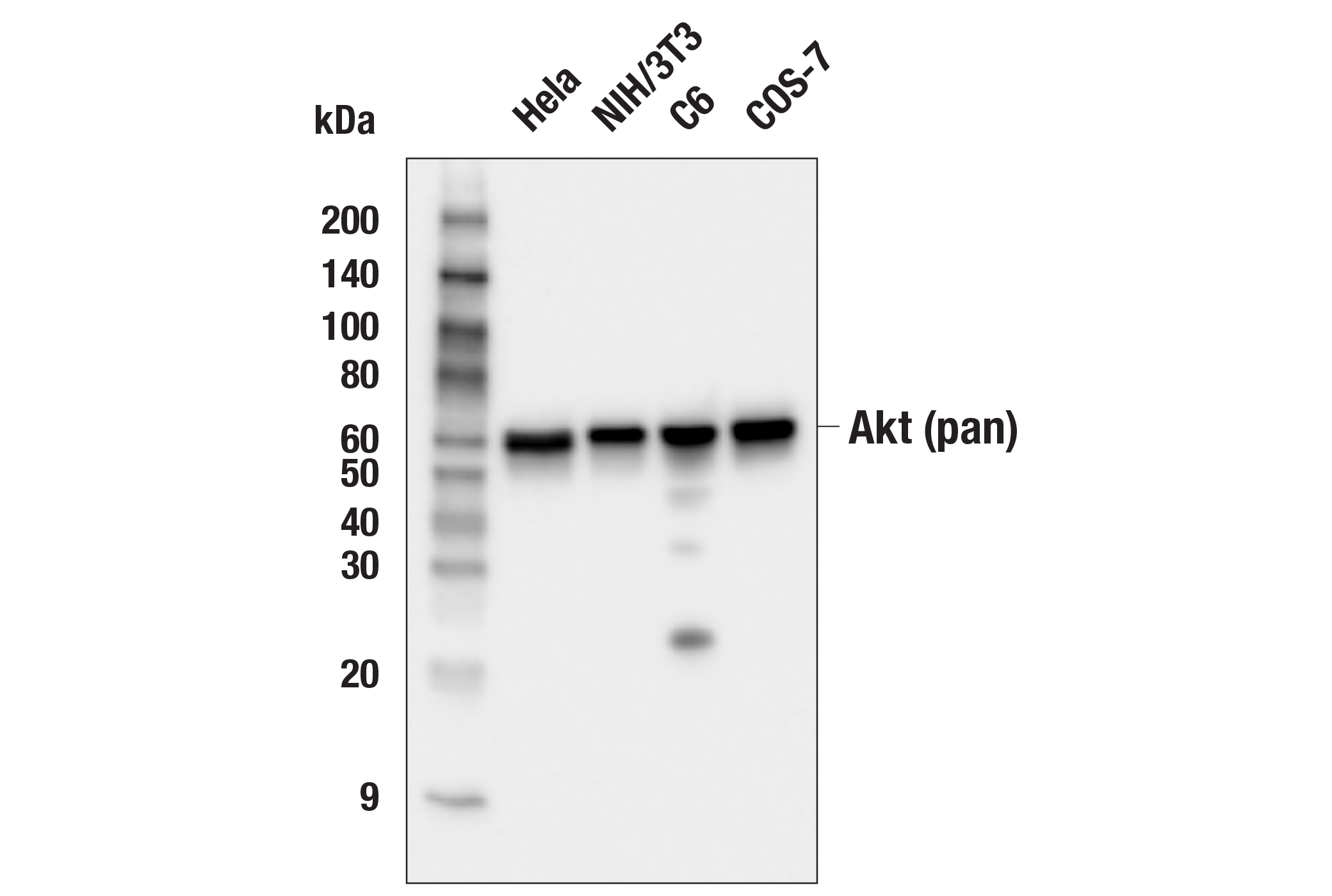
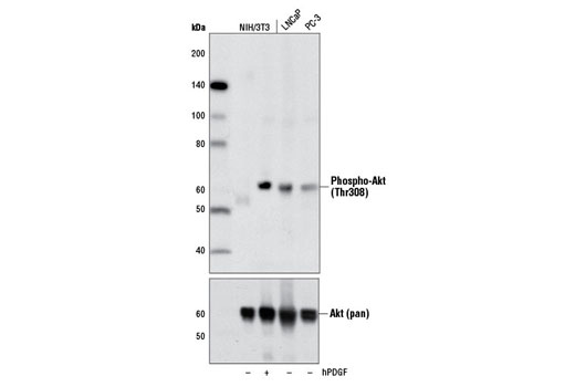
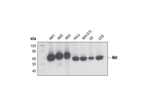
| Akt (pan) (C67E7) Rabbit mAb | 4691 | 20 µl | 60 kDa | Rabbit IgG |
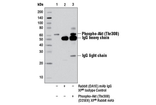
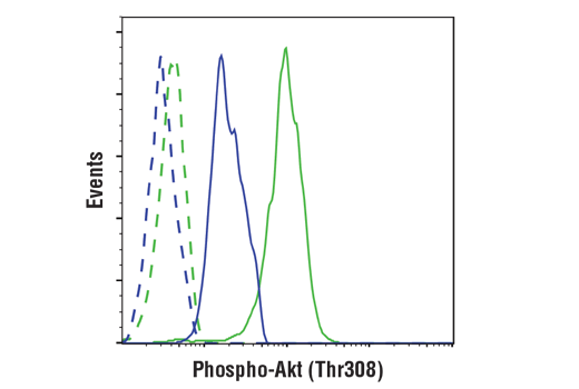
| Phospho-Akt (Thr308) (D25E6) XP® Rabbit mAb | 13038 | 20 µl | 60 kDa | Rabbit IgG |
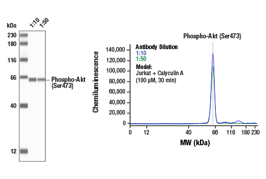
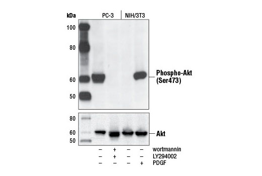
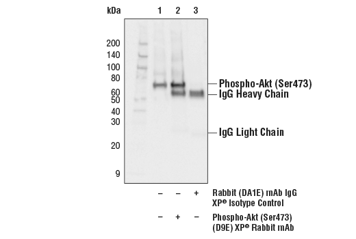
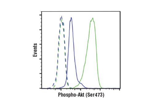
| Phospho-Akt (Ser473) (D9E) XP® Rabbit mAb | 4060 | 20 µl | 60 kDa | Rabbit IgG |
| Anti-rabbit IgG, HRP-linked Antibody | 7074 | 100 µl | Goat |
Please visit cellsignal.com for individual component applications, species cross-reactivity, dilutions, protocols, and additional product information.
Description
The AS160 Signaling Antibody Sampler Kit provides an economical means of detecting select components involved in the AS160 signaling pathway. The kit contains enough primary antibodies to perform at least two western blot experiments per antibody.
Storage
Background
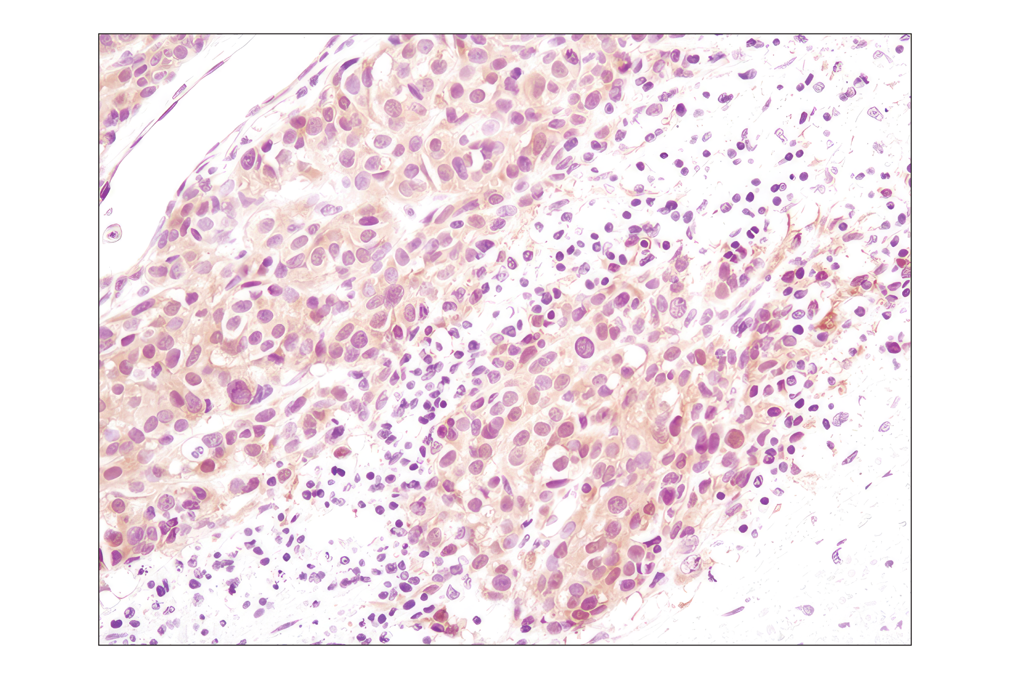
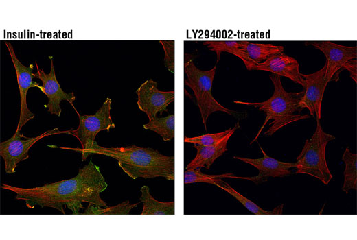
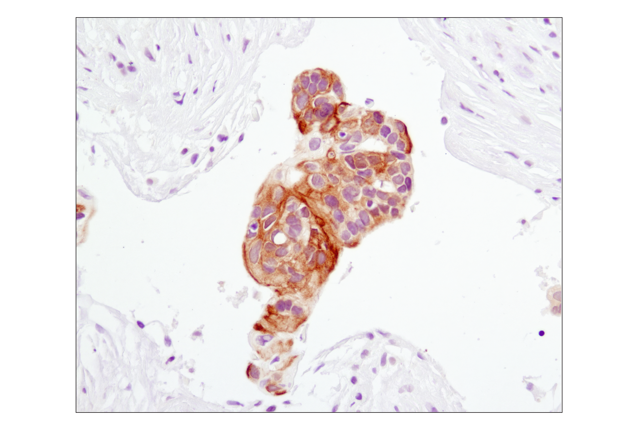
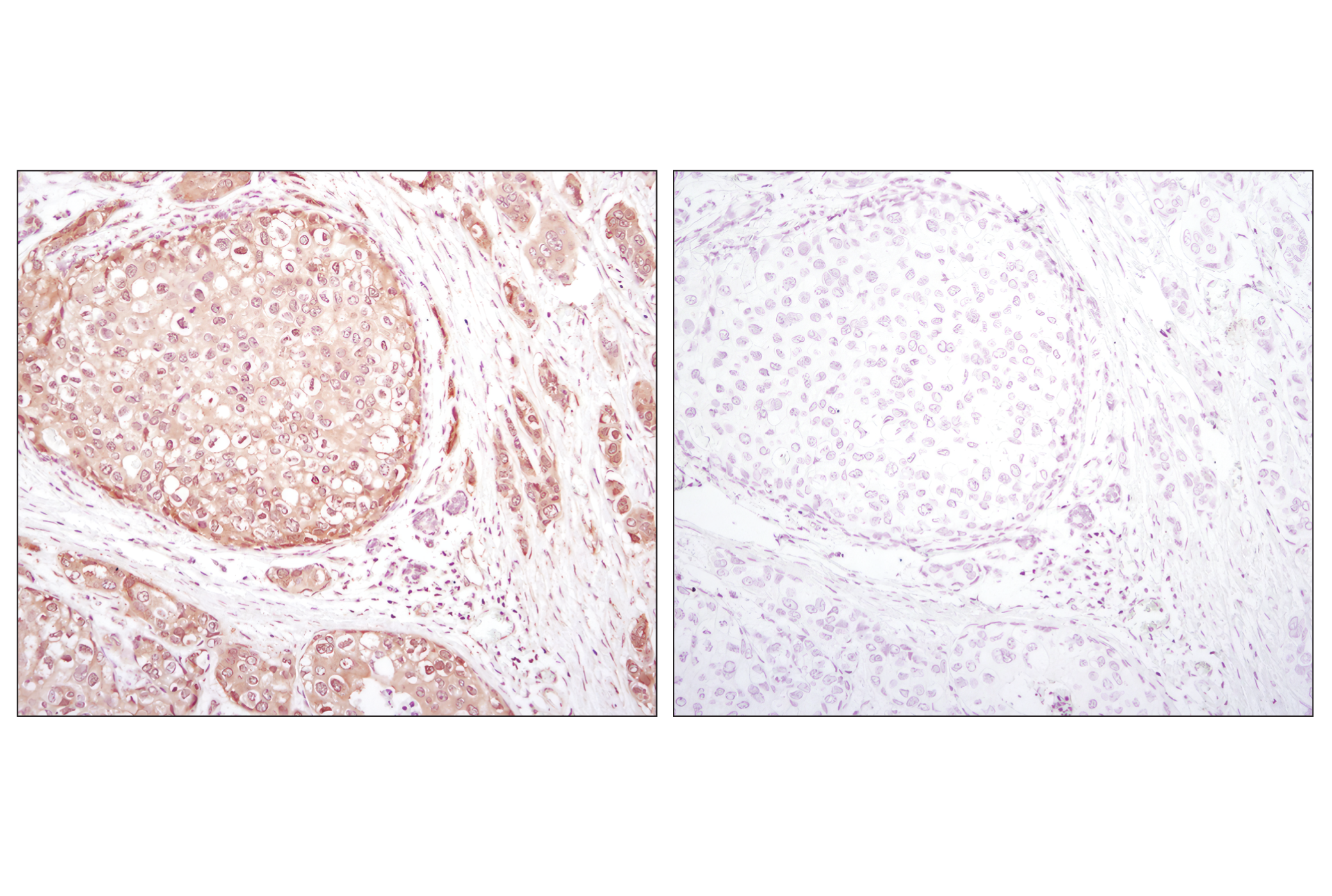
Insulin is a major hormone controlling critical energy functions, such as glucose and lipid metabolism. Insulin binds to and activates the insulin receptor (IR) tyrosine kinase, which phosphorylates and recruits adaptor proteins. The signaling pathway initiated by insulin and its receptor stimulates glucose uptake in muscle cells and adipocytes through translocation of the Glut4 glucose transporter from the cytoplasm to the plasma membrane (1). A 160 kDa substrate of the Akt Ser/Thr kinase (AS160, TBC1D4) is a Rab GTPase-activating protein that regulates insulin-stimulated Glut4 trafficking. AS160 is expressed in many tissues including brain, kidney, liver, and brown and white fat (2). Multiple Akt phosphorylation sites have been identified on AS160 in vivo, with five sites (Ser318, Ser570, Ser588, Thr642, and Thr751) showing increased phosphorylation following insulin treatment (2,3). Studies using recombinant AS160 demonstrate that insulin-stimulated phosphorylation of AS160 is a crucial step in Glut4 translocation (3) and is reduced in some patients with type 2 diabetes (4). The interaction of 14-3-3 regulatory proteins with AS160 phosphorylated at Thr642 is a necessary step for Glut4 translocation (5). Phosphorylation of AS160 by AMPK is involved in the regulation of contraction-stimulated Glut4 translocation (6).
Akt, also referred to as PKB or Rac, plays a critical role in controlling survival and apoptosis (7-9). This protein kinase is activated by insulin and various growth and survival factors to function in a wortmannin-sensitive pathway involving PI3 kinase (8,9). Akt is activated by phospholipid binding and activation loop phosphorylation at Thr308 by PDK1 (10) and by phosphorylation within the carboxy terminus at Ser473. The previously elusive PDK2 responsible for phosphorylation of Akt at Ser473 has been identified as the mammalian target of rapamycin (mTOR) in a rapamycin-insensitive complex with rictor and Sin1 (11,12).
- Watson, R.T. and Pessin, J.E. (2006) Trends Biochem. Sci. 31, 215-22.
- Kane, S. et al. (2002) J. Biol. Chem. 277, 22115-8.
- Sano, H. et al. (2003) J. Biol. Chem. 278, 14599-602.
- Karlsson, H.K. et al. (2005) Diabetes 54, 1692-7.
- Ramm, G. et al. (2006) J. Biol. Chem. 281, 29174-80.
- Kramer, H.F. et al. (2006) J. Biol. Chem. 281, 31478-85.
- Franke, T.F. et al. (1997) Cell 88, 435-7.
- Burgering, B.M. and Coffer, P.J. (1995) Nature 376, 599-602.
- Franke, T.F. et al. (1995) Cell 81, 727-36.
- Alessi, D.R. et al. (1996) EMBO J 15, 6541-51.
- Sarbassov, D.D. et al. (2005) Science 307, 1098-101.
- Jacinto, E. et al. (2006) Cell 127, 125-37.
Background References
Trademarks and Patents
使用に関する制限
法的な権限を与えられたCSTの担当者が署名した書面によって別途明示的に合意された場合を除き、 CST、その関連会社または代理店が提供する製品には以下の条件が適用されます。お客様が定める条件でここに定められた条件に含まれるものを超えるもの、 または、ここに定められた条件と異なるものは、法的な権限を与えられたCSTの担当者が別途書面にて受諾した場合を除き、拒絶され、 いかなる効力も効果も有しません。
研究専用 (For Research Use Only) またはこれに類似する表示がされた製品は、 いかなる目的についても FDA または外国もしくは国内のその他の規制機関により承認、認可または許可を受けていません。 お客様は製品を診断もしくは治療目的で使用してはならず、また、製品に表示された内容に違反する方法で使用してはなりません。 CST が販売または使用許諾する製品は、エンドユーザーであるお客様に対し、使途を研究および開発のみに限定して提供されるものです。 診断、予防もしくは治療目的で製品を使用することまたは製品を再販売 (単独であるか他の製品等の一部であるかを問いません) もしくはその他の商業的利用の目的で購入することについては、CST から別途許諾を得る必要があります。 お客様は以下の事項を遵守しなければなりません。(a) CST の製品 (単独であるか他の資材と一緒であるかを問いません) を販売、使用許諾、貸与、寄付もしくはその他の態様で第三者に譲渡したり使用させたりしてはなりません。また、商用の製品を製造するために CST の製品を使用してはなりません。(b) 複製、改変、リバースエンジニアリング、逆コンパイル、 分解または他の方法により製品の構造または技術を解明しようとしてはなりません。また、 CST の製品またはサービスと競合する製品またはサービスを開発する目的で CST の製品を使用してはなりません。(c) CST の製品の商標、商号、ロゴ、特許または著作権に関する通知または表示を除去したり改変したりしてはなりません。(d) CST の製品をCST 製品販売条件(CST’s Product Terms of Sale) および該当する書面のみに従って使用しなければなりません。(e) CST の製品に関連してお客様が使用する第三者の製品またはサービスに関する使用許諾条件、 サービス提供条件またはこれに類する合意事項を遵守しなければなりません。