| Product Includes | Product # | Quantity | Mol. Wt | Isotype/Source |
|---|---|---|---|---|
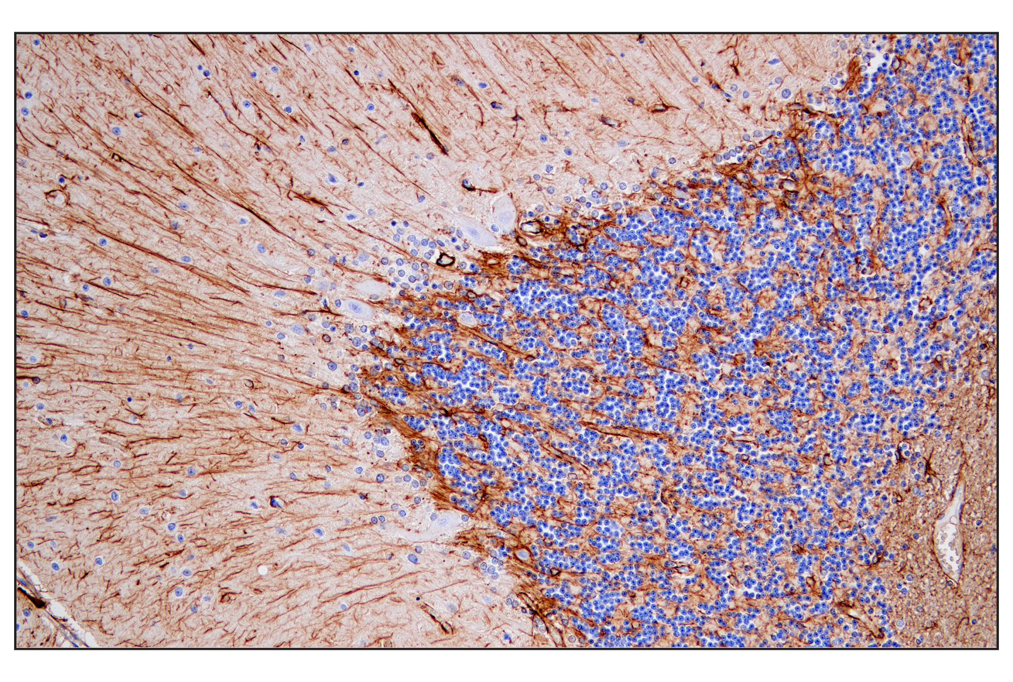
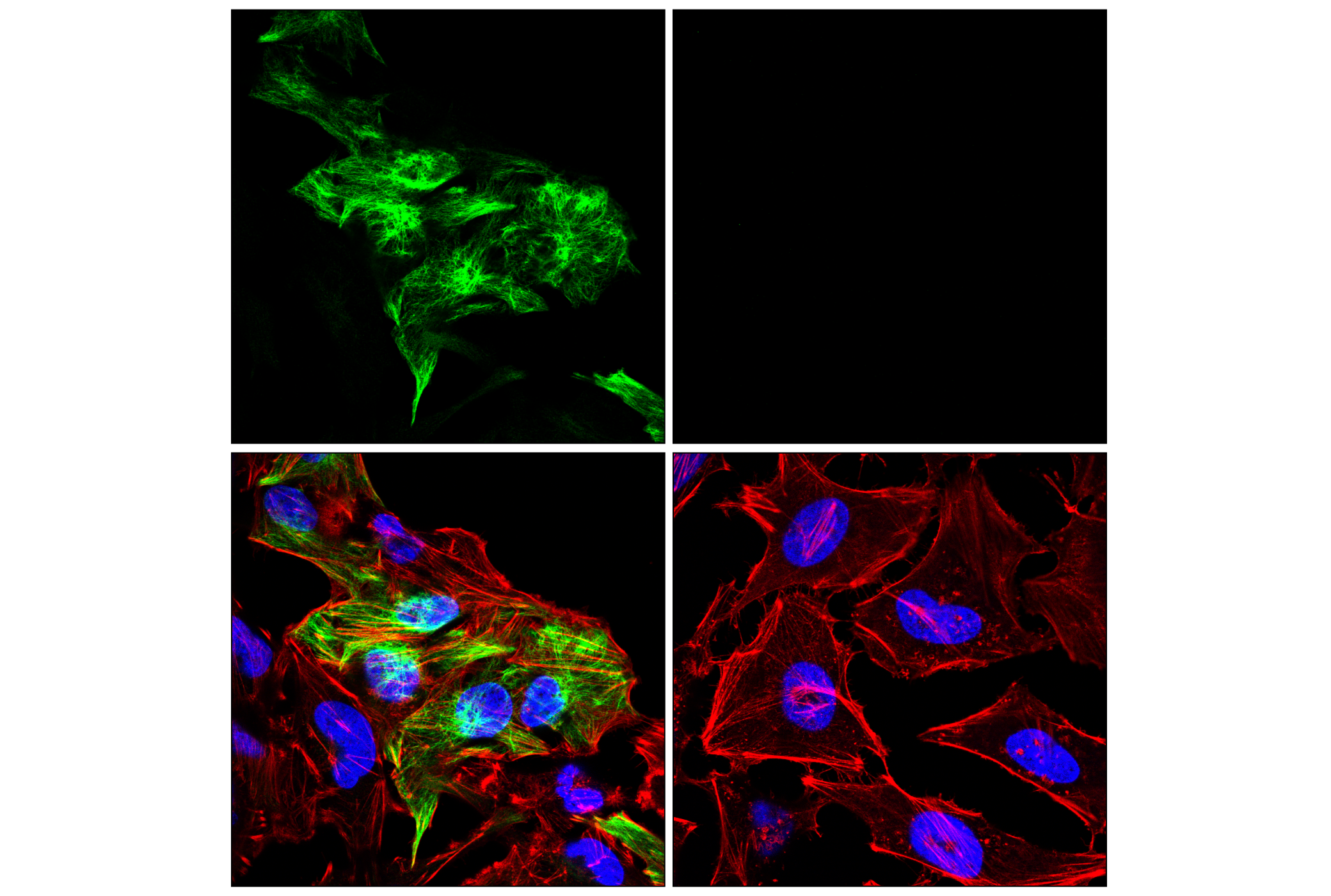
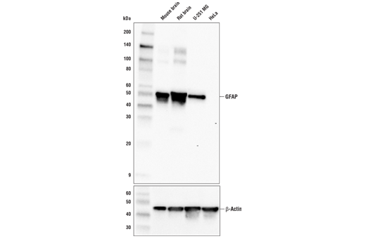
| GFAP (E4L7M) XP® Rabbit mAb | 80788 | 20 µl | 50 kDa | Rabbit IgG |
| EAAT1 (D44E2) XP® Rabbit mAb | 5684 | 20 µl | 58 kDa | Rabbit IgG |
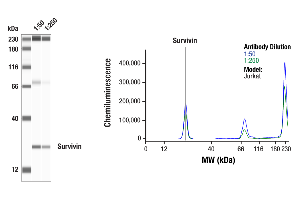
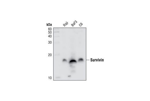
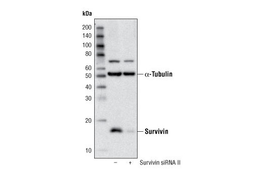
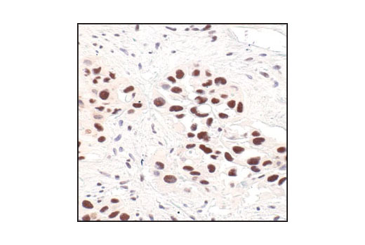
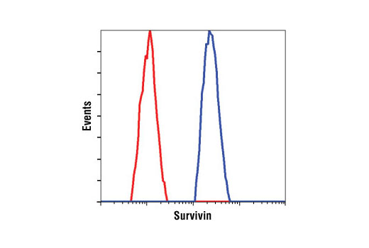
| Survivin (71G4B7) Rabbit mAb | 2808 | 20 µl | 16 kDa | Rabbit IgG |
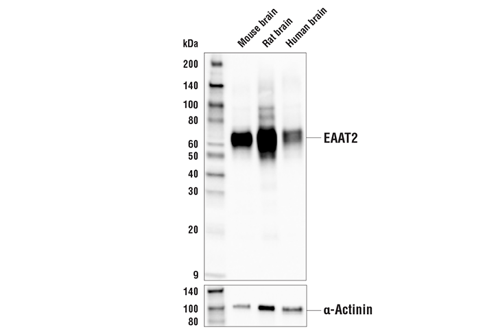
| EAAT2 (E3P5K) Rabbit mAb | 20848 | 20 µl | 65 kDa | Rabbit IgG |
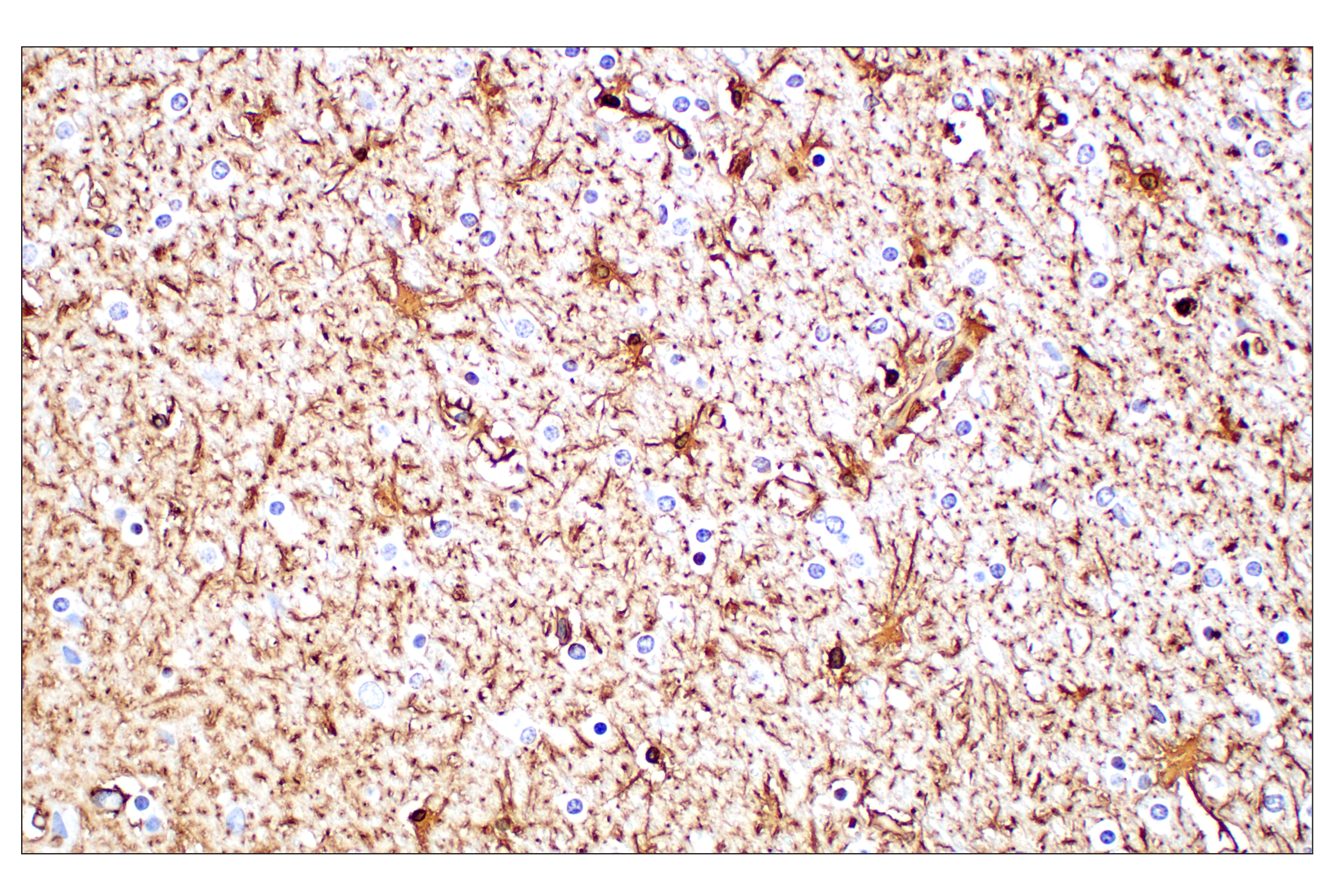
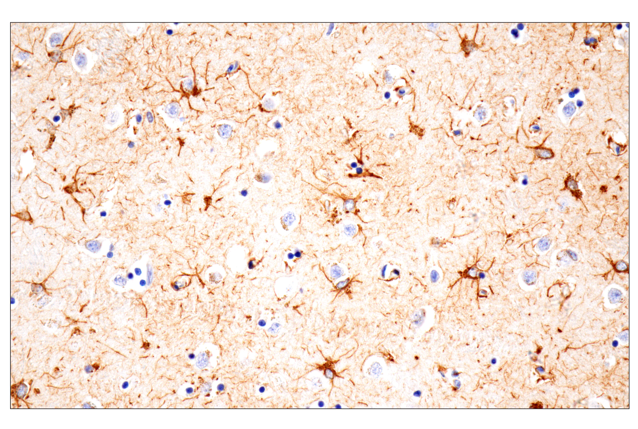
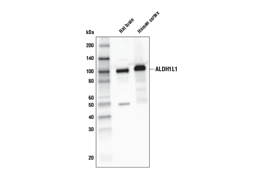
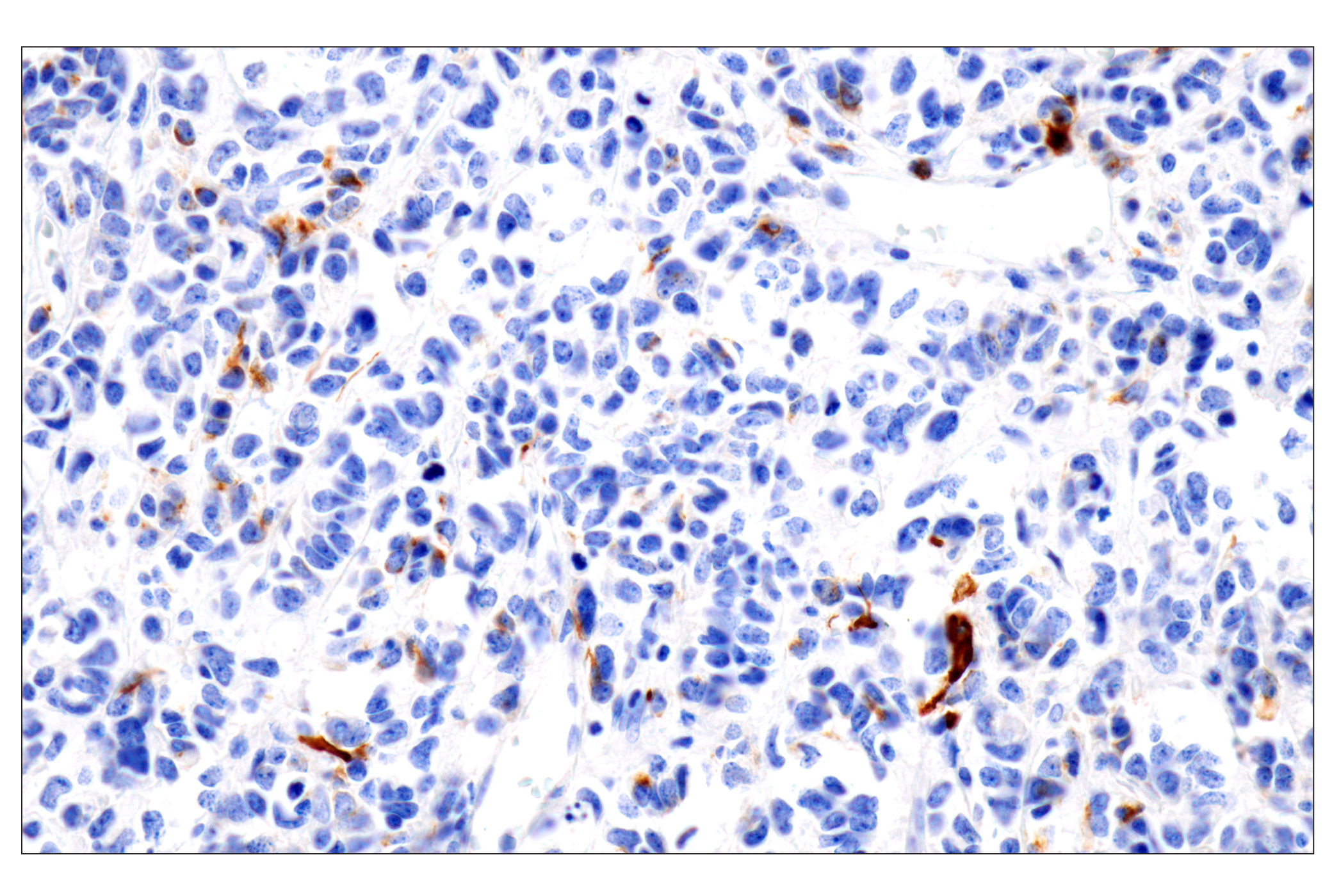
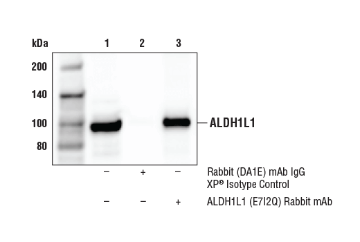
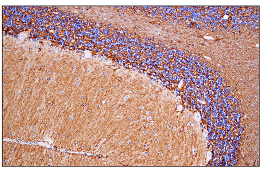
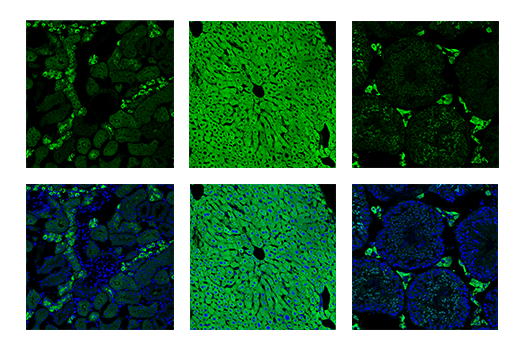
| ALDH1L1 (E7I2Q) Rabbit mAb | 85828 | 20 µl | 98 kDa | Rabbit IgG |
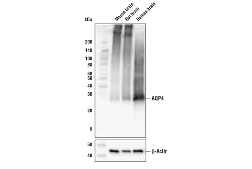
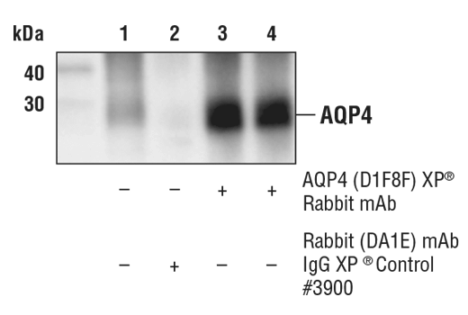
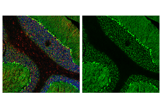
| AQP4 (D1F8E) XP® Rabbit mAb | 59678 | 20 µl | 28 kDa | Rabbit IgG |
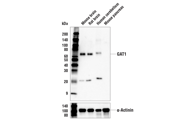
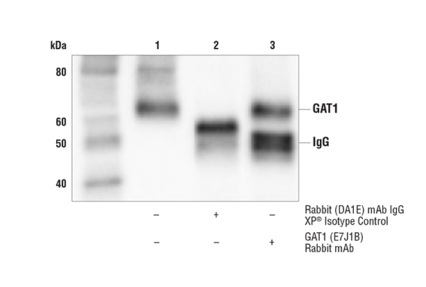
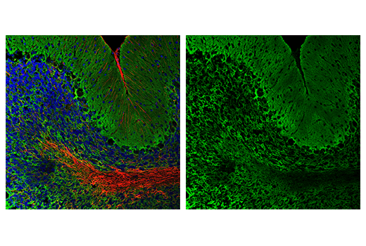
| GAT1 (E7J1B) Rabbit mAb | 37342 | 20 µl | 65 kDa | Rabbit IgG |
| Anti-rabbit IgG, HRP-linked Antibody | 7074 | 100 µl | Goat |
Please visit cellsignal.com for individual component applications, species cross-reactivity, dilutions, protocols, and additional product information.
Description
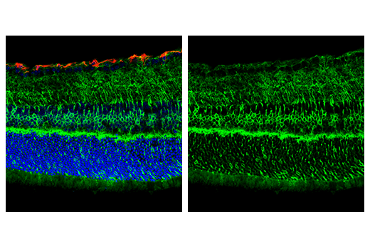
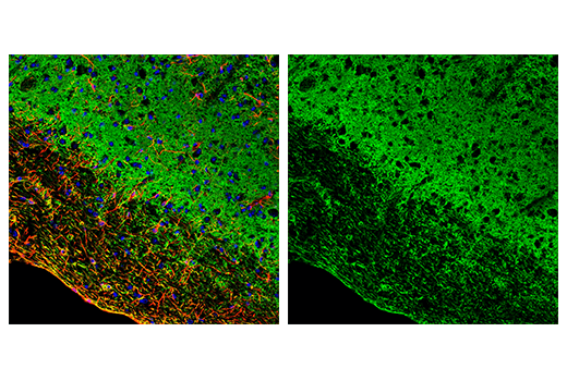
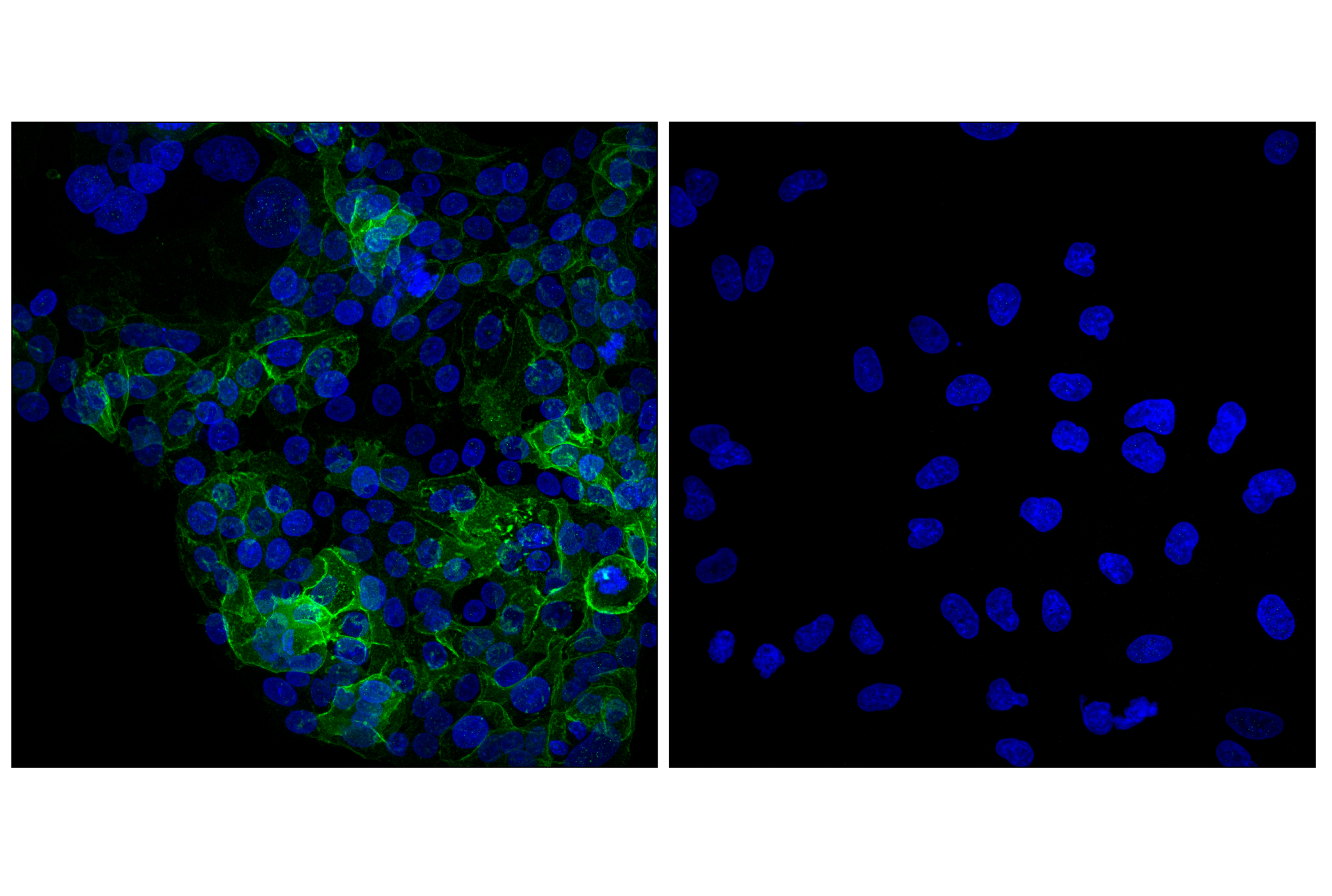
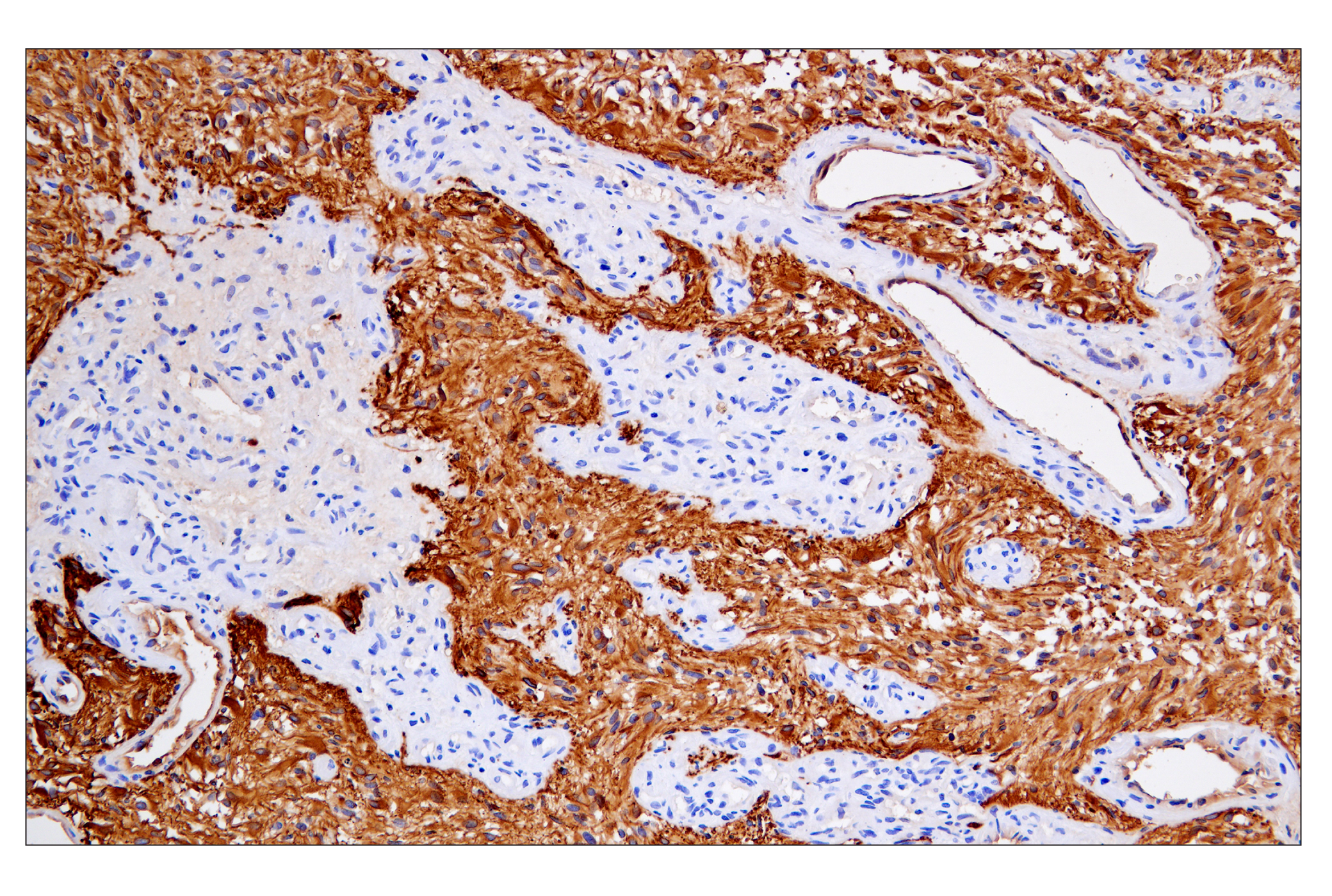
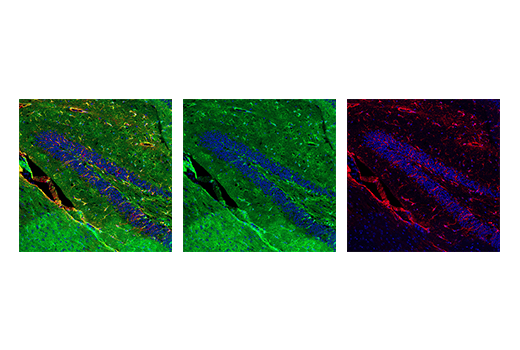
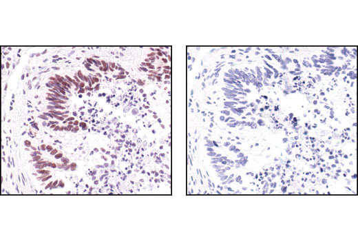
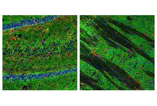
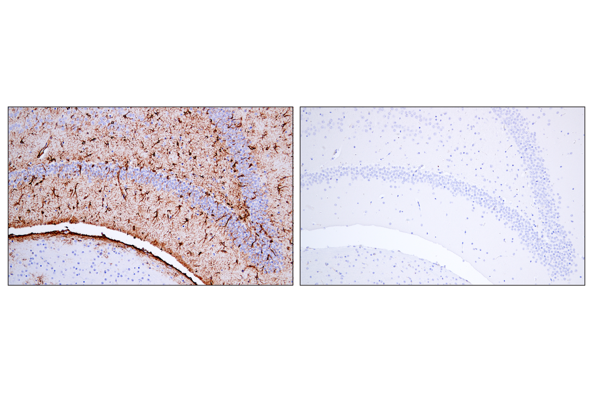
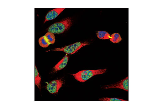
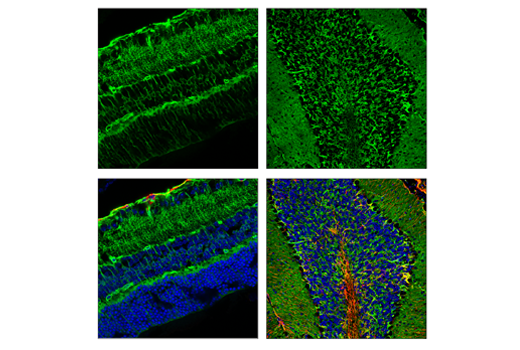
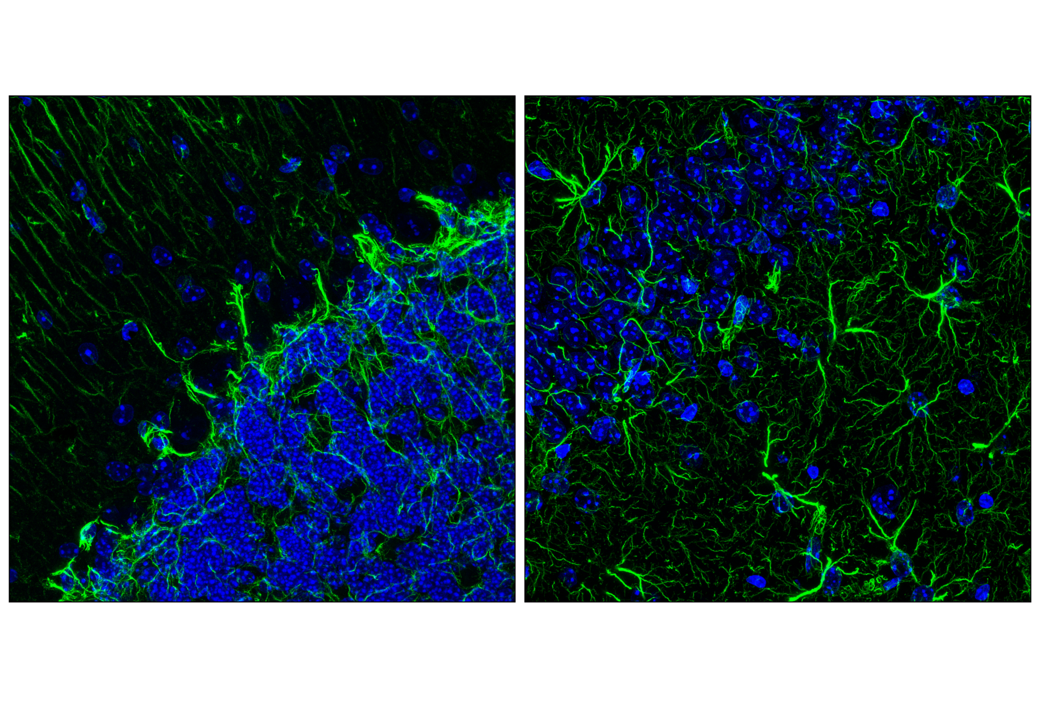
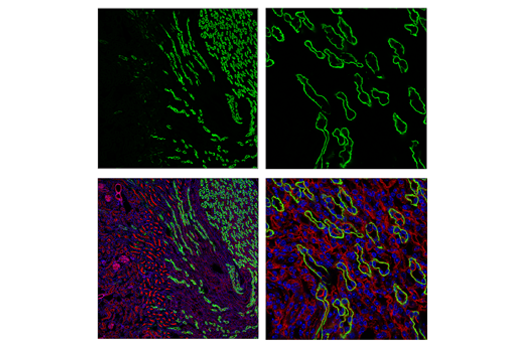
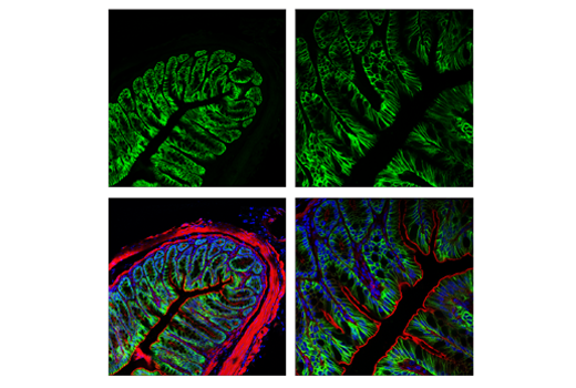
The Astrocyte Markers Antibody Sampler Kit provides an economical means of detecting astrocyte markers by Immunofluorescence or Western Blot. The kit includes enough antibodies to perform at least two western blot or twenty IF tests with each primary antibody.
Storage
Background
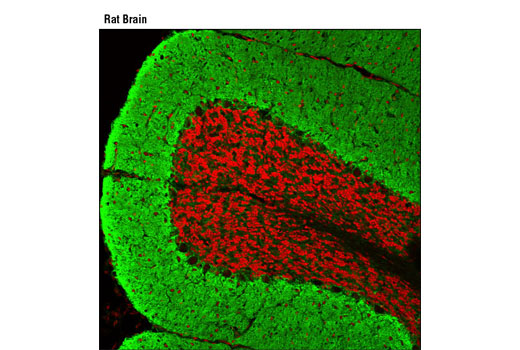
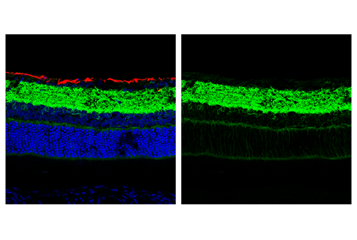
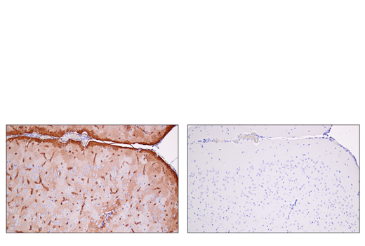
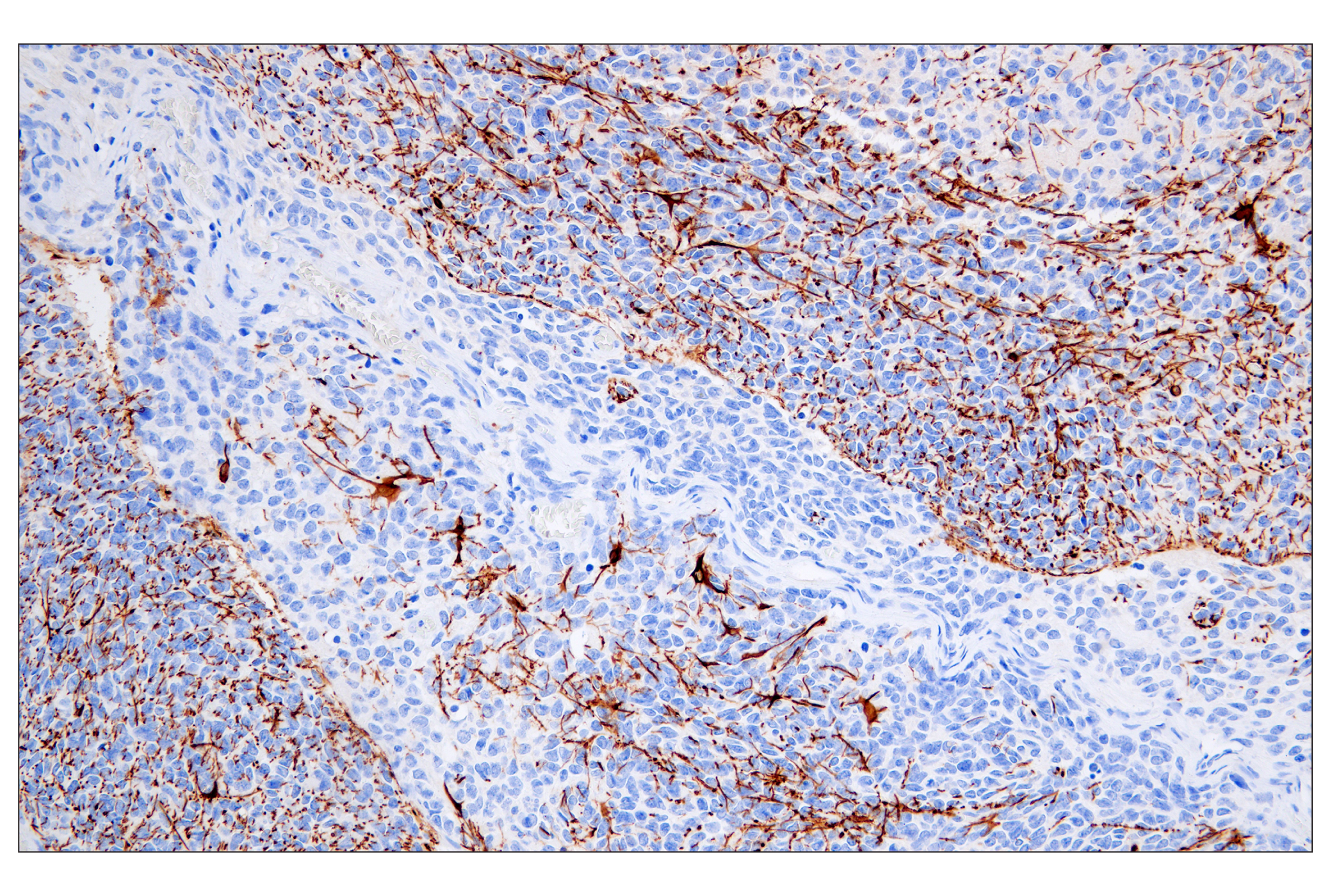
Astrocytes are a population of cells with distinctive morphological and functional characteristics that differ within specific areas of the brain. Postnatally, astrocyte progenitors migrate to reach their brain area and related properties. They have a regulatory role in brain functions that are implicated in neurogenesis and synaptogenesis, controlling blood-brain barrier permeability and maintaining extracellular homeostasis. Mature astrocytes also express some genes enriched in cell progenitors, suggesting they can retain proliferative potential (1). Astrocytes in the human brain are characterized by a heavy expression of the glial fibrillary acidic protein (GFAP), comprised of interlaminar that are located in layers I and II, protoplasmic in layers III and IV, varicose projections in layers V and VI, and fibrous astroglia in white matter (2,3).
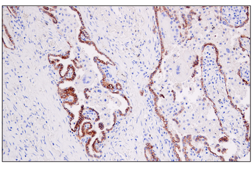
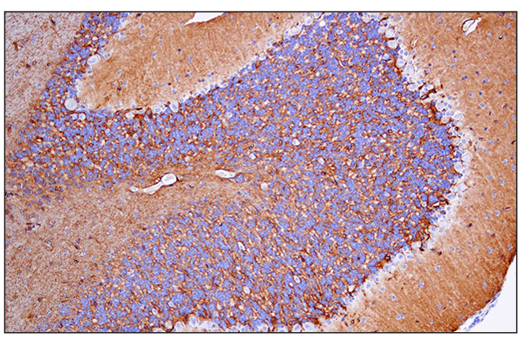
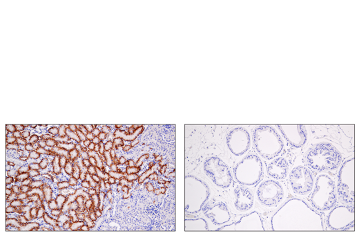
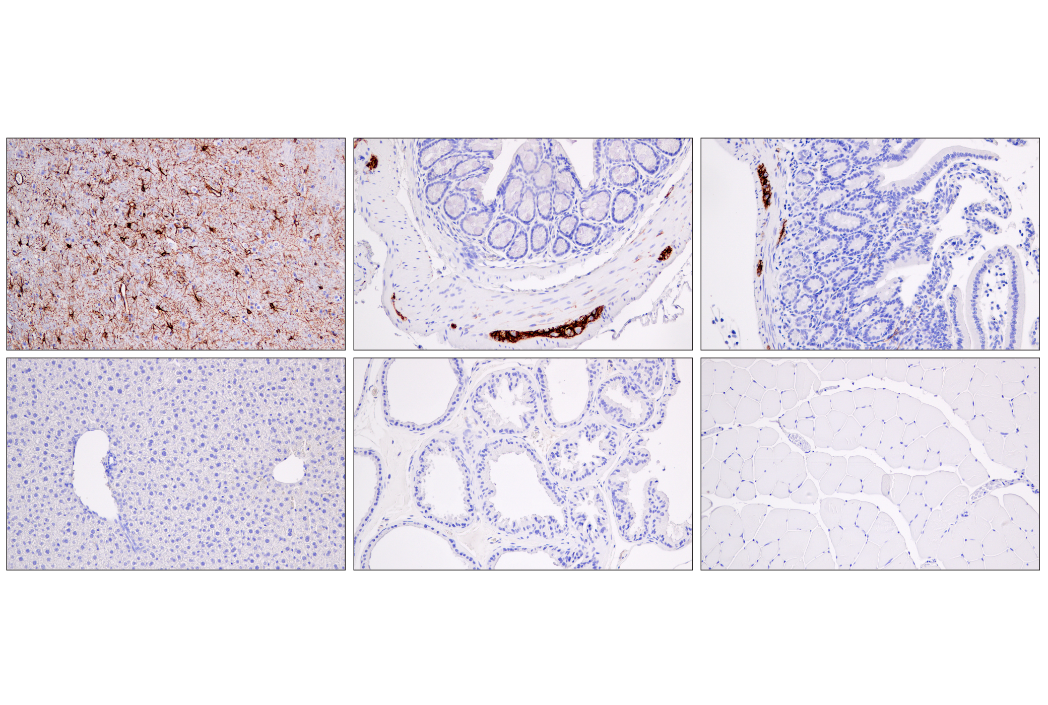
Aquaporins (AQP) are integral membrane proteins that serve as channels in the transfer of water and small solutes across the membrane. AQP4 is present in the brain and it is enriched in astrocytes to regulate water homeostasis, preventing cerebral edema caused by solute imbalance (4,5). Excitatory amino acid transporters are members of sodium-dependent, high-affinity transporters that mediate the uptake of L-glutamate and D-aspartate (6). GABA transmitters are Na+/Cl--dependent transporters that regulate neurotransmitter transport, including GAT1 (SLC6), GAT2, GAT3, and BGT1 (7). GAT1 expresses in the brain, preferentially in glial cells, but is also found in neurons, regulating the uptake and release of neurotransmitters in terminal clefts (8-10). EAAT2, also known as GLT-1, is primarily expressed in astrocytes accounting for up to 90% of the total glutamate transport in the brain. EAAT1 upregulates increased concentrations of glutamate in astrocytes and has a neuroprotective potential following ischemia since reactive astrocytes and activated microglia express EAAT1 but not EAAT2 (11-13).
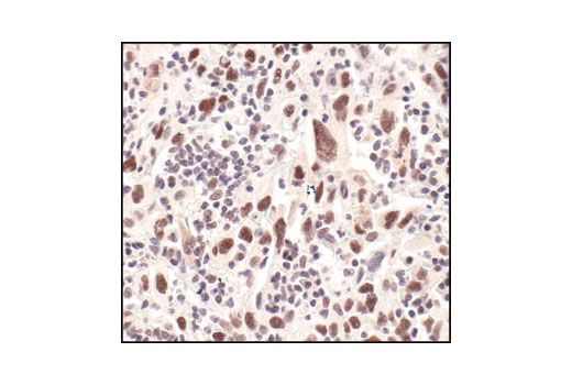
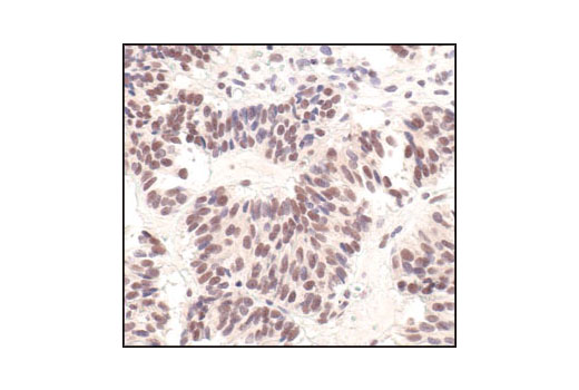
Aldehyde dehydrogenase 1 family member L1 (ALDH1L1) is a member of the aldehyde dehydrogenase super family (14,15). ALDH1L1 has also been shown to be a useful astrocyte marker throughout the grey and white matter of the brain, labeling both the cell body and processes of astrocytes. ALDH1L1 does not label neurons or oligodendrocytes (16). It has been shown that expression of ALDH1L1 in astrocytes is stably expressed through normal tissue and during astrogliosis (17). Survivin is a 16 kDa anti-apoptotic protein highly expressed during fetal development and cancer malignancy. It binds and inhibits caspase-3, controlling the checkpoint in the G2/M-phase of the cell cycle by inhibiting apoptosis and promoting cell division (18). Survivin expression is associated with malignant phenotypes and prognosis of glioma (19).
- Siracusa, R. et al. (2019) Front Pharmacol 10, 1114.
- Vasile, F. et al. (2017) Brain Struct Funct 222, 2017-2029.
- Eng, L.F. et al. (2000) Neurochem Res 25, 1439-51.
- Takata, K. et al. (2004) Prog Histochem Cytochem 39, 1-83.
- Kobayashi, H. et al. (2004) J Pharmacol Sci 96, 264-70.
- Amara, S.G. and Fontana, A.C. (2002) Neurochem Int 41, 313-8.
- Kristensen, A.S. et al. (2011) Pharmacol Rev 63, 585-640.
- Borden, L.A. (1996) Neurochem Int 29, 335-56.
- Moldavan, M. et al. (2017) J Neurophysiol 118, 3092-3106.
- Lorenz-Guertin, J.M. and Jacob, T.C. (2018) Dev Neurobiol 78, 238-270.
- Hediger, M.A. (1999) Am J Physiol 277, F487-92.
- Gegelashvili, G. et al. (1996) Neuroreport 8, 261-5.
- Beschorner, R. et al. (2007) Histopathology 50, 897-910.
- Krupenko, S.A. (2009) Chem Biol Interact 178, 84-93.
- Krupenko, S.A. and Oleinik, N.V. (2002) Cell Growth Differ 13, 227-36.
- Cahoy, J.D. et al. (2008) J Neurosci 28, 264-78.
- Michalovicz, L.T. et al. (2019) J Neurochem 150, 420-440.
- Reed, J.C. and Reed, S.I. (1999) Nat Cell Biol 1, E199-200.
- Tong, X. et al. (2019) Oncol Lett 18, 359-367.
Background References
Trademarks and Patents
使用に関する制限
法的な権限を与えられたCSTの担当者が署名した書面によって別途明示的に合意された場合を除き、 CST、その関連会社または代理店が提供する製品には以下の条件が適用されます。お客様が定める条件でここに定められた条件に含まれるものを超えるもの、 または、ここに定められた条件と異なるものは、法的な権限を与えられたCSTの担当者が別途書面にて受諾した場合を除き、拒絶され、 いかなる効力も効果も有しません。
研究専用 (For Research Use Only) またはこれに類似する表示がされた製品は、 いかなる目的についても FDA または外国もしくは国内のその他の規制機関により承認、認可または許可を受けていません。 お客様は製品を診断もしくは治療目的で使用してはならず、また、製品に表示された内容に違反する方法で使用してはなりません。 CST が販売または使用許諾する製品は、エンドユーザーであるお客様に対し、使途を研究および開発のみに限定して提供されるものです。 診断、予防もしくは治療目的で製品を使用することまたは製品を再販売 (単独であるか他の製品等の一部であるかを問いません) もしくはその他の商業的利用の目的で購入することについては、CST から別途許諾を得る必要があります。 お客様は以下の事項を遵守しなければなりません。(a) CST の製品 (単独であるか他の資材と一緒であるかを問いません) を販売、使用許諾、貸与、寄付もしくはその他の態様で第三者に譲渡したり使用させたりしてはなりません。また、商用の製品を製造するために CST の製品を使用してはなりません。(b) 複製、改変、リバースエンジニアリング、逆コンパイル、 分解または他の方法により製品の構造または技術を解明しようとしてはなりません。また、 CST の製品またはサービスと競合する製品またはサービスを開発する目的で CST の製品を使用してはなりません。(c) CST の製品の商標、商号、ロゴ、特許または著作権に関する通知または表示を除去したり改変したりしてはなりません。(d) CST の製品をCST 製品販売条件(CST’s Product Terms of Sale) および該当する書面のみに従って使用しなければなりません。(e) CST の製品に関連してお客様が使用する第三者の製品またはサービスに関する使用許諾条件、 サービス提供条件またはこれに類する合意事項を遵守しなければなりません。