#O75143
9776
| Product Includes | Quantity | Reactivity | MW(kDa) | Isotype | |
|---|---|---|---|---|---|
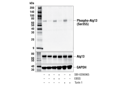
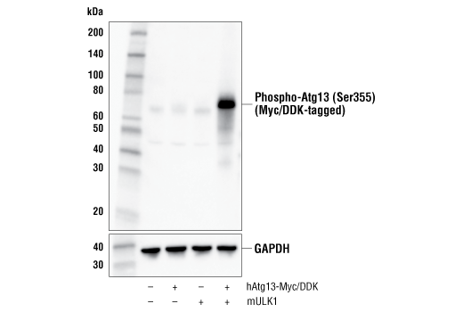
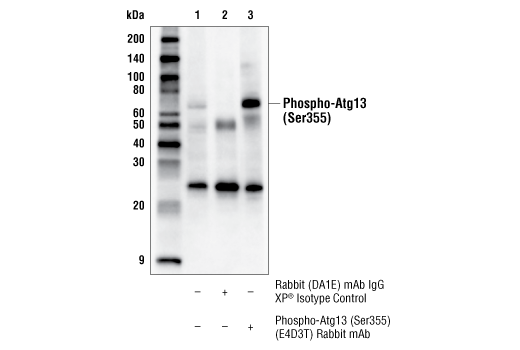
| Phospho-Atg13 (Ser355) (E4D3T) Rabbit mAb 46329 | 100 µl | H M R | 72 | Rabbit IgG | |
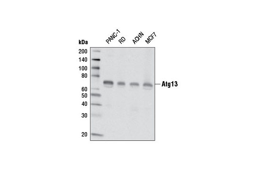
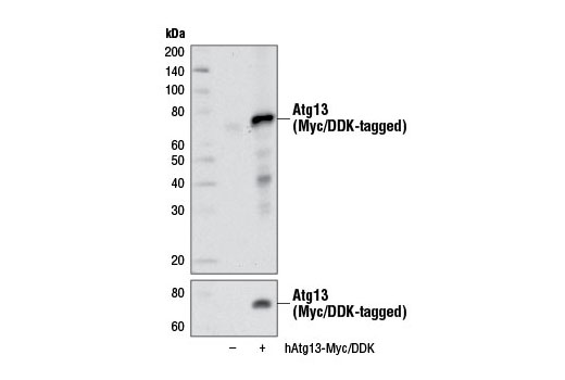
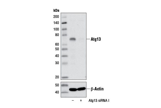
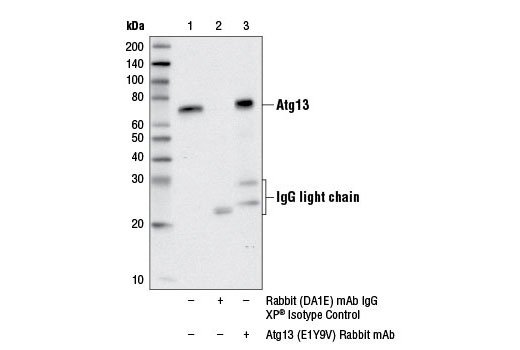
| Atg13 (E1Y9V) Rabbit mAb 13468 | 100 µl | H | 72 | Rabbit IgG |
Please visit cellsignal.com for individual component applications, species cross-reactivity, dilutions, protocols, and additional product information.
Description
PhosphoPlus® Duets from Cell Signaling Technology (CST) provide a means to assess protein activation status. Each Duet contains an activation-state and total protein antibody to your target of interest. These antibodies have been selected from CST's product offering based upon superior performance in specified applications.
Storage
Background
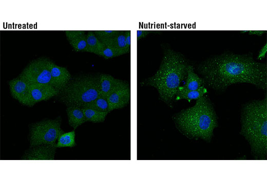
Autophagy is a catabolic process for the autophagosomic-lysosomal degradation of bulk cytoplasmic contents (1,2). Autophagy is generally activated by conditions of nutrient deprivation but has also been associated with a number of physiological processes including development, differentiation, neurodegeneration, infection, and cancer (3). The molecular machinery of autophagy was largely discovered in yeast and referred to as autophagy-related (Atg) genes.
Atg13/Apg13 was originally identified in yeast as a constitutively expressed protein that was genetically linked to Atg1/Apg1, a protein kinase required for autophagy (4). Overexpression of Atg1 suppresses the defects in autophagy observed in Atg13 mutants (4). Autophagy requires a direct association between Atg1 and Atg13, and is inhibited by TOR-dependent phosphorylation of Atg13 under high-nutrient conditions (5). Similarly, mammalian Atg13 forms a complex with the Atg1 homologues ULK1/2, along with FIP200, which localizes to autophagic isolation membranes and regulates autophagosome biogenesis (6-8). mTOR phosphorylates both Atg13 and ULK1, suppressing ULK1 kinase activity and autophagy (7-9). ULK1 can directly phosphorylate Atg13 at a yet unidentified site, presumably to promote autophagy (7,8). Additional studies suggest that Atg13 and FIP200 can function independently of ULK1 and ULK2 to induce autophagy through an unknown mechanism (10).
ULK1-dependent phosphorylation of Atg13 at Ser355, which corresponds to Ser318 of isoform 2 of Atg13, leads to the recruitment of Atg13 to damaged mitochondria, enabling efficient mitophagy (11).
- Reggiori, F. and Klionsky, D.J. (2002) Eukaryot Cell 1, 11-21.
- Codogno, P. and Meijer, A.J. (2005) Cell Death Differ 12 Suppl 2, 1509-18.
- Levine, B. and Yuan, J. (2005) J Clin Invest 115, 2679-88.
- Funakoshi, T. et al. (1997) Gene 192, 207-13.
- Kamada, Y. et al. (2000) J Cell Biol 150, 1507-13.
- Ganley, I.G. et al. (2009) J Biol Chem 284, 12297-305.
- Hosokawa, N. et al. (2009) Mol Biol Cell 20, 1981-91.
- Jung, C.H. et al. (2009) Mol Biol Cell 20, 1992-2003.
- Kim, J. et al. (2011) Nat Cell Biol 13, 132-41.
- Alers, S. et al. (2011) Autophagy 7, 1423-33.
- Joo, J.H. et al. (2011) Mol Cell 43, 572-85.
Background References
Trademarks and Patents
使用に関する制限
法的な権限を与えられたCSTの担当者が署名した書面によって別途明示的に合意された場合を除き、 CST、その関連会社または代理店が提供する製品には以下の条件が適用されます。お客様が定める条件でここに定められた条件に含まれるものを超えるもの、 または、ここに定められた条件と異なるものは、法的な権限を与えられたCSTの担当者が別途書面にて受諾した場合を除き、拒絶され、 いかなる効力も効果も有しません。
研究専用 (For Research Use Only) またはこれに類似する表示がされた製品は、 いかなる目的についても FDA または外国もしくは国内のその他の規制機関により承認、認可または許可を受けていません。 お客様は製品を診断もしくは治療目的で使用してはならず、また、製品に表示された内容に違反する方法で使用してはなりません。 CST が販売または使用許諾する製品は、エンドユーザーであるお客様に対し、使途を研究および開発のみに限定して提供されるものです。 診断、予防もしくは治療目的で製品を使用することまたは製品を再販売 (単独であるか他の製品等の一部であるかを問いません) もしくはその他の商業的利用の目的で購入することについては、CST から別途許諾を得る必要があります。 お客様は以下の事項を遵守しなければなりません。(a) CST の製品 (単独であるか他の資材と一緒であるかを問いません) を販売、使用許諾、貸与、寄付もしくはその他の態様で第三者に譲渡したり使用させたりしてはなりません。また、商用の製品を製造するために CST の製品を使用してはなりません。(b) 複製、改変、リバースエンジニアリング、逆コンパイル、 分解または他の方法により製品の構造または技術を解明しようとしてはなりません。また、 CST の製品またはサービスと競合する製品またはサービスを開発する目的で CST の製品を使用してはなりません。(c) CST の製品の商標、商号、ロゴ、特許または著作権に関する通知または表示を除去したり改変したりしてはなりません。(d) CST の製品をCST 製品販売条件(CST’s Product Terms of Sale) および該当する書面のみに従って使用しなければなりません。(e) CST の製品に関連してお客様が使用する第三者の製品またはサービスに関する使用許諾条件、 サービス提供条件またはこれに類する合意事項を遵守しなければなりません。