| Product Includes | Product # | Quantity | Mol. Wt | Isotype/Source |
|---|---|---|---|---|
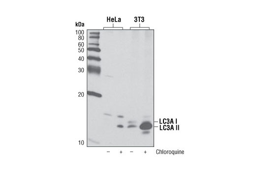
| LC3A (D50G8) XP® Rabbit mAb | 4599 | 20 µl | 14, 16 kDa | Rabbit IgG |
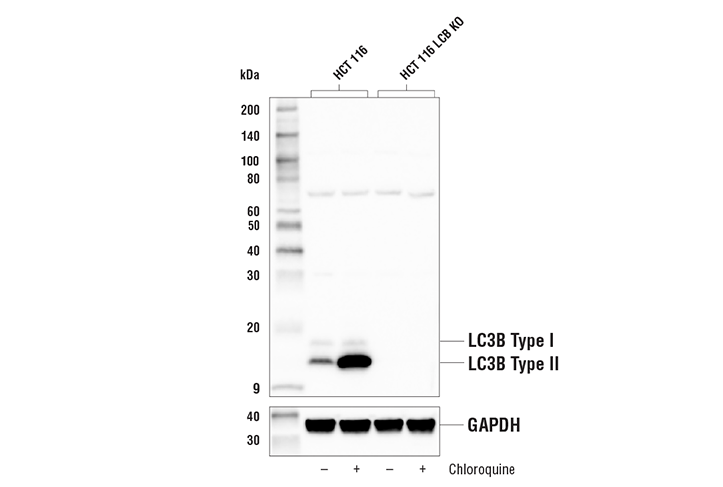
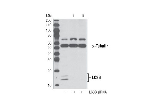
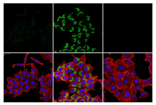
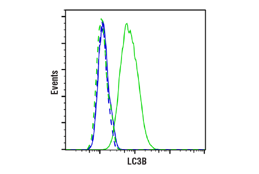
| LC3B (D11) XP® Rabbit mAb | 3868 | 20 µl | 14, 16 kDa | Rabbit IgG |
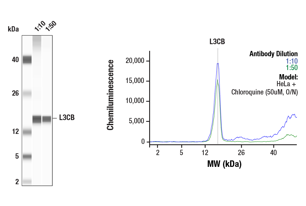
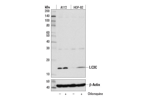
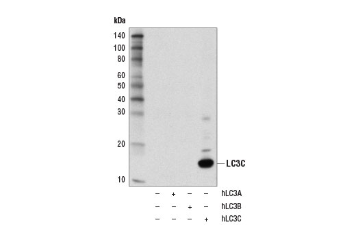
| LC3C (D3O6P) Rabbit mAb | 14736 | 20 µl | 14 kDa | Rabbit IgG |
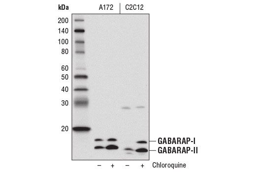
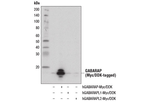
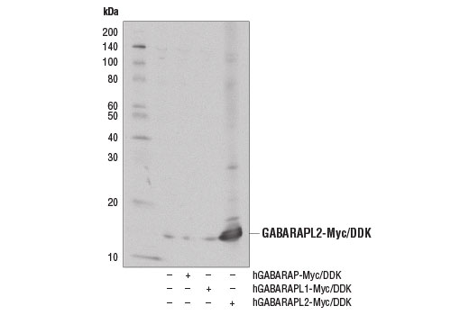
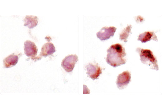
| GABARAP (E1J4E) Rabbit mAb | 13733 | 20 µl | 14, 16 kDa | Rabbit IgG |
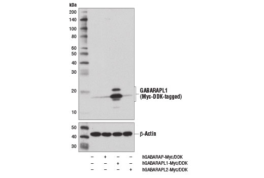
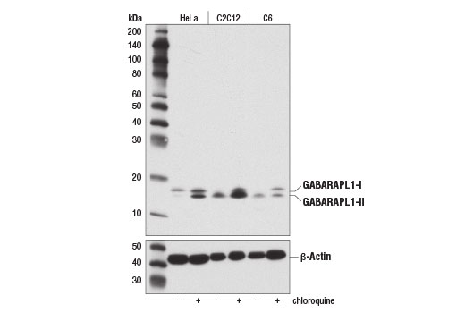
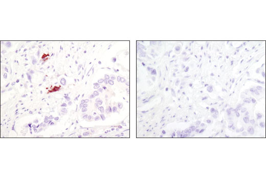
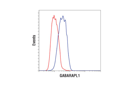
| GABARAPL1 (D5R9Y) XP® Rabbit mAb | 26632 | 20 µl | 14, 16 kDa | Rabbit IgG |
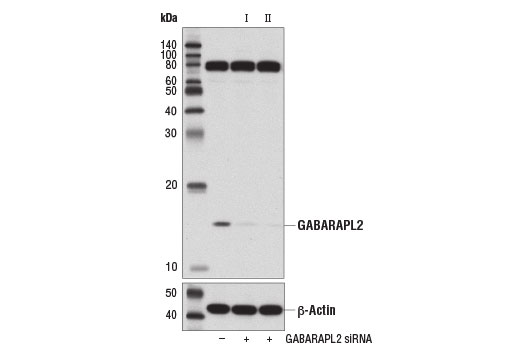
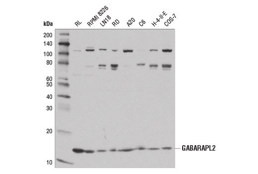
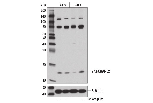
| GABARAPL2 (D1W9T) Rabbit mAb | 14256 | 20 µl | 14 kDa | Rabbit IgG |
| Anti-rabbit IgG, HRP-linked Antibody | 7074 | 100 µl | Goat |
Please visit cellsignal.com for individual component applications, species cross-reactivity, dilutions, protocols, and additional product information.
Description
The Autophagy Atg8 Family Antibody Sampler Kit provides an economical means of detecting each of the Atg8 family members. The kit contains enough primary antibody to perform at least two western blot experiments.
Storage
Background
Autophagy is a catabolic process for the autophagosomic-lysosomal degradation of bulk cytoplasmic contents (1,2). Autophagy is generally activated by conditions of nutrient deprivation, but it has also been associated with a number of physiological processes, including development, differentiation, neurodegenerative diseases, infection, and cancer (3).
Atg8 is a ubiquitin-like protein that is critical for autophagosome formation. Atg8 is synthesized as a precursor protein that is processed by the cysteine protease Atg4, followed by lipidation with phosphatidylethanolamine (PE) in a ubiqutin-like conjugation pathway involving Atg7 and Atg3 (4). This processing of Atg8, which is described as a conversion from type-I to type-II forms, is frequently described as a marker for autophagy. The type-II form of Atg8 is incorporated into maturing autophagosomes and leads to the recruitment of additional autophagy components, including cargo receptors like SQSTM1/p62. While yeast has a single Atg8 gene, many eukaryotes have at least six orthologs, including three microtubule-associated protein 1 light chain 3 (MAP1LC3/LC3) family members (LC3A, LC3B, and LC3C) and three GABAA receptor associated protein (GABARAP) family members (GABARAP, GABARAPL1/GEC1, and GABARAPL2/GATE-16). While highly conserved, these various family members can have important differences in their post-translational processing, expression profile, and protein interactions including distinct cargo receptor. This complexity within the Atg8 family is critical for selective mechanisms of autophagy that have been reported (5, 6).
- Reggiori, F. and Klionsky, D.J. (2002) Eukaryot Cell 1, 11-21.
- Codogno, P. and Meijer, A.J. (2005) Cell Death Differ 12 Suppl 2, 1509-18.
- Levine, B. and Yuan, J. (2005) J Clin Invest 115, 2679-88.
- Ichimura, Y. et al. (2000) Nature 408, 488-92.
- Slobodkin, M.R. and Elazar, Z. (2013) Essays Biochem 55, 51-64.
- Schaaf, M.B. et al. (2016) FASEB J 30, 3961-3978.
Background References
Trademarks and Patents
使用に関する制限
法的な権限を与えられたCSTの担当者が署名した書面によって別途明示的に合意された場合を除き、 CST、その関連会社または代理店が提供する製品には以下の条件が適用されます。お客様が定める条件でここに定められた条件に含まれるものを超えるもの、 または、ここに定められた条件と異なるものは、法的な権限を与えられたCSTの担当者が別途書面にて受諾した場合を除き、拒絶され、 いかなる効力も効果も有しません。
研究専用 (For Research Use Only) またはこれに類似する表示がされた製品は、 いかなる目的についても FDA または外国もしくは国内のその他の規制機関により承認、認可または許可を受けていません。 お客様は製品を診断もしくは治療目的で使用してはならず、また、製品に表示された内容に違反する方法で使用してはなりません。 CST が販売または使用許諾する製品は、エンドユーザーであるお客様に対し、使途を研究および開発のみに限定して提供されるものです。 診断、予防もしくは治療目的で製品を使用することまたは製品を再販売 (単独であるか他の製品等の一部であるかを問いません) もしくはその他の商業的利用の目的で購入することについては、CST から別途許諾を得る必要があります。 お客様は以下の事項を遵守しなければなりません。(a) CST の製品 (単独であるか他の資材と一緒であるかを問いません) を販売、使用許諾、貸与、寄付もしくはその他の態様で第三者に譲渡したり使用させたりしてはなりません。また、商用の製品を製造するために CST の製品を使用してはなりません。(b) 複製、改変、リバースエンジニアリング、逆コンパイル、 分解または他の方法により製品の構造または技術を解明しようとしてはなりません。また、 CST の製品またはサービスと競合する製品またはサービスを開発する目的で CST の製品を使用してはなりません。(c) CST の製品の商標、商号、ロゴ、特許または著作権に関する通知または表示を除去したり改変したりしてはなりません。(d) CST の製品をCST 製品販売条件(CST’s Product Terms of Sale) および該当する書面のみに従って使用しなければなりません。(e) CST の製品に関連してお客様が使用する第三者の製品またはサービスに関する使用許諾条件、 サービス提供条件またはこれに類する合意事項を遵守しなければなりません。