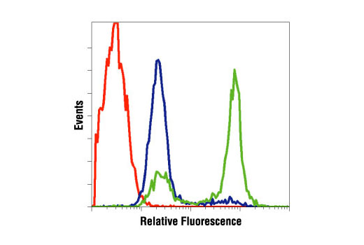
| Product Includes | Product # | Quantity | Mol. Wt | Isotype/Source |
|---|---|---|---|---|
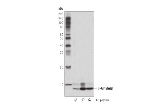
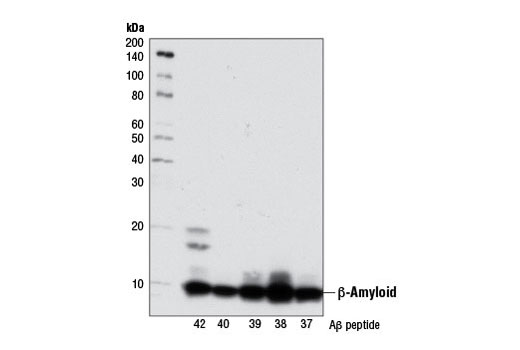
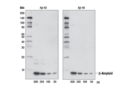
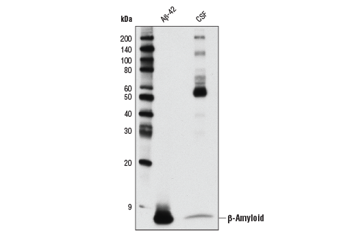
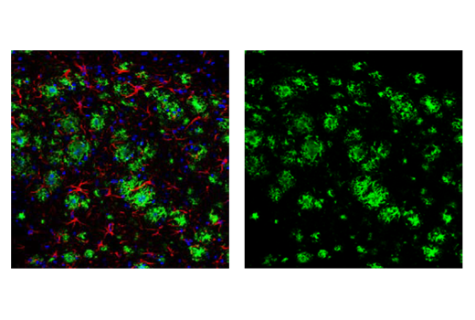
| β-Amyloid (D54D2) XP® Rabbit mAb | 8243 | 20 µl | 5 kDa | Rabbit IgG |
| β-Amyloid (D3D2N) Mouse mAb | 15126 | 20 µl | 5 kDa | Mouse IgG1 |
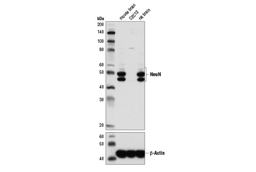
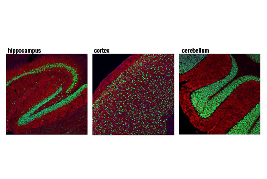
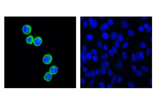
| NeuN (D4G4O) XP® Rabbit mAb | 24307 | 20 µl | 46-55 kDa | Rabbit IgG |
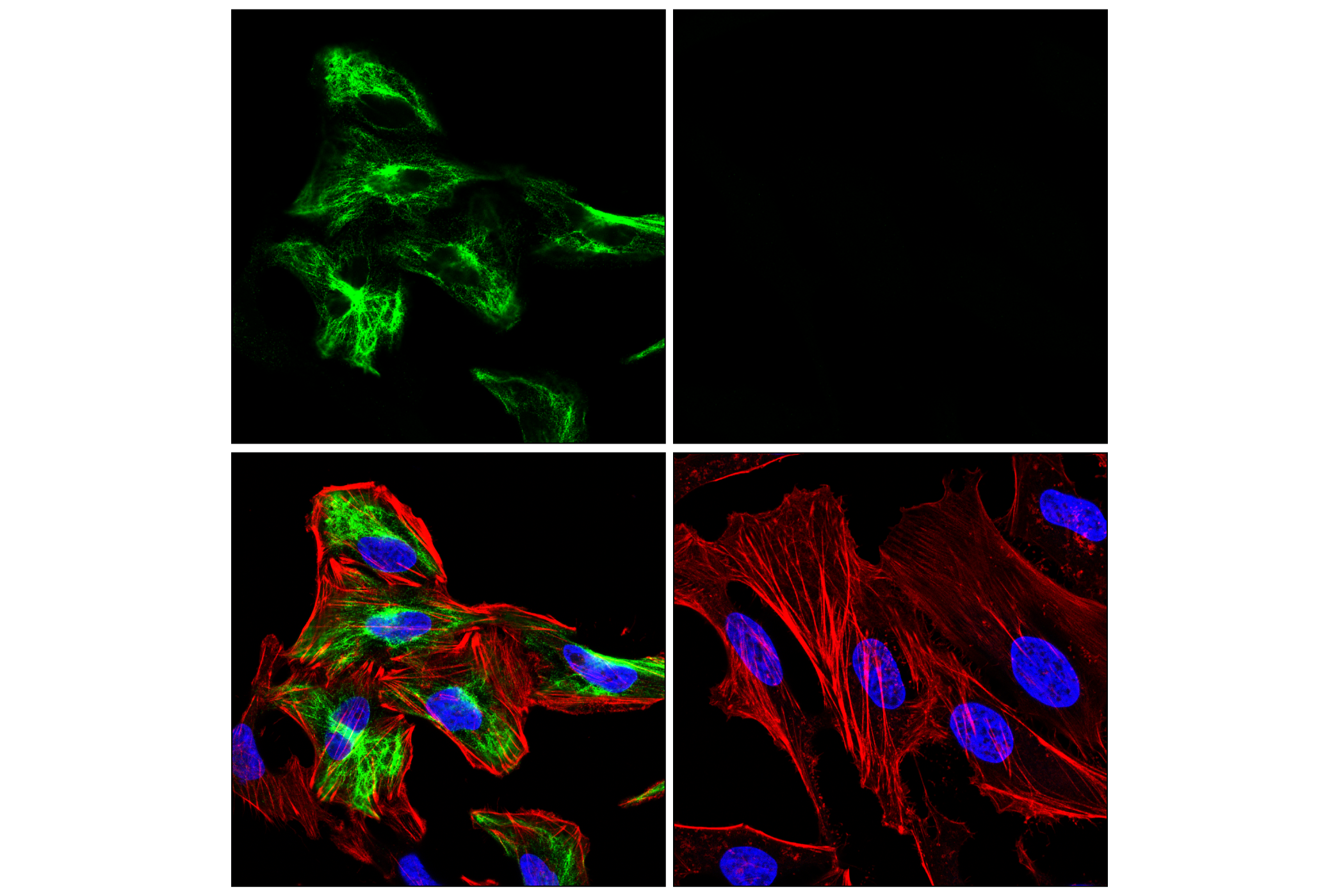
| Synaptophysin (7H12) Mouse mAb (IF Formulated) | 9020 | 20 µl | Mouse IgG1 | |
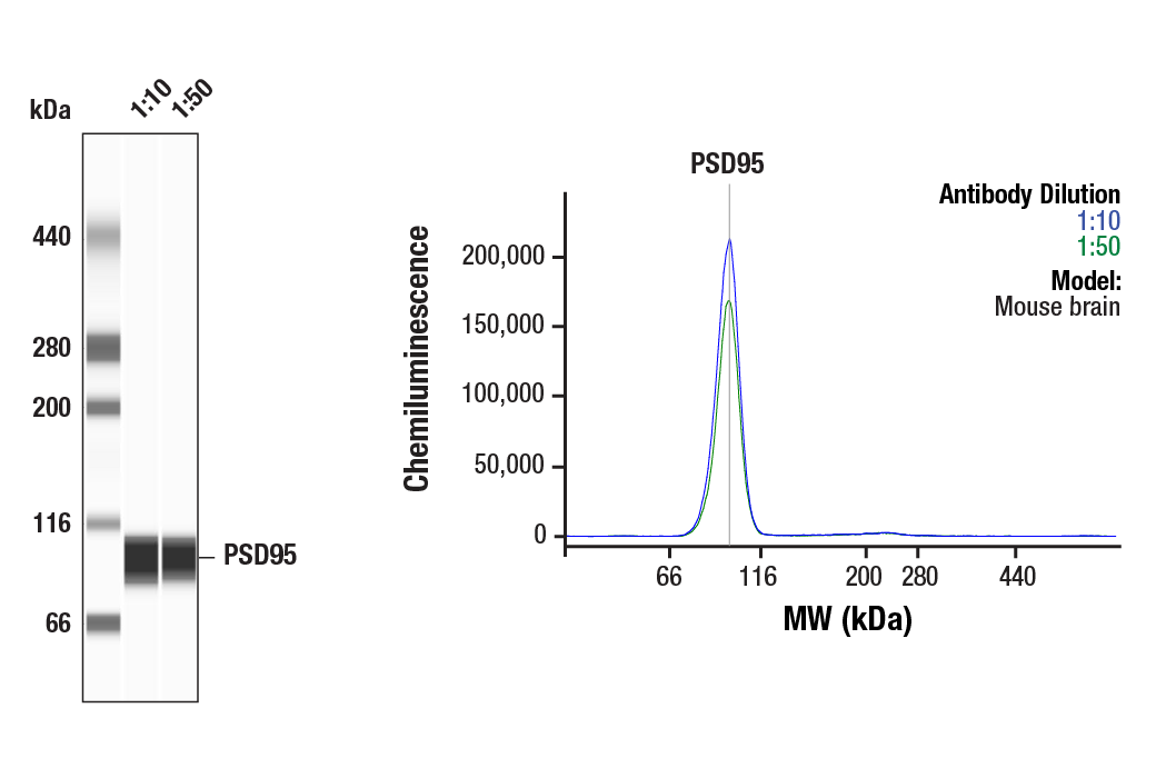
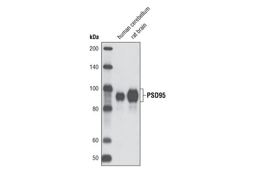
| PSD95 (D27E11) XP® Rabbit mAb | 3450 | 20 µl | 95 kDa | Rabbit IgG |
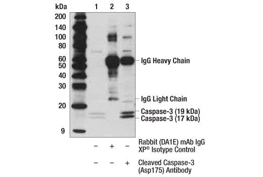
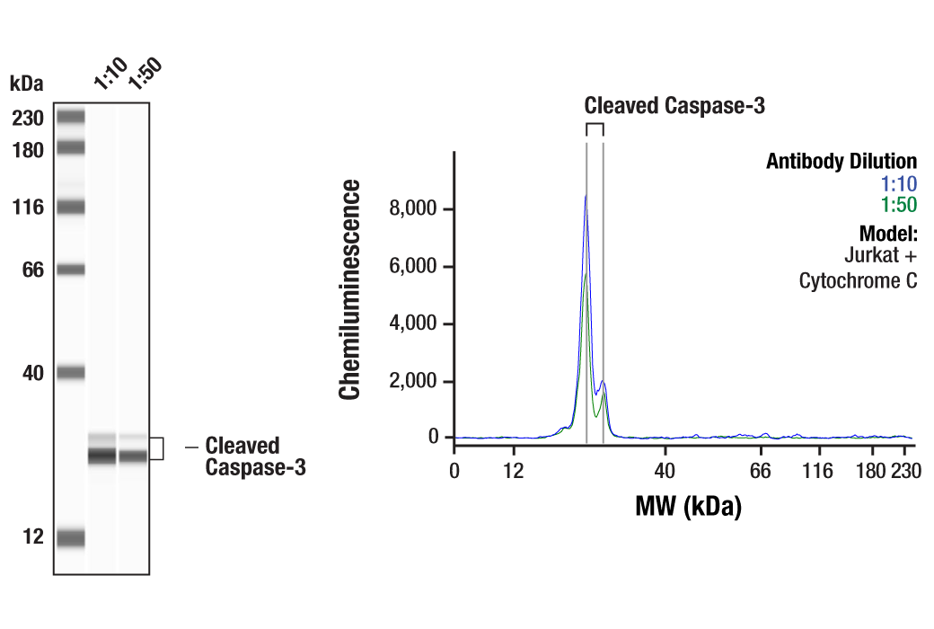
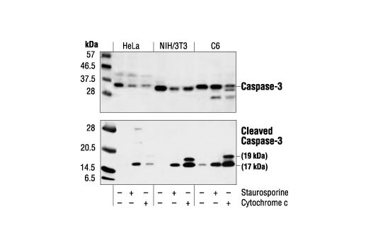
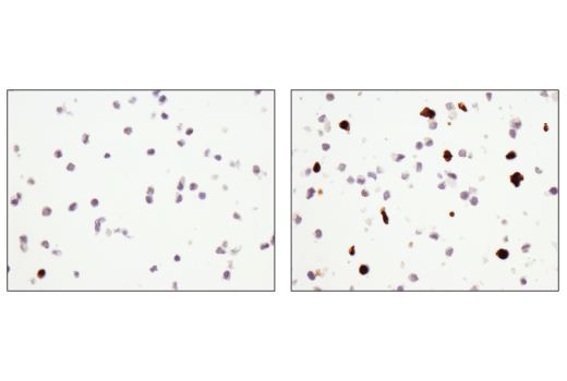
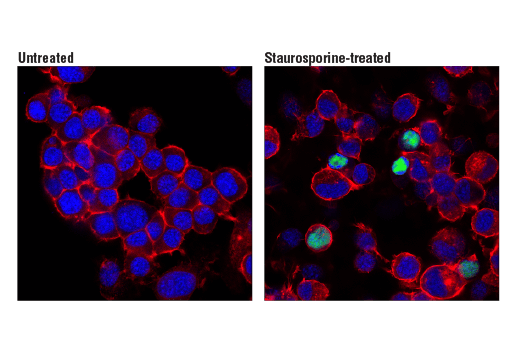
| Cleaved Caspase-3 (Asp175) Antibody | 9661 | 20 µl | 17, 19 kDa | Rabbit |
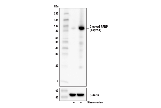
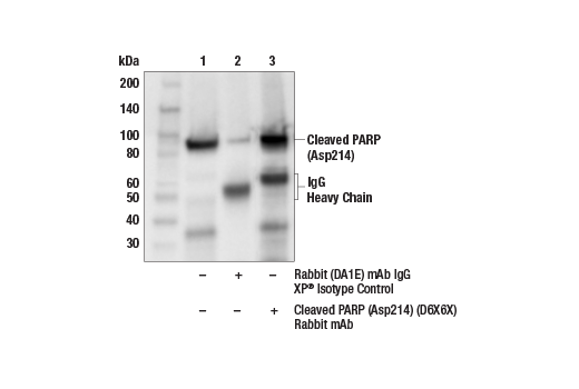
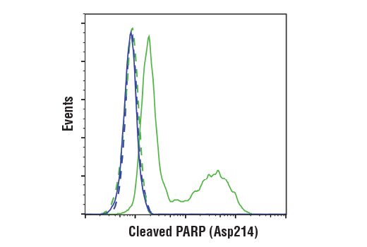
| Cleaved PARP (Asp214) (D6X6X) Rabbit mAb | 94885 | 20 µl | 89 kDa | Rabbit IgG |
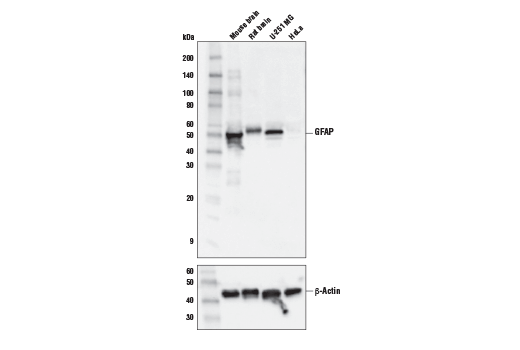
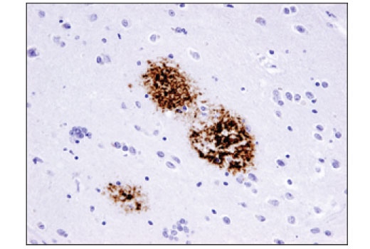
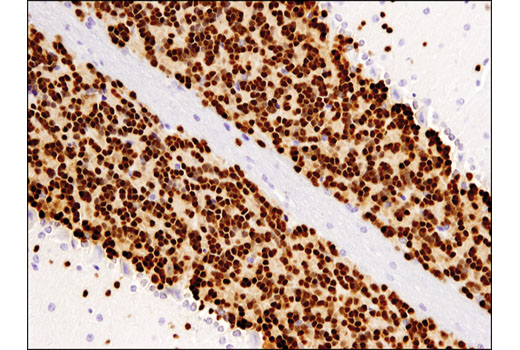
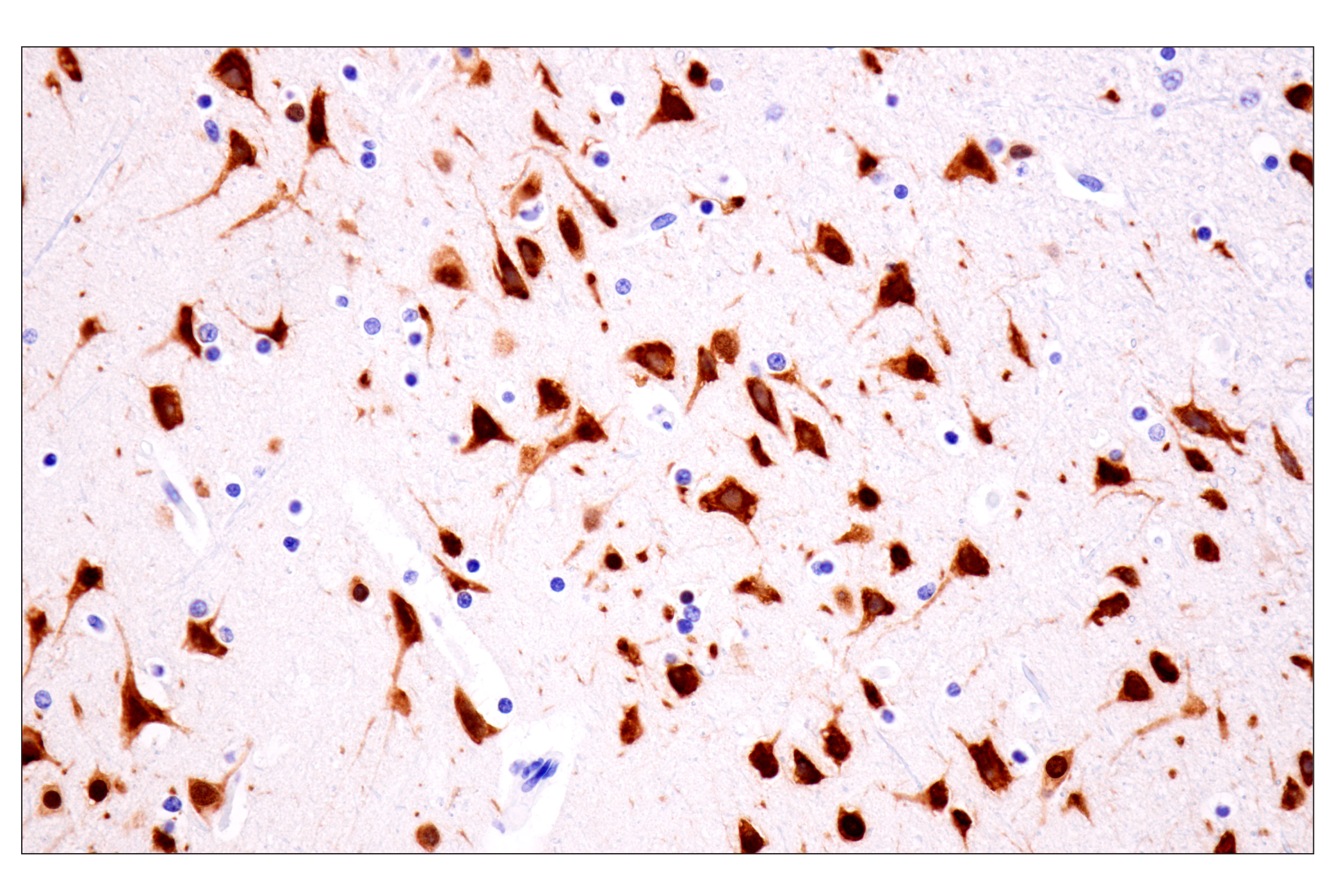
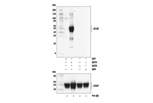
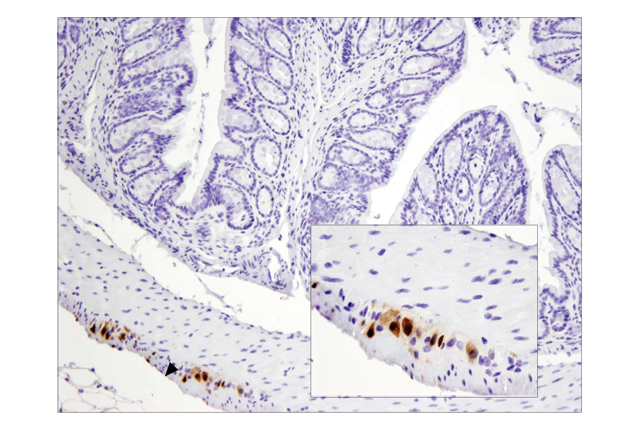
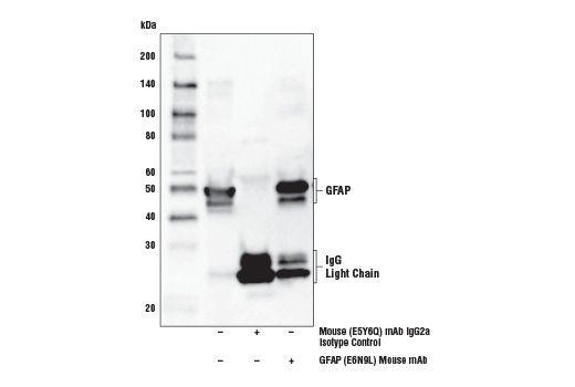
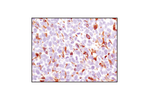
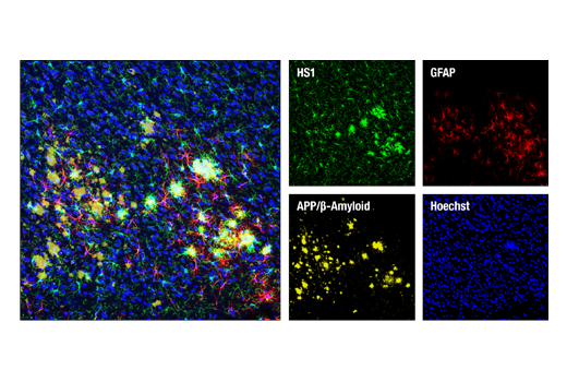
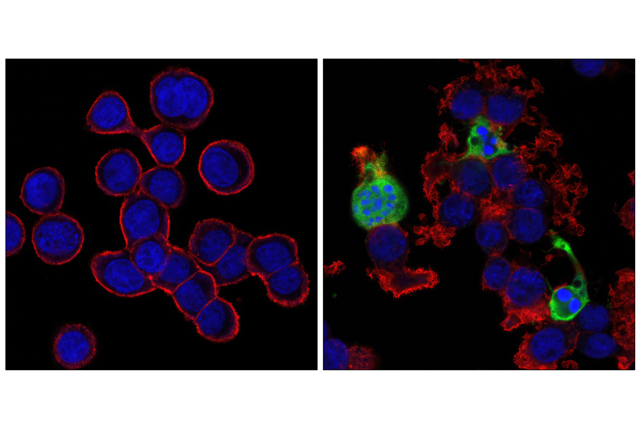
| GFAP (E6N9L) Mouse mAb | 34001 | 20 µl | 50 kDa | Mouse IgG2a |
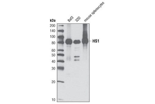
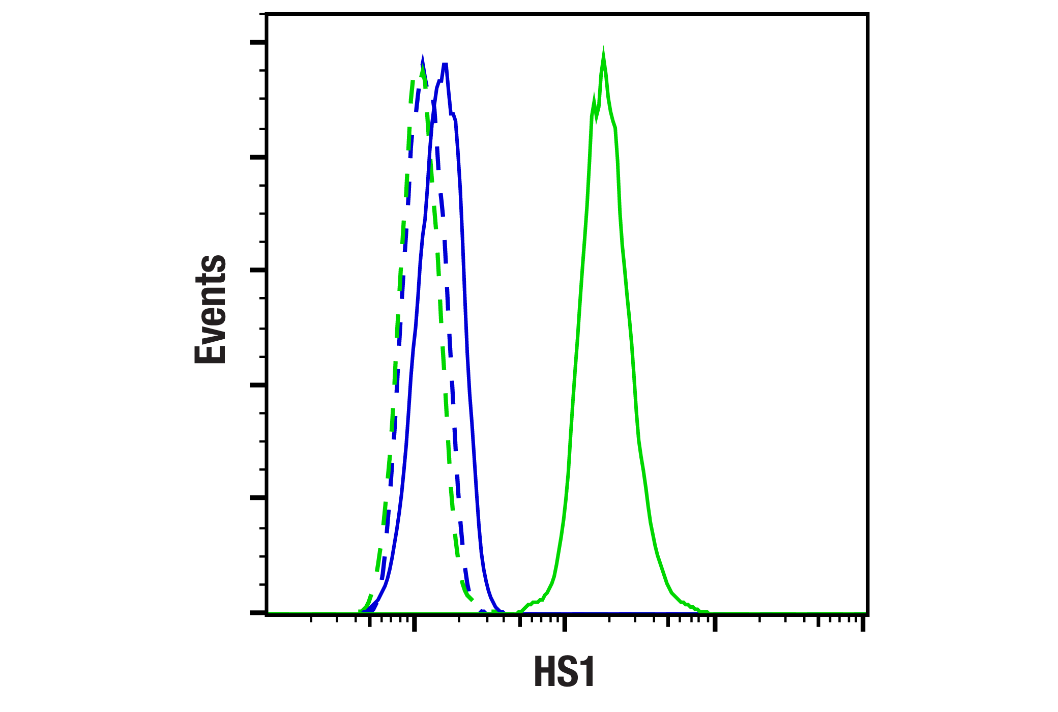
| HS1 (D5A9) XP® Rabbit mAb | 3892 | 20 µl | 80 kDa | Rabbit IgG |
Please visit cellsignal.com for individual component applications, species cross-reactivity, dilutions, protocols, and additional product information.
Description
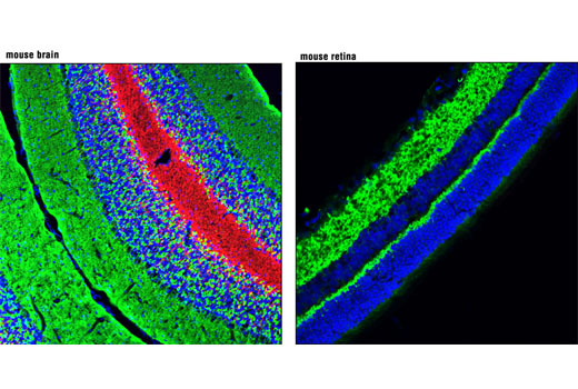
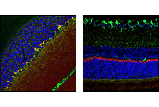
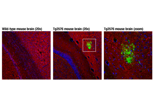
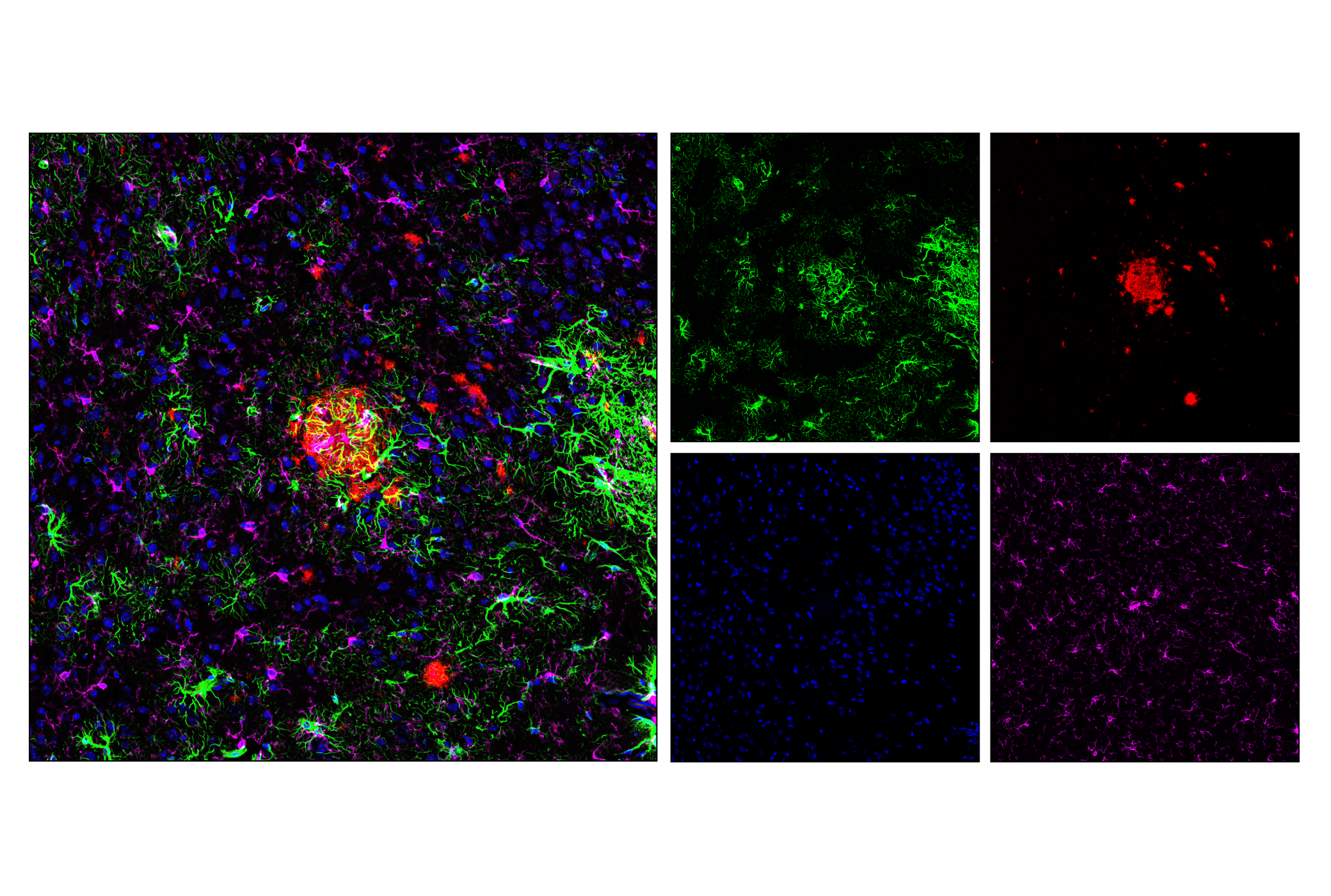
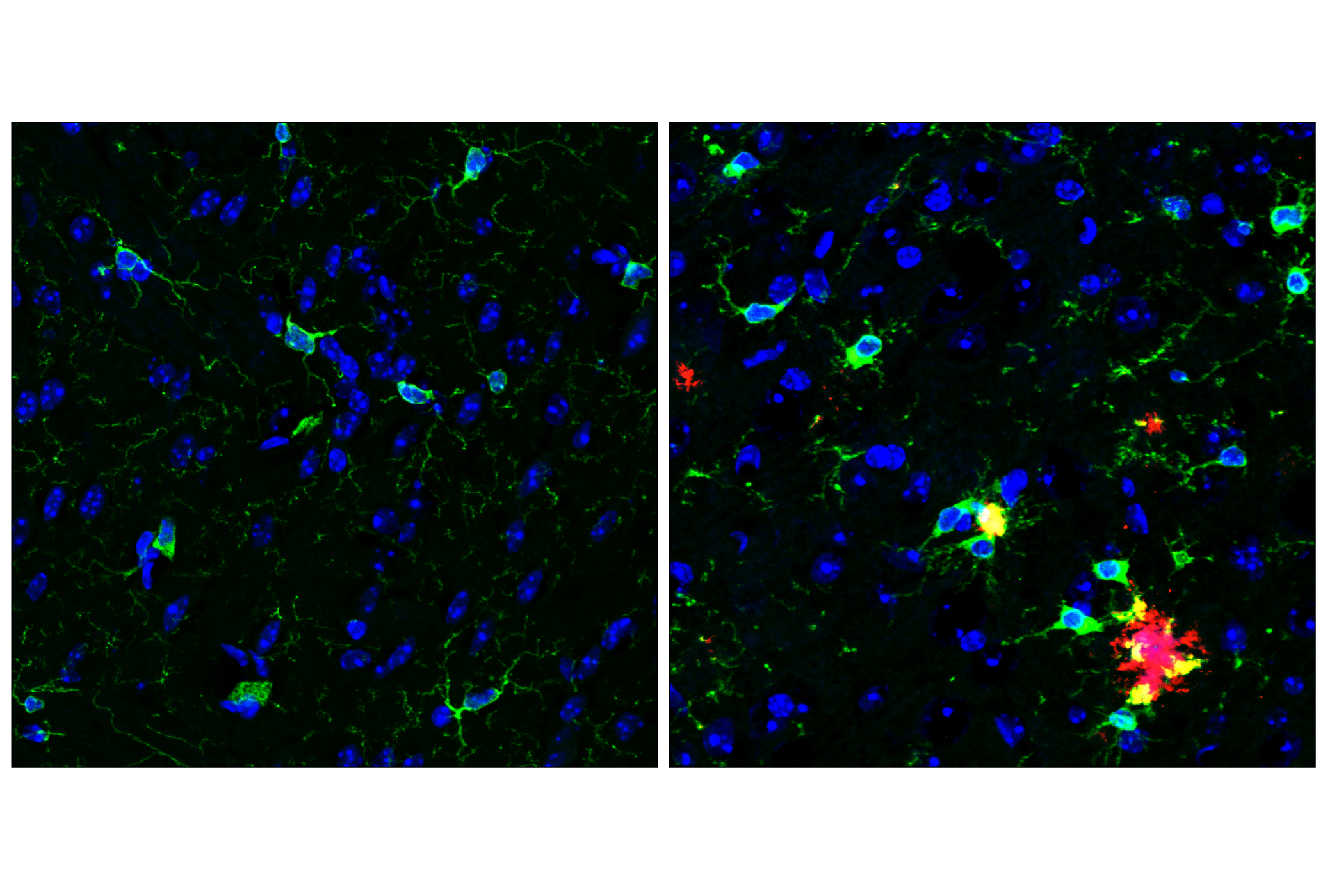
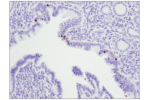
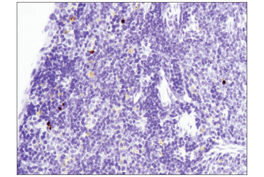
The β-Amyloid Mouse Model Neuronal Viability IF Antibody Sampler Kit provides an economical means of detecting proteins to confirm neuronal viability and surrounding astrocytes and microglia in mouse models by immunofluorescence.
Storage
Background
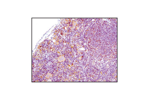
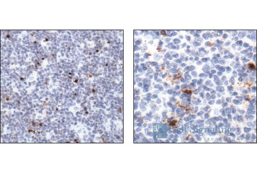
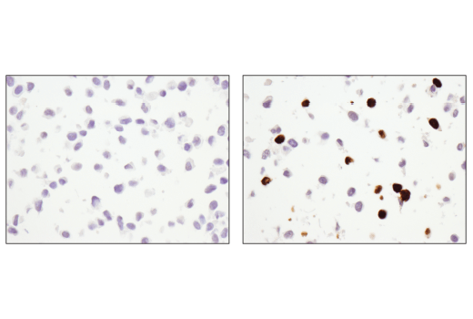
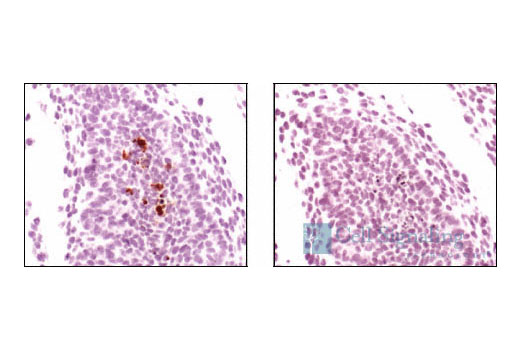
Amyloid β (Aβ) precursor protein (APP) is a 100-140 kDa transmembrane glycoprotein that exists as several isoforms. The amino acid sequence of APP contains the amyloid domain, which can be released by a two-step proteolytic cleavage. The extracellular deposition and accumulation of the released Aβ fragments form the main components of amyloid plaques, a major pathological hallmark of Alzheimer’s disease (1). Neuronal nuclei (NeuN, Fox-3, RBFOX3) is a nuclear protein expressed in most post-mitotic neurons of the central and peripheral nervous systems. NeuN is not detected in Purkinje cells, sympathetic ganglion cells, Cajal-Retzius cells, INL retinal cells, inferior olivary, or dentate nucleus neurons (2). Glial fibrillary acidic protein (GFAP) is the main intermediate filament in mature brain astroglial and radial glial cells and GFAP also plays an important role in modulating astroglial motility and shape (3). HS1 is a protein kinase substrate that is expressed only in tissues and cells of hematopoietic origin (4). Previous work identifying markers of specific brain cell types using RNA-seq has shown HS1 to be a useful and specific tool to study microglia (5). Synaptophysin (SYP) is a neuronal synaptic vesicle glycoprotein that occurs in presynaptic vesicles of neurons (6). Postsynaptic Density protein 95 (PSD95) is a member of the membrane-associated guanylate kinase (MAGUK) family of proteins. PSD95 is a scaffolding protein involved in the assembly and function of the postsynaptic density complex (7,8). Caspase-3 (CPP-32, Apoptain, Yama, SCA-1) is a critical executioner of apoptosis, as it is either partially or totally responsible for the proteolytic cleavage of many key proteins, including nuclear enzyme poly (ADP-ribose) polymerase (PARP) (9). PARP, a 116 kDa nuclear poly (ADP-ribose) polymerase, appears to be involved in DNA repair in response to environmental stress (10). PARP helps cells to maintain their viability; cleavage of PARP facilitates cellular disassembly and serves as a marker of cells undergoing apoptosis (11).
- Selkoe, D.J. (1996) J Biol Chem 271, 18295-8.
- Mullen, R.J. et al. (1992) Development 116, 201-11.
- Eng, L.F. et al. (2000) Neurochem Res 25, 1439-51.
- Kitamura, D. et al. (1995) Biochem Biophys Res Commun 208, 1137-46.
- Zhang, Y. et al. (2014) J Neurosci 34, 11929-47.
- Wiedenmann, B. et al. (1986) Proc Natl Acad Sci U S A 83, 3500-4.
- Cao, J. et al. (2005) J Cell Biol 168, 117-26.
- Chetkovich, D.M. et al. (2002) J Neurosci 22, 6415-25.
- Fernandes-Alnemri, T. et al. (1994) J Biol Chem 269, 30761-4.
- Satoh, M.S. and Lindahl, T. (1992) Nature 356, 356-8.
- Oliver, F.J. et al. (1998) J Biol Chem 273, 33533-9.
Background References
Trademarks and Patents
使用に関する制限
法的な権限を与えられたCSTの担当者が署名した書面によって別途明示的に合意された場合を除き、 CST、その関連会社または代理店が提供する製品には以下の条件が適用されます。お客様が定める条件でここに定められた条件に含まれるものを超えるもの、 または、ここに定められた条件と異なるものは、法的な権限を与えられたCSTの担当者が別途書面にて受諾した場合を除き、拒絶され、 いかなる効力も効果も有しません。
研究専用 (For Research Use Only) またはこれに類似する表示がされた製品は、 いかなる目的についても FDA または外国もしくは国内のその他の規制機関により承認、認可または許可を受けていません。 お客様は製品を診断もしくは治療目的で使用してはならず、また、製品に表示された内容に違反する方法で使用してはなりません。 CST が販売または使用許諾する製品は、エンドユーザーであるお客様に対し、使途を研究および開発のみに限定して提供されるものです。 診断、予防もしくは治療目的で製品を使用することまたは製品を再販売 (単独であるか他の製品等の一部であるかを問いません) もしくはその他の商業的利用の目的で購入することについては、CST から別途許諾を得る必要があります。 お客様は以下の事項を遵守しなければなりません。(a) CST の製品 (単独であるか他の資材と一緒であるかを問いません) を販売、使用許諾、貸与、寄付もしくはその他の態様で第三者に譲渡したり使用させたりしてはなりません。また、商用の製品を製造するために CST の製品を使用してはなりません。(b) 複製、改変、リバースエンジニアリング、逆コンパイル、 分解または他の方法により製品の構造または技術を解明しようとしてはなりません。また、 CST の製品またはサービスと競合する製品またはサービスを開発する目的で CST の製品を使用してはなりません。(c) CST の製品の商標、商号、ロゴ、特許または著作権に関する通知または表示を除去したり改変したりしてはなりません。(d) CST の製品をCST 製品販売条件(CST’s Product Terms of Sale) および該当する書面のみに従って使用しなければなりません。(e) CST の製品に関連してお客様が使用する第三者の製品またはサービスに関する使用許諾条件、 サービス提供条件またはこれに類する合意事項を遵守しなければなりません。