WB, IP, IF-IC
H M R Mk
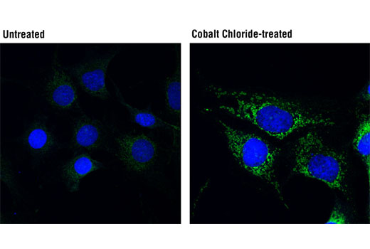
Endogenous
38, 76
Rabbit IgG
#O60238
665
Product Information
Product Usage Information
| Application | Dilution |
|---|---|
| Western Blotting | 1:1000 |
| Immunoprecipitation | 1:100 |
| Immunofluorescence (Immunocytochemistry) | 1:100 - 1:400 |
Storage
Specificity / Sensitivity
Species Reactivity:
Human, Mouse, Rat, Monkey
Source / Purification
Monoclonal antibody is produced by immunizing animals with a synthetic peptide corresponding to residues surrounding Glu128 of human BNIP3L/Nix protein.
Background
BCL2/Adenovirus E1B 19 kDa protein-interacting protein 3-like (BNIP3L) (1), also termed BNIP3α (2), B5 (3), and Nix (4), is a member of the Bcl-2 family of apoptotic regulators with highest homology to BNIP3. BNIP3L can bind BNIP3, Bcl-xL, and Bcl-2 (1-5). BNIP3L forms homodimers that withstand denaturing by SDS and reducing conditions (5). BNIP3L is a mitochondrial protein and knockout studies suggest that BNIP3L regulates autophagic clearance of damaged mitochondria during erythroid maturation via mitochondrial autophagy (6,7). It has been shown that the expression of BNIP3L is up-regulated during terminal erythroid differentiation (6-8), as well as in tumor cell lines during hypoxia (9-11). BNIP3L directly regulates the elimination of mitochondria through its ability to bind to and recruit important components of the autophagic machinery, including LC3/Atg8 and GABARAP proteins, via its amino-terminal LC3-interacting region (LIR) (12). BNIP3L may also indirectly activate phagophore formation either via the recruitment of autophagy proteins or by binding Bcl-xL, which in turn releases Beclin-1 (13). BNIP3L/Nix also plays a pivotal role in Parkin-mediated mitochondrial autophagy via its ability to mediate the mitochondrial translocation of Parkin (14). Activated BNIP3L can promote the opening of mitochondrial permeability transition pores resulting in mitochondrial depolarization, generation of reactive oxygen species, and induction of necrosis. Due to its involvement in cell death and autophagy, research scientists have implicated BNIP3L in heart disease and cancer (13).
- Matsushima, M. et al. (1998) Genes Chromosomes Cancer 21, 230-5.
- Yasuda, M. et al. (1999) Cancer Res 59, 533-7.
- Ohi, N. et al. (1999) Cell Death Differ 6, 314-25.
- Chen, G. et al. (1999) J Biol Chem 274, 7-10.
- Imazu, T. et al. (1999) Oncogene 18, 4523-9.
- Schweers, R.L. et al. (2007) Proc Natl Acad Sci USA 104, 19500-5.
- Sandoval, H. et al. (2008) Nature 454, 232-5.
- Aerbajinai, W. et al. (2003) Blood 102, 712-7.
- Sowter, H.M. et al. (2001) Cancer Res 61, 6669-73.
- Bruick, R.K. (2000) Proc Natl Acad Sci USA 97, 9082-7.
- Fei, P. et al. (2004) Cancer Cell 6, 597-609.
- Novak, I. et al. (2010) EMBO Rep 11, 45-51.
- Zhang, J. and Ney, P.A. (2009) Cell Death Differ 16, 939-46.
- Ding, W.X. et al. (2010) J Biol Chem 285, 27879-90.
Species Reactivity
Species reactivity is determined by testing in at least one approved application (e.g., western blot).
Western Blot Buffer
IMPORTANT: For western blots, incubate membrane with diluted primary antibody in 5% w/v BSA, 1X TBS, 0.1% Tween® 20 at 4°C with gentle shaking, overnight.
Applications Key
WB: Western Blotting IP: Immunoprecipitation IF-IC: Immunofluorescence (Immunocytochemistry)
Cross-Reactivity Key
H: human M: mouse R: rat Hm: hamster Mk: monkey Vir: virus Mi: mink C: chicken Dm: D. melanogaster X: Xenopus Z: zebrafish B: bovine Dg: dog Pg: pig Sc: S. cerevisiae Ce: C. elegans Hr: horse GP: Guinea Pig Rab: rabbit All: all species expected
Trademarks and Patents
使用に関する制限
法的な権限を与えられたCSTの担当者が署名した書面によって別途明示的に合意された場合を除き、 CST、その関連会社または代理店が提供する製品には以下の条件が適用されます。お客様が定める条件でここに定められた条件に含まれるものを超えるもの、 または、ここに定められた条件と異なるものは、法的な権限を与えられたCSTの担当者が別途書面にて受諾した場合を除き、拒絶され、 いかなる効力も効果も有しません。
研究専用 (For Research Use Only) またはこれに類似する表示がされた製品は、 いかなる目的についても FDA または外国もしくは国内のその他の規制機関により承認、認可または許可を受けていません。 お客様は製品を診断もしくは治療目的で使用してはならず、また、製品に表示された内容に違反する方法で使用してはなりません。 CST が販売または使用許諾する製品は、エンドユーザーであるお客様に対し、使途を研究および開発のみに限定して提供されるものです。 診断、予防もしくは治療目的で製品を使用することまたは製品を再販売 (単独であるか他の製品等の一部であるかを問いません) もしくはその他の商業的利用の目的で購入することについては、CST から別途許諾を得る必要があります。 お客様は以下の事項を遵守しなければなりません。(a) CST の製品 (単独であるか他の資材と一緒であるかを問いません) を販売、使用許諾、貸与、寄付もしくはその他の態様で第三者に譲渡したり使用させたりしてはなりません。また、商用の製品を製造するために CST の製品を使用してはなりません。(b) 複製、改変、リバースエンジニアリング、逆コンパイル、 分解または他の方法により製品の構造または技術を解明しようとしてはなりません。また、 CST の製品またはサービスと競合する製品またはサービスを開発する目的で CST の製品を使用してはなりません。(c) CST の製品の商標、商号、ロゴ、特許または著作権に関する通知または表示を除去したり改変したりしてはなりません。(d) CST の製品をCST 製品販売条件(CST’s Product Terms of Sale) および該当する書面のみに従って使用しなければなりません。(e) CST の製品に関連してお客様が使用する第三者の製品またはサービスに関する使用許諾条件、 サービス提供条件またはこれに類する合意事項を遵守しなければなりません。