| Product Includes | Product # | Quantity | Mol. Wt | Isotype/Source |
|---|---|---|---|---|
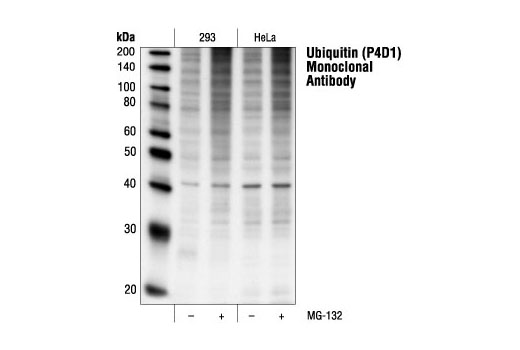
| Ubiquitin (P4D1) Mouse mAb | 3936 | 20 µl | Mouse IgG1 | |
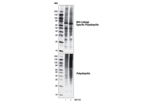
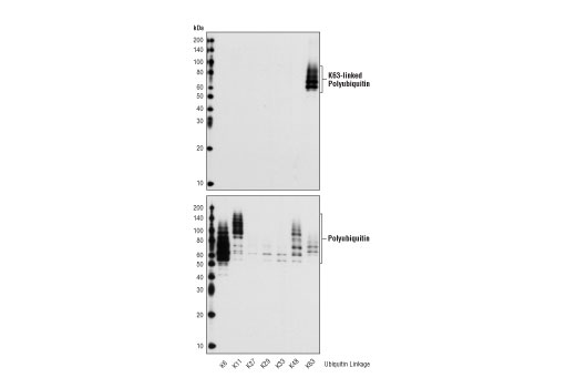
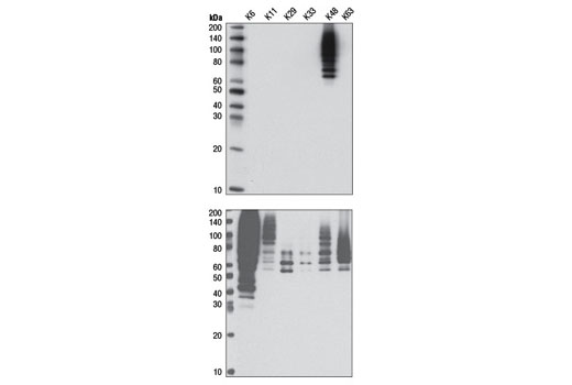
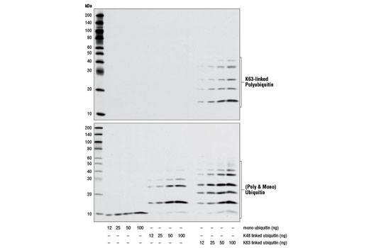
| K63-linkage Specific Polyubiquitin (D7A11) Rabbit mAb | 5621 | 20 µl | Rabbit IgG | |
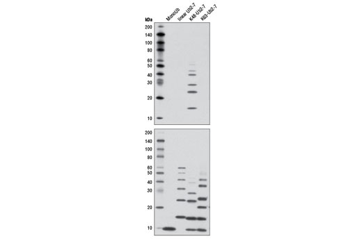
| K48-linkage Specific Polyubiquitin (D9D5) Rabbit mAb | 8081 | 20 µl | Rabbit IgG | |
| Anti-rabbit IgG, HRP-linked Antibody | 7074 | 100 µl | Goat | |
| Anti-mouse IgG, HRP-linked Antibody | 7076 | 100 µl | Horse |
Please visit cellsignal.com for individual component applications, species cross-reactivity, dilutions, protocols, and additional product information.
Description
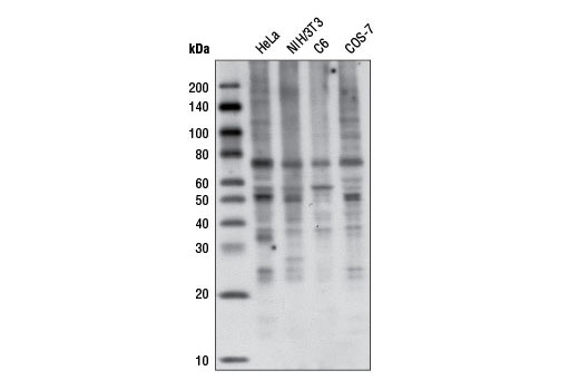
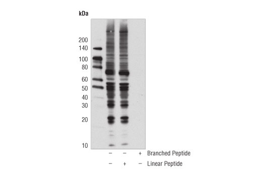
Branched Ubiquitin Antibody Sampler Kit provides an economical means of detecting total and common branch specific forms of ubiquitin. The kit includes enough antibody to perform two western blot experiments with each primary antibody.
Storage
Background
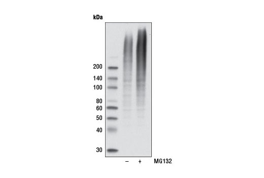
Ubiquitin is a conserved polypeptide unit that plays an important role in the ubiquitin-proteasome pathway. Ubiquitin can be covalently linked to many cellular proteins by the ubiquitination process, which targets proteins for degradation by the 26S proteasome. Three components are involved in the target protein-ubiquitin conjugation process. Ubiquitin is first activated by forming a thiolester complex with the activation component E1; the activated ubiquitin is subsequently transferred to the ubiquitin-carrier protein E2, then from E2 to ubiquitin ligase E3 for final delivery to the epsilon-NH2 of the target protein lysine residue (1-3). The ubiquitin-proteasome pathway has been implicated in a wide range of normal biological processes and in disease-related abnormalities. Several proteins such as IκB, p53, cdc25A, and Bcl-2 have been shown to be targets for the ubiquitin-proteasome process as part of regulation of cell cycle progression, differentiation, cell stress response, and apoptosis (4-7).
Substrate proteins are linked to ubiquitin using seven distinct ubiquitin lysine residues (Lys6, Lys11, Lys27, Lys29, Lys33, Lys48 and Lys63). Formation of a polyubiquitin chain occurs when a lysine residue of ubiquitin is linked to the carboxy-terminal glycine of another ubiquitin. Proteins polyubiquinated at specific lysine residues display a tendency to be targeted for different processes; K48-linked polyubiquitin chains mainly target proteins for proteasomal degradation while K63-linked polyubiquitin regulates protein function, subcellular localization, or protein-protein interactions (8). K63-linked polyubiquitin chains exert nonproteolytic functions in vivo, such as protein trafficking, kinase/phosphatase activation, and DNA damage control, all of which might be important in regulation of cancer survival and development (9,10).
- Ciechanover, A. (1998) EMBO J 17, 7151-60.
- Hochstrasser, M. (2000) Nat Cell Biol 2, E153-7.
- Hochstrasser, M. (2000) Science 289, 563-4.
- Bernardi, R. et al. (2000) Oncogene 19, 2447-54.
- Aberle, H. et al. (1997) EMBO J 16, 3797-804.
- Salomoni, P. and Pandolfi, P.P. (2002) Nat Cell Biol 4, E152-3.
- Jesenberger, V. and Jentsch, S. (2002) Nat Rev Mol Cell Biol 3, 112-21.
- Komander, D. (2009) Biochem Soc Trans 37, 937-53.
- Chen, Z.J. and Sun, L.J. (2009) Mol Cell 33, 275-86.
- Yang, W.L. et al. (2010) Oncogene 29, 4493-503.
Background References
Trademarks and Patents
使用に関する制限
法的な権限を与えられたCSTの担当者が署名した書面によって別途明示的に合意された場合を除き、 CST、その関連会社または代理店が提供する製品には以下の条件が適用されます。お客様が定める条件でここに定められた条件に含まれるものを超えるもの、 または、ここに定められた条件と異なるものは、法的な権限を与えられたCSTの担当者が別途書面にて受諾した場合を除き、拒絶され、 いかなる効力も効果も有しません。
研究専用 (For Research Use Only) またはこれに類似する表示がされた製品は、 いかなる目的についても FDA または外国もしくは国内のその他の規制機関により承認、認可または許可を受けていません。 お客様は製品を診断もしくは治療目的で使用してはならず、また、製品に表示された内容に違反する方法で使用してはなりません。 CST が販売または使用許諾する製品は、エンドユーザーであるお客様に対し、使途を研究および開発のみに限定して提供されるものです。 診断、予防もしくは治療目的で製品を使用することまたは製品を再販売 (単独であるか他の製品等の一部であるかを問いません) もしくはその他の商業的利用の目的で購入することについては、CST から別途許諾を得る必要があります。 お客様は以下の事項を遵守しなければなりません。(a) CST の製品 (単独であるか他の資材と一緒であるかを問いません) を販売、使用許諾、貸与、寄付もしくはその他の態様で第三者に譲渡したり使用させたりしてはなりません。また、商用の製品を製造するために CST の製品を使用してはなりません。(b) 複製、改変、リバースエンジニアリング、逆コンパイル、 分解または他の方法により製品の構造または技術を解明しようとしてはなりません。また、 CST の製品またはサービスと競合する製品またはサービスを開発する目的で CST の製品を使用してはなりません。(c) CST の製品の商標、商号、ロゴ、特許または著作権に関する通知または表示を除去したり改変したりしてはなりません。(d) CST の製品をCST 製品販売条件(CST’s Product Terms of Sale) および該当する書面のみに従って使用しなければなりません。(e) CST の製品に関連してお客様が使用する第三者の製品またはサービスに関する使用許諾条件、 サービス提供条件またはこれに類する合意事項を遵守しなければなりません。