| Product Includes | Product # | Quantity | Mol. Wt | Isotype/Source |
|---|---|---|---|---|
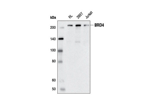
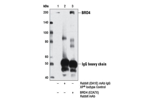
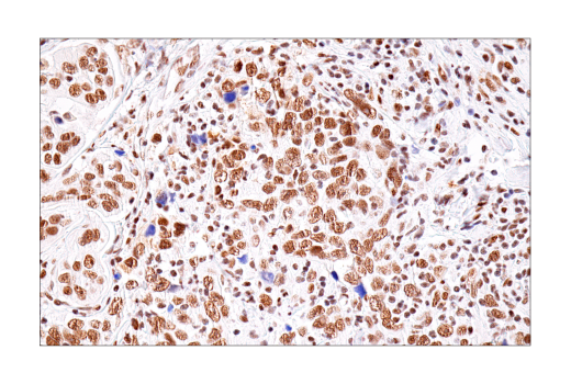
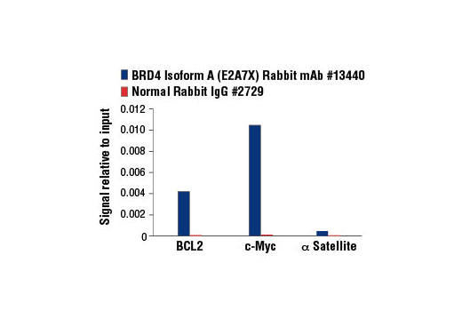
| BRD4 (E2A7X) Rabbit mAb | 13440 | 20 µl | 200 kDa | Rabbit IgG |
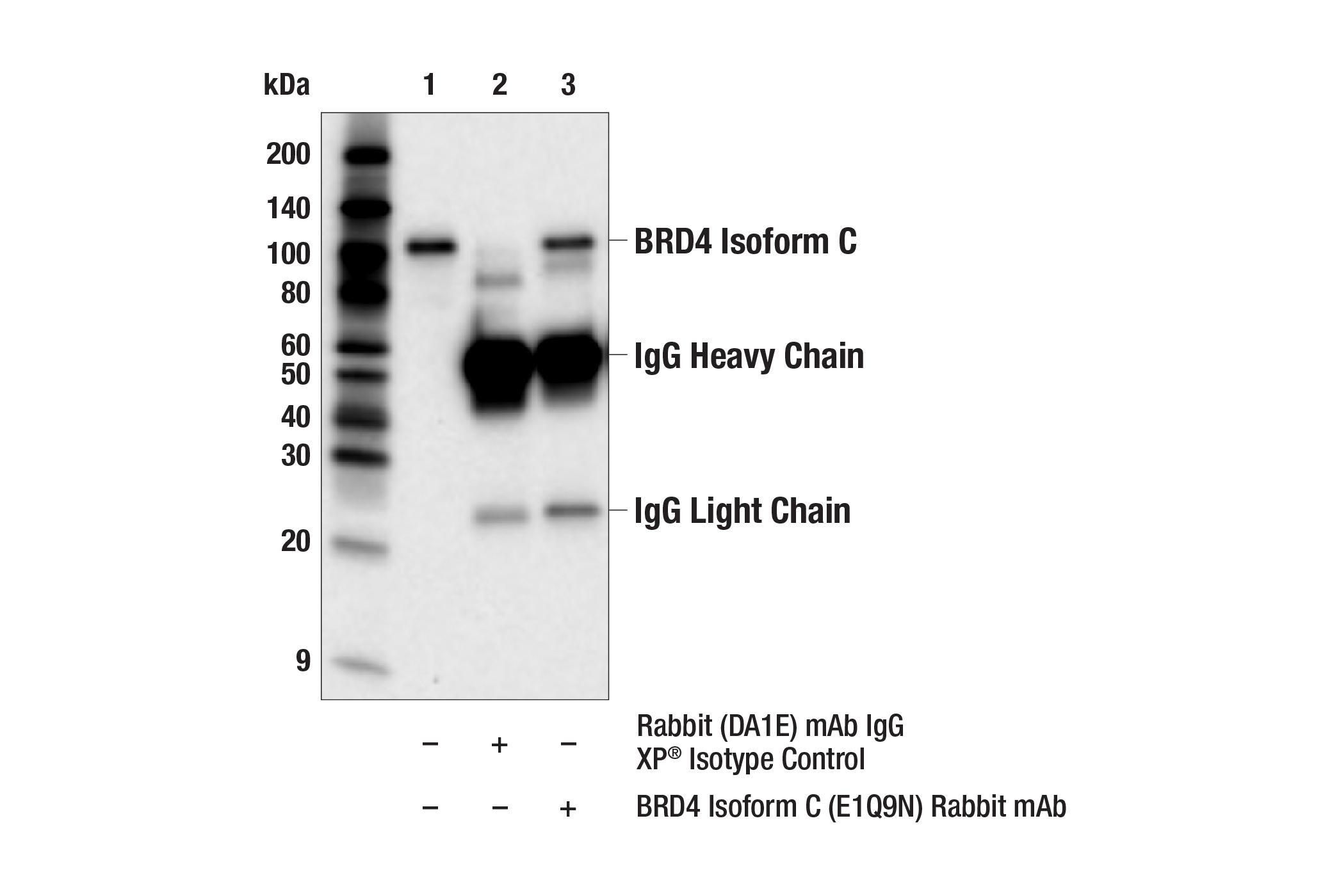
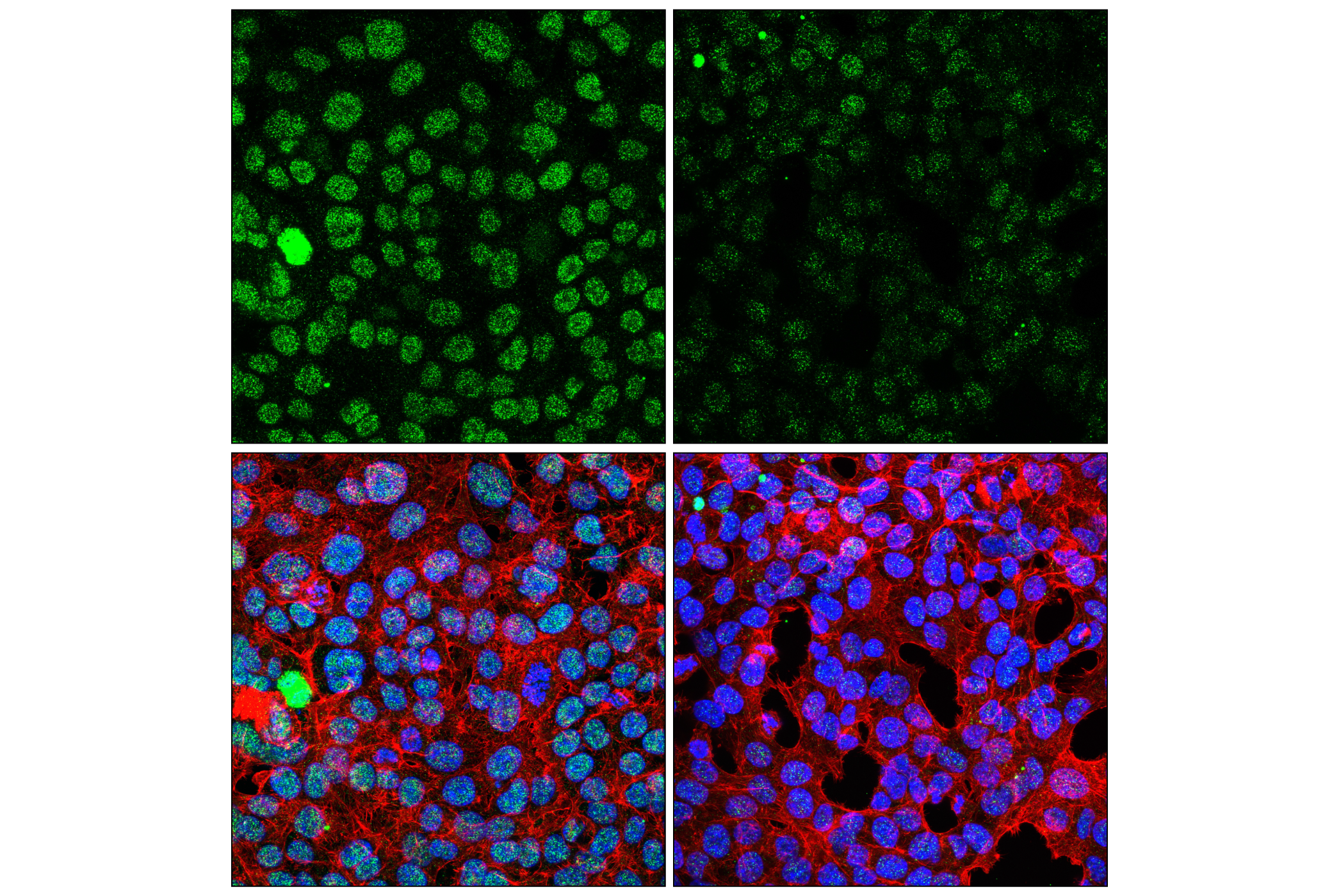
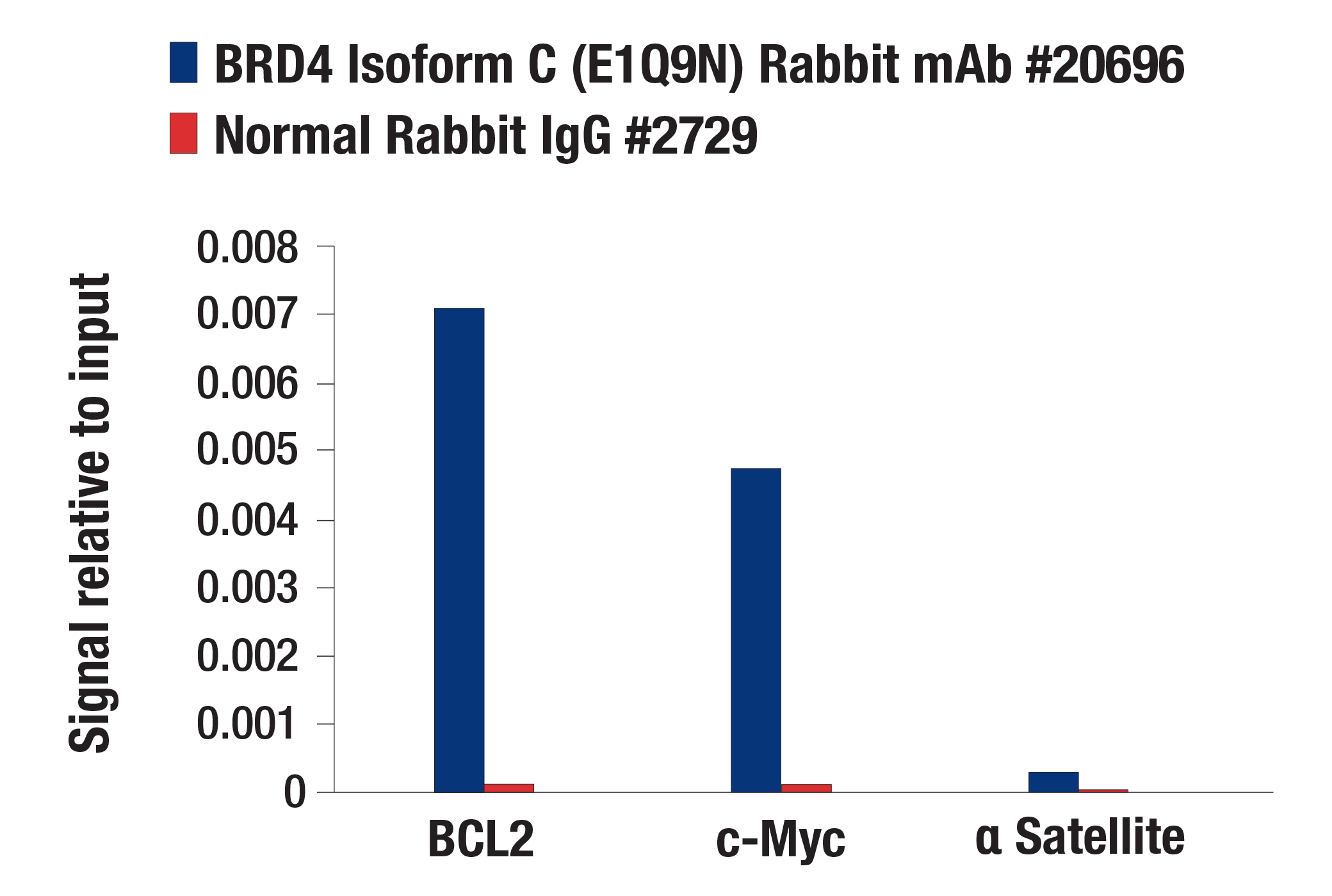
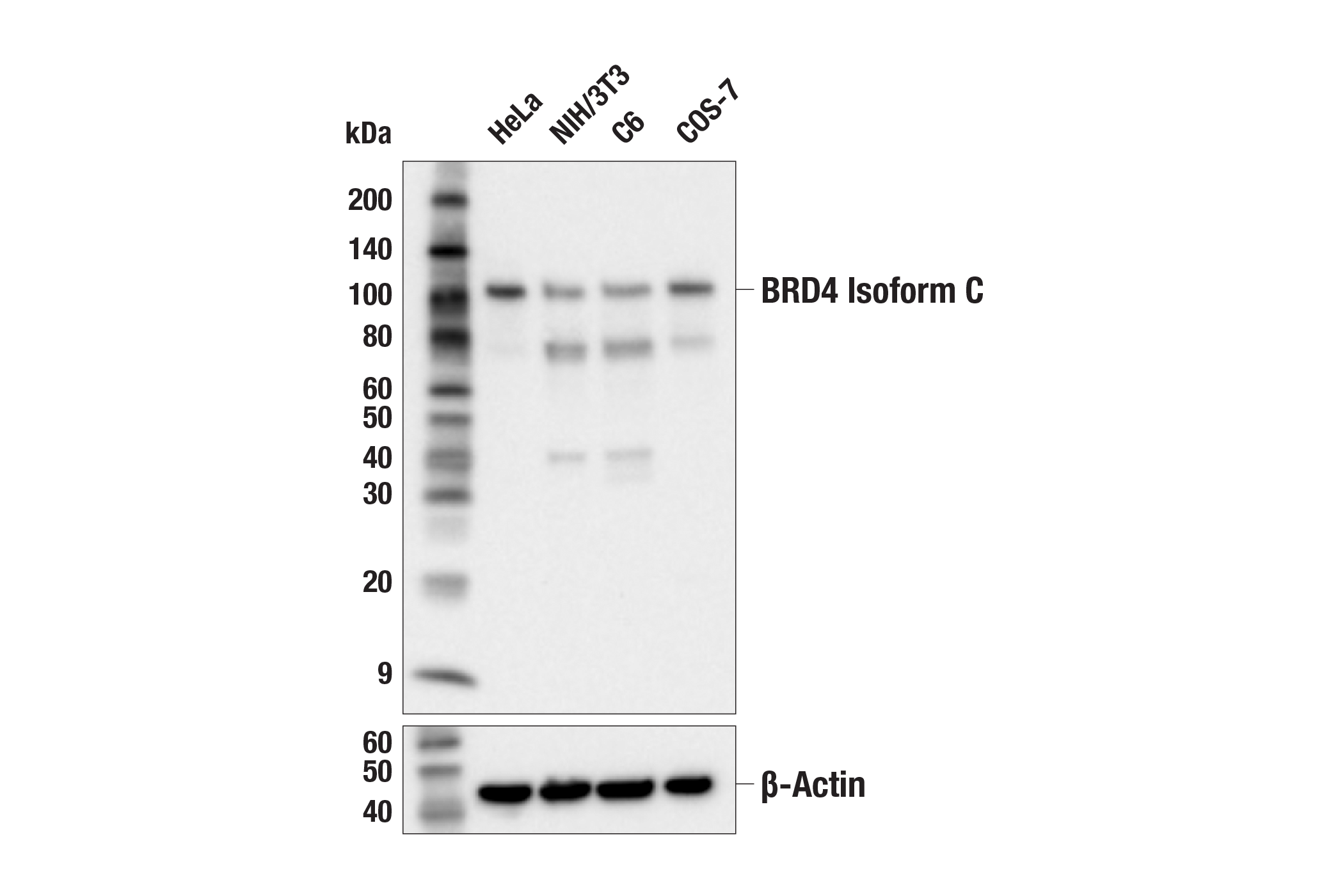
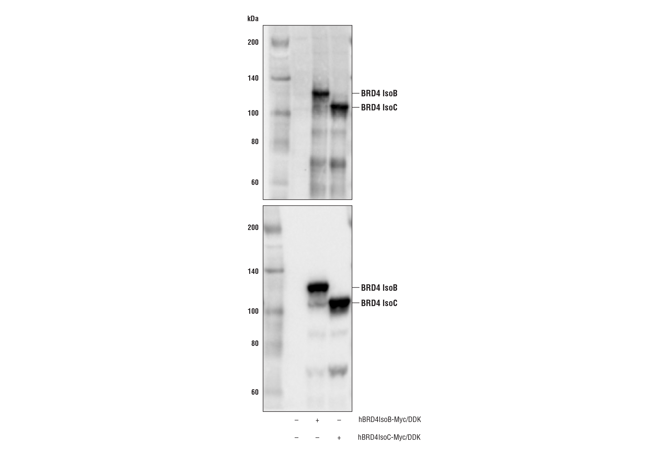
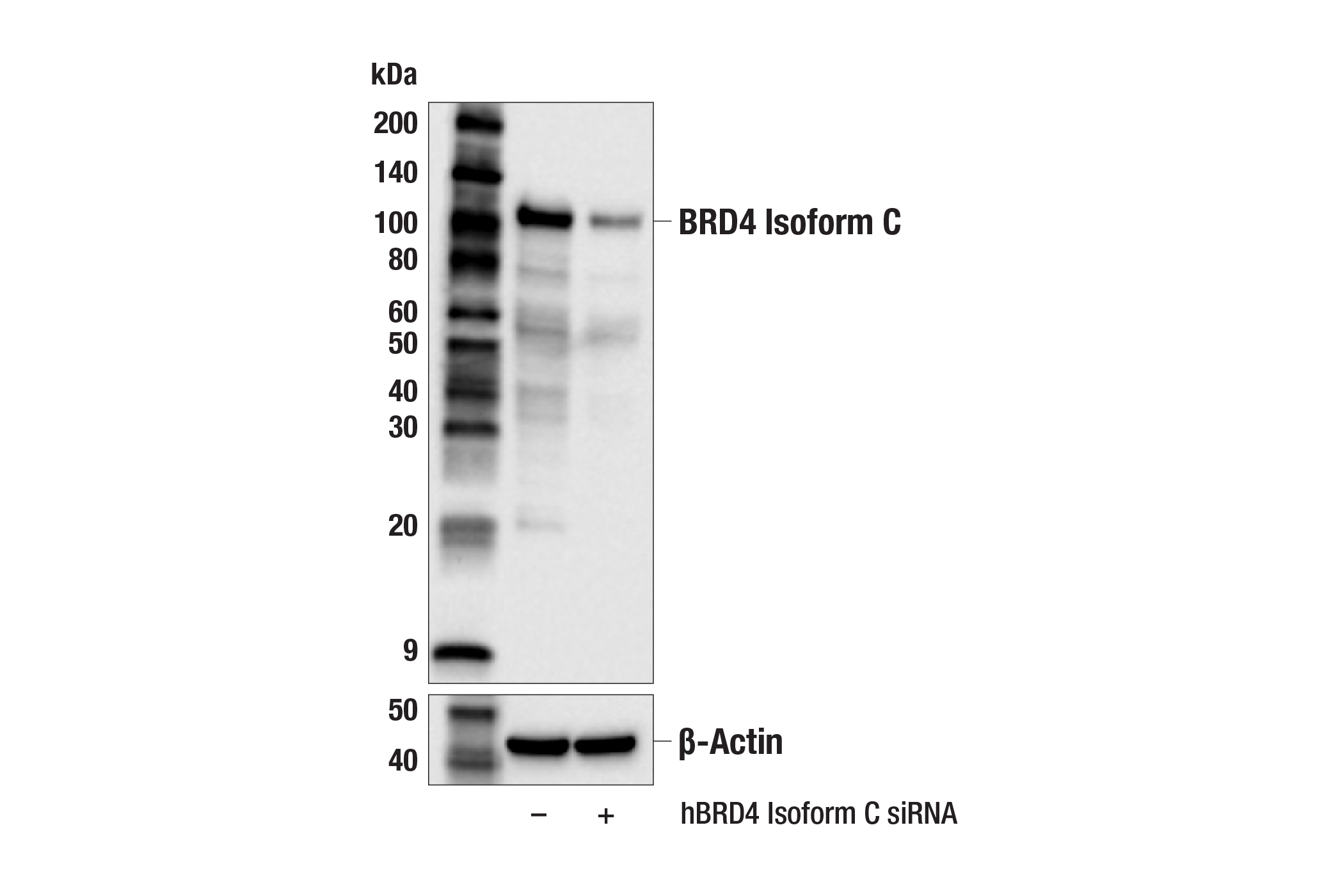
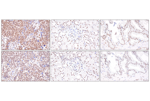
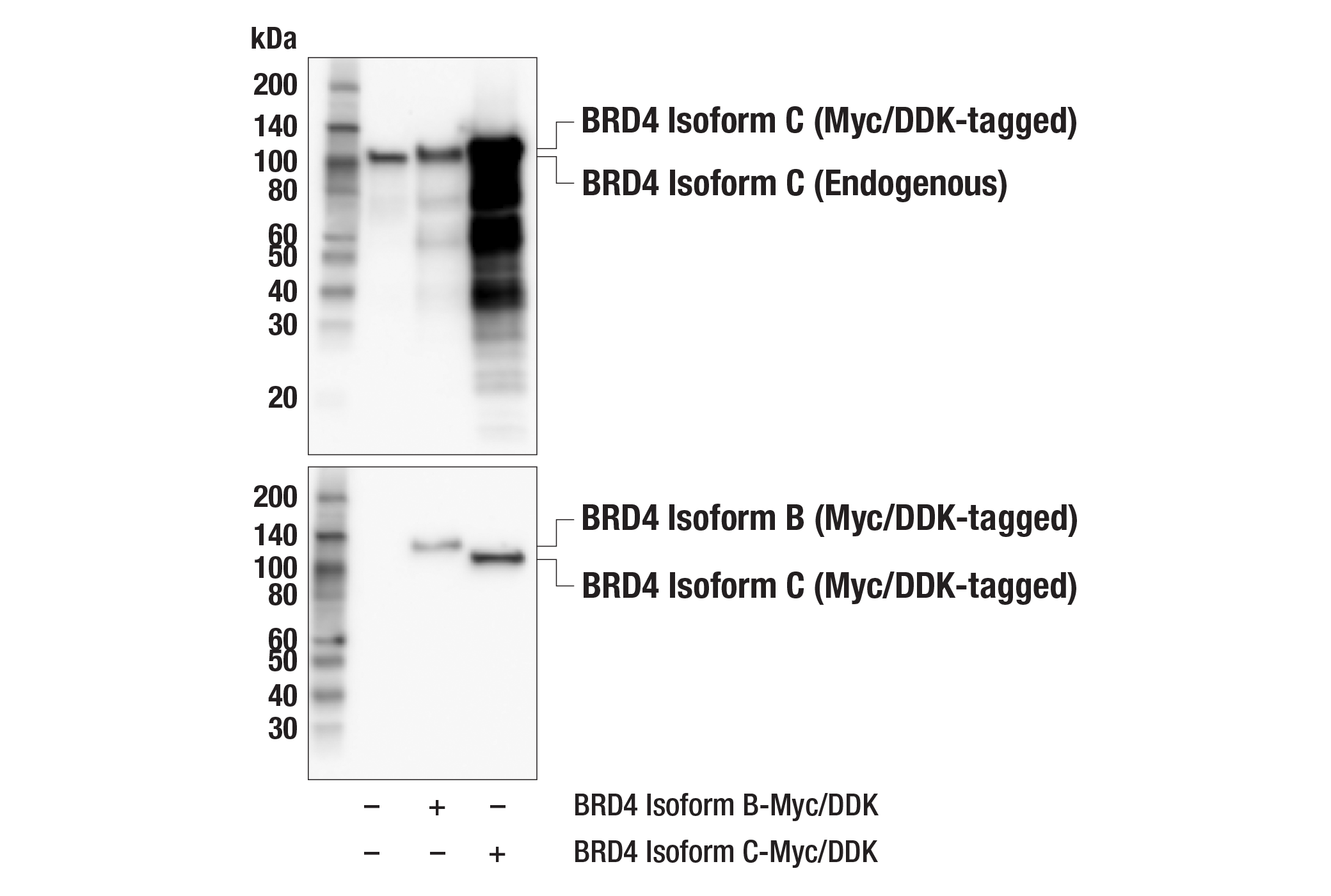
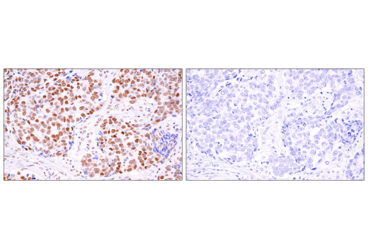
| BRD4 Isoform C (E1Q9N) Rabbit mAb | 20696 | 20 µl | 105 kDa | Rabbit IgG |
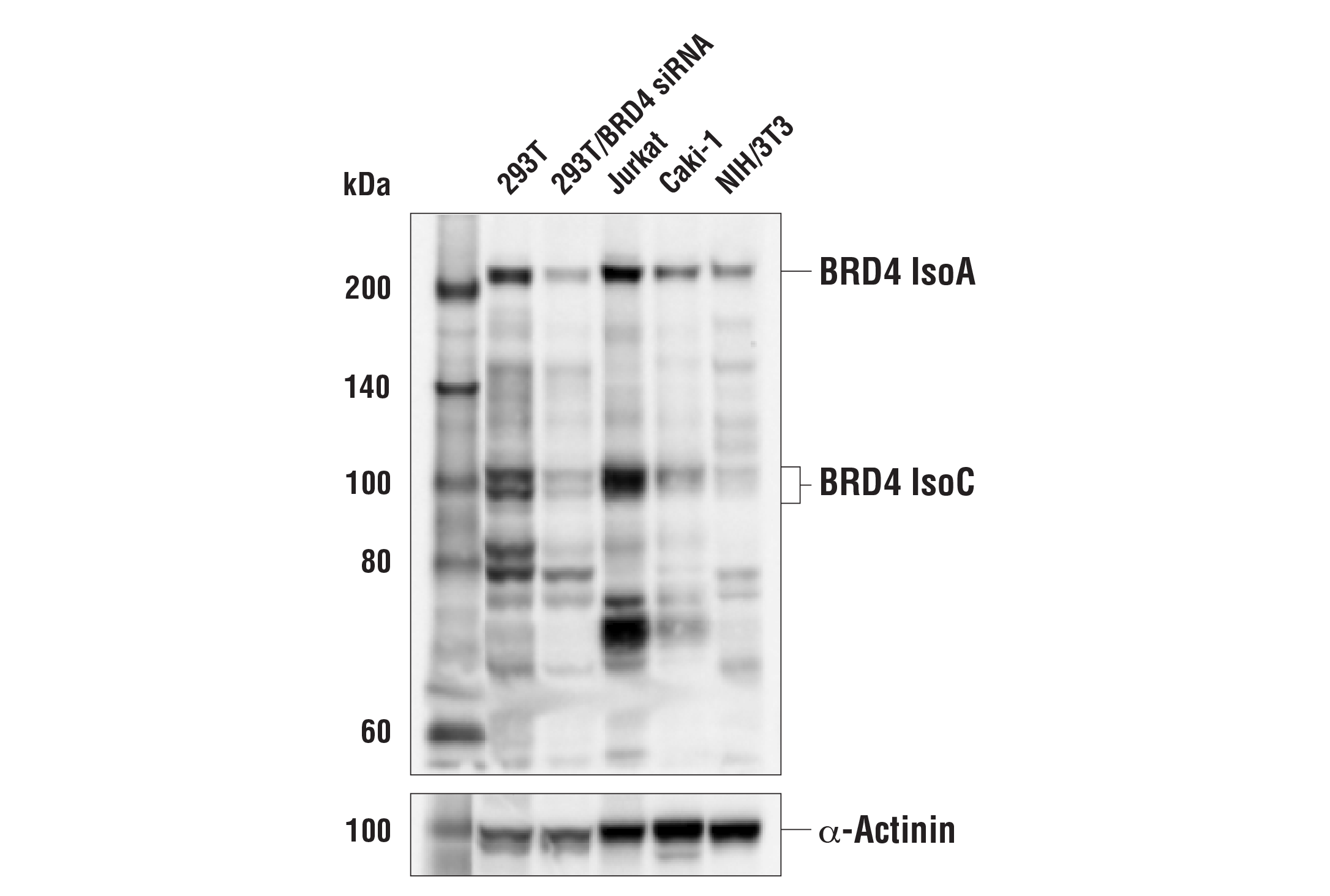
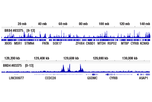
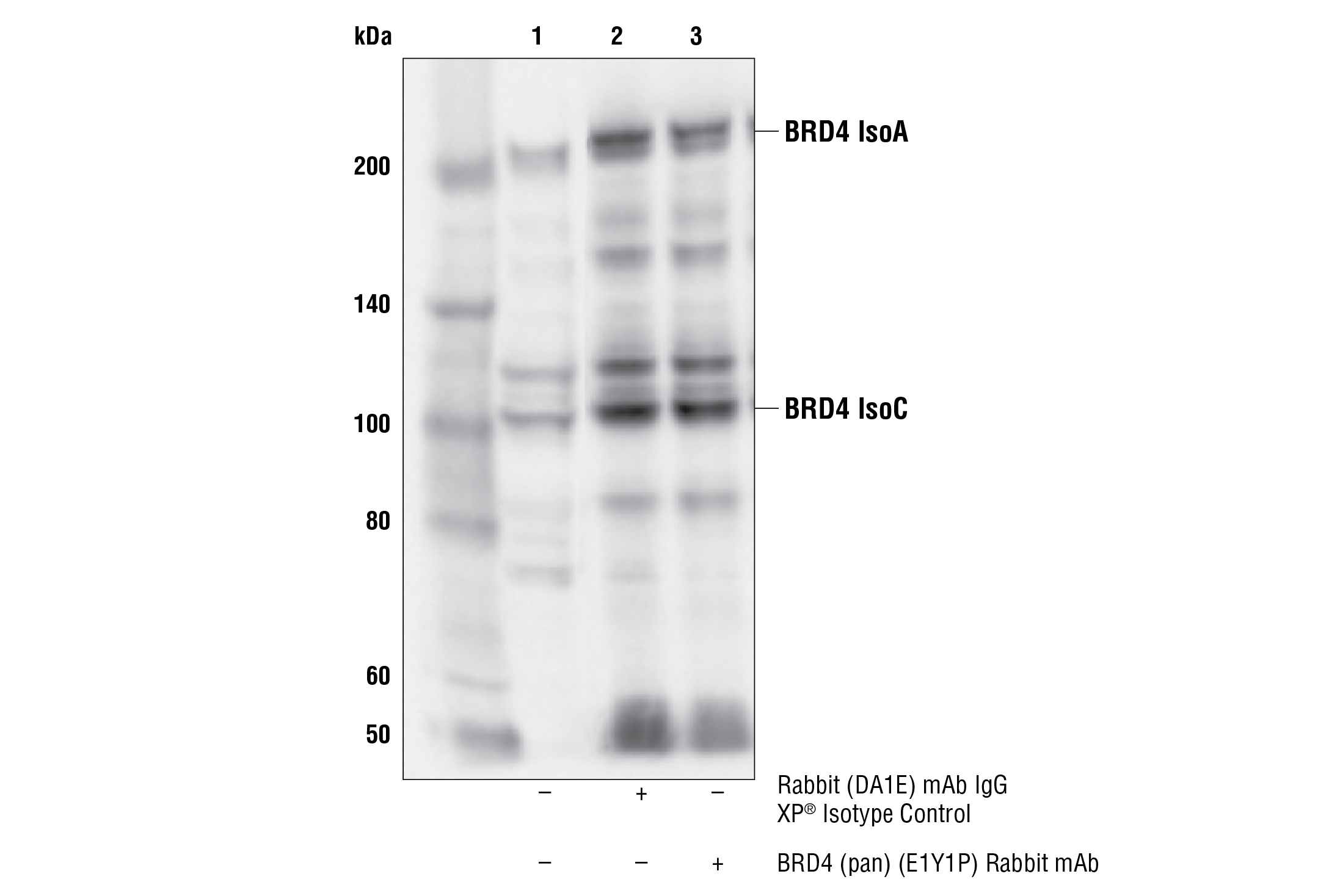
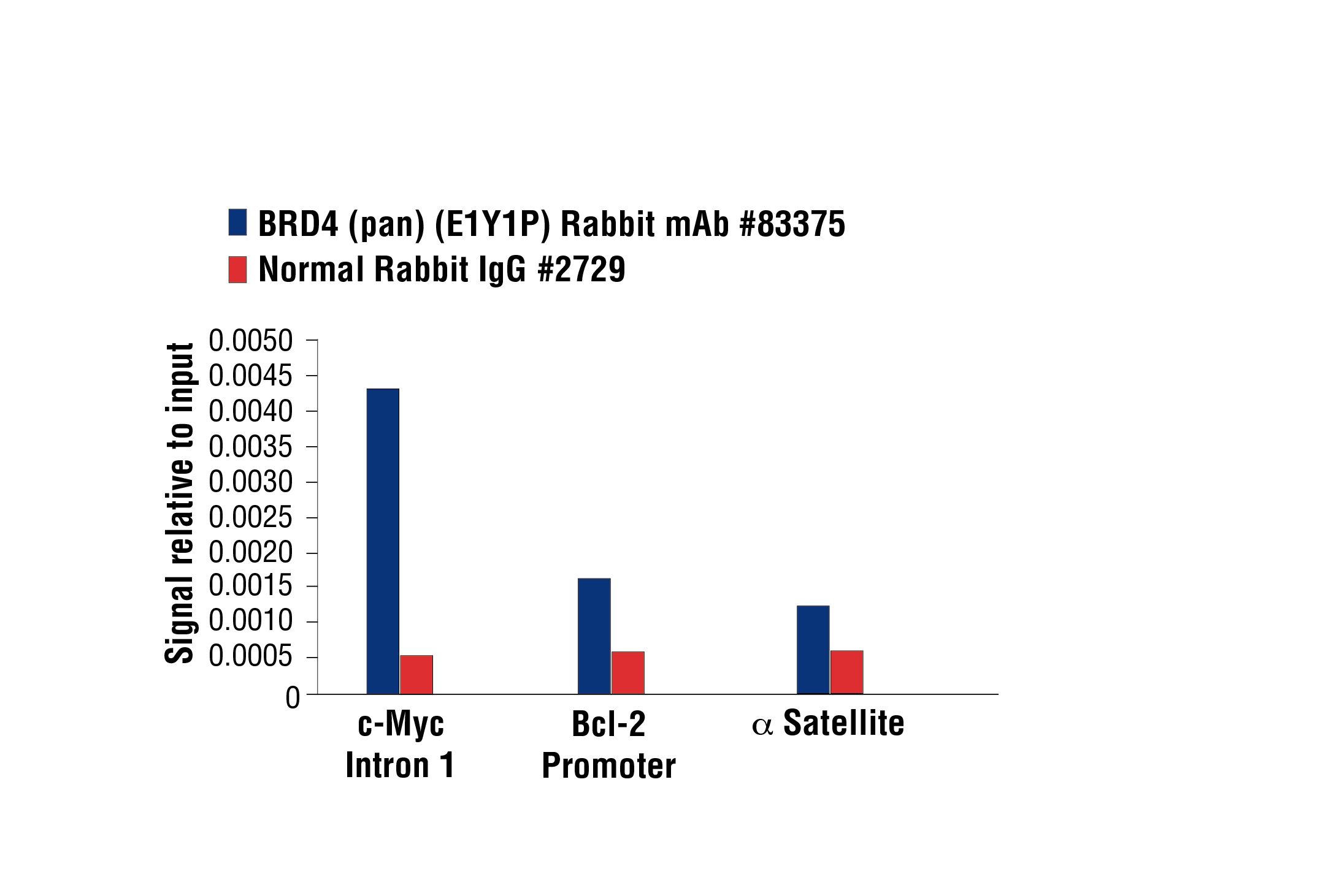
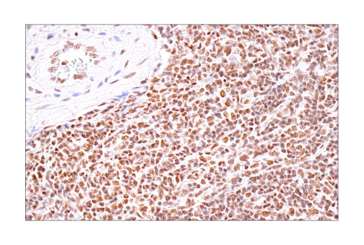
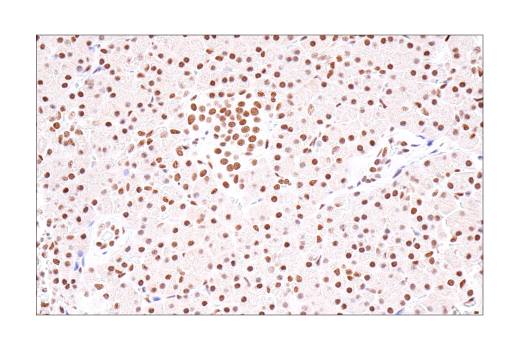
| BRD4 (pan) (E1Y1P) Rabbit mAb | 83375 | 20 µl | 200, 110 kDa | Rabbit IgG |
| Anti-rabbit IgG, HRP-linked Antibody | 7074 | 100 µl | Goat |
Please visit cellsignal.com for individual component applications, species cross-reactivity, dilutions, protocols, and additional product information.
Description
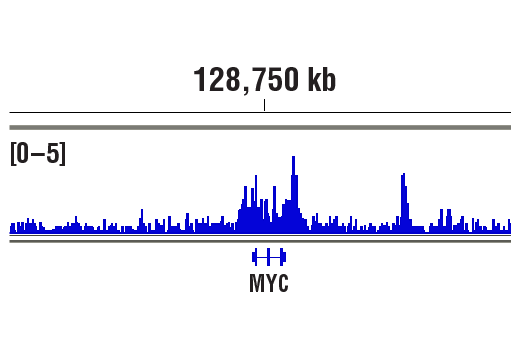
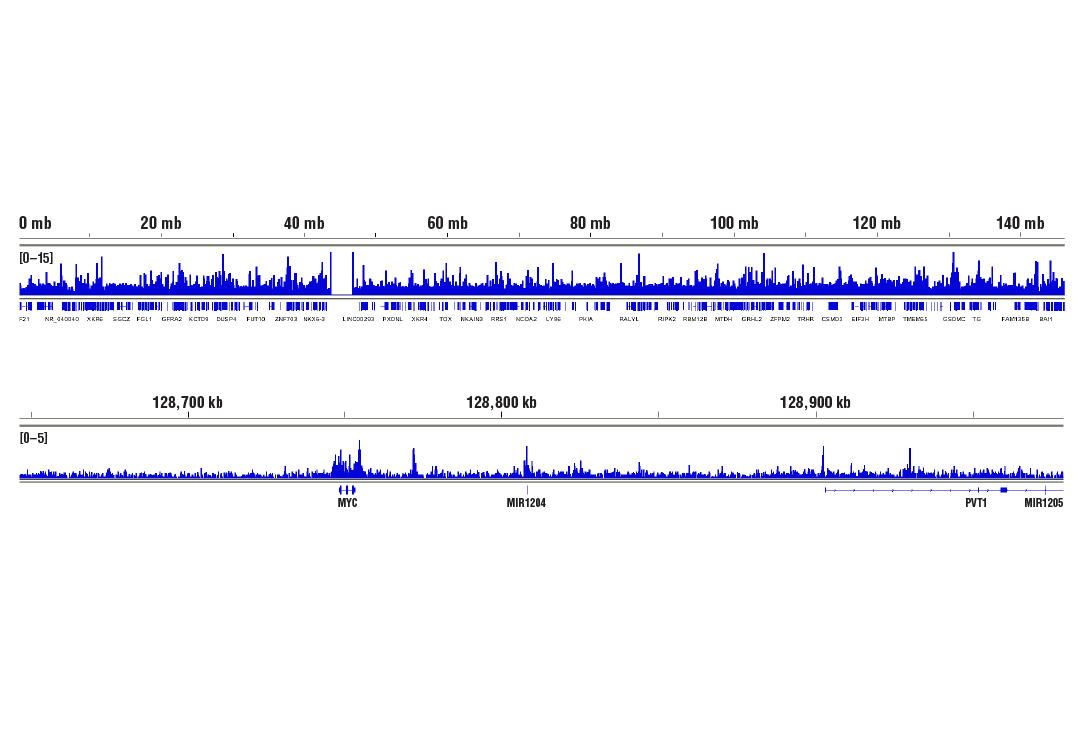
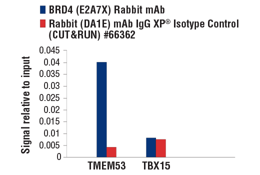
The BRD4 Antibody Sampler Kit provides an economical means of detecting various BRD4 isoforms. The kit includes enough antibodies to perform two western blot experiments with each primary antibody.
Storage
Background
Bromodomain-containing protein 4 (BRD4) is a member of the bromodomains and extra terminal (BET) family of proteins, which also includes BRD2, BRD3, and BRDT (1-3). BET family proteins contain two tandem bromodomains and an extra terminal (ET) domain, and bind acetyl lysine residues (3). BRD4 is a chromatin-binding protein with a preference for Lys14 on histone H3 as well as Lys5 and Lys12 on histone H4 (4). BRD4 chromatin binding occurs throughout the cell cycle, including condensed mitotic chromosomes, when the majority of genes are silenced (5). BRD4 association with chromatin during mitosis is thought to be an important part of the bookmarking mechanism to accelerate re-activation of the silenced genes upon exit from mitosis (2,6). BRD4 has been shown to facilitate transcription by recruiting the positive transcription elongation factor b (pTEFb) complex that phosphorylates Ser2 of the heptapeptide repeat of the carboxy-terminal domain of RNA polymerase II, promoting transcription elongation (3,7,8). In addition, BRD4 has been found to be part of the super elongation complex and the polymerase associated factor complex (PAFc) in MLL-fusion derived leukemia cell lines, demonstrating a role for BRD4 in the regulation of transcription elongation (9). Research studies have shown that BRD4 (and BET family proteins) may be promising therapeutic targets for various Myc-driven cancers, such as Burkitt’s lymphoma and certain acute myeloid leukemias (1,10,11). Investigators have found molecular inhibition of BET proteins to be effective in inducing apoptosis in various MLL-fusion driven leukemic cell lines by competing BRD3 and BRD4 from chromatin, leading to reduced expression of Bcl-2, Myc, and CDK6 (9). BET inhibition has also been shown to have antitumor activities against nuclear protein in testis (NUT) midline carcinoma cell lines and xenografts in mice where BRD4 is found to be a frequent translocation partner of the NUT protein (12). In addition, BRD4 regulates the expression of some inflammatory genes, and inhibition of BRD4 (and BET family proteins) chromatin binding causes reduced expression of a subset of inflammatory genes in macrophages, leading to protection against endotoxic shock and sepsis (13).
The expression of BRD4 isoform C, also known as the short isoform (BRD4-S), is similar to the long isoform (BRD4-L). The balance of expression between the two isoforms has been shown to contribute to disease states, where BRD4-S has shown to be oncogenic in a few contexts (14-16). BRD4-S is the predominant isoform that binds to modified histones and has a stronger affinity than BRD4-L (17,18). BRD4-S also plays a role in DNA damage, where it recruits the condensin II complex to enhance DNA damage induced cell death (18).
- Belkina, A.C. and Denis, G.V. (2012) Nat Rev Cancer 12, 465-77.
- Voigt, P. and Reinberg, D. (2011) Genome Biol 12, 133.
- Wu, S.Y. and Chiang, C.M. (2007) J Biol Chem 282, 13141-5.
- Dey, A. et al. (2003) Proc Natl Acad Sci U S A 100, 8758-63.
- Dey, A. et al. (2009) Mol Biol Cell 20, 4899-909.
- Zhao, R. et al. (2011) Nat Cell Biol 13, 1295-304.
- Jang, M.K. et al. (2005) Mol Cell 19, 523-34.
- Yang, Z. et al. (2005) Mol Cell 19, 535-45.
- Dawson, M.A. et al. (2011) Nature 478, 529-33.
- Muller, S. et al. (2011) Expert Rev Mol Med 13, e29.
- Mertz, J.A. et al. (2011) Proc Natl Acad Sci U S A 108, 16669-74.
- Filippakopoulos, P. et al. (2010) Nature 468, 1067-73.
- Nicodeme, E. et al. (2010) Nature 468, 1119-23.
- Wu, S.Y. et al. (2020) Mol Cell 78, 1114-1132.e10.
- Crawford, N.P. et al. (2008) Proc Natl Acad Sci USA 105, 6380-5.
- Fernandez, P. et al. (2014) Cell Rep 9, 248-260.
- Wang, R. et al. (2012) J Biol Chem 287, 10738-52.
- Han, X. et al. (2020) Nat Struct Mol Biol 27, 333-341.
Background References
Trademarks and Patents
使用に関する制限
法的な権限を与えられたCSTの担当者が署名した書面によって別途明示的に合意された場合を除き、 CST、その関連会社または代理店が提供する製品には以下の条件が適用されます。お客様が定める条件でここに定められた条件に含まれるものを超えるもの、 または、ここに定められた条件と異なるものは、法的な権限を与えられたCSTの担当者が別途書面にて受諾した場合を除き、拒絶され、 いかなる効力も効果も有しません。
研究専用 (For Research Use Only) またはこれに類似する表示がされた製品は、 いかなる目的についても FDA または外国もしくは国内のその他の規制機関により承認、認可または許可を受けていません。 お客様は製品を診断もしくは治療目的で使用してはならず、また、製品に表示された内容に違反する方法で使用してはなりません。 CST が販売または使用許諾する製品は、エンドユーザーであるお客様に対し、使途を研究および開発のみに限定して提供されるものです。 診断、予防もしくは治療目的で製品を使用することまたは製品を再販売 (単独であるか他の製品等の一部であるかを問いません) もしくはその他の商業的利用の目的で購入することについては、CST から別途許諾を得る必要があります。 お客様は以下の事項を遵守しなければなりません。(a) CST の製品 (単独であるか他の資材と一緒であるかを問いません) を販売、使用許諾、貸与、寄付もしくはその他の態様で第三者に譲渡したり使用させたりしてはなりません。また、商用の製品を製造するために CST の製品を使用してはなりません。(b) 複製、改変、リバースエンジニアリング、逆コンパイル、 分解または他の方法により製品の構造または技術を解明しようとしてはなりません。また、 CST の製品またはサービスと競合する製品またはサービスを開発する目的で CST の製品を使用してはなりません。(c) CST の製品の商標、商号、ロゴ、特許または著作権に関する通知または表示を除去したり改変したりしてはなりません。(d) CST の製品をCST 製品販売条件(CST’s Product Terms of Sale) および該当する書面のみに従って使用しなければなりません。(e) CST の製品に関連してお客様が使用する第三者の製品またはサービスに関する使用許諾条件、 サービス提供条件またはこれに類する合意事項を遵守しなければなりません。