| Product Includes | Product # | Quantity | Mol. Wt | Isotype/Source |
|---|---|---|---|---|
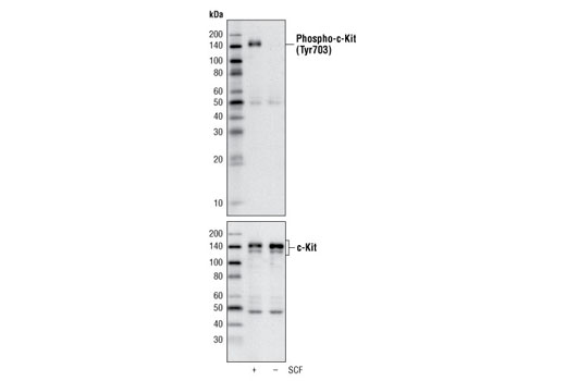
| Phospho-c-Kit (Tyr703) (D12E12) Rabbit mAb | 3073 | 20 µl | 145 kDa | Rabbit IgG |
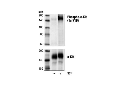
| Phospho-c-Kit (Tyr719) Antibody | 3391 | 20 µl | 120, 145 kDa | Rabbit |
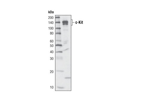
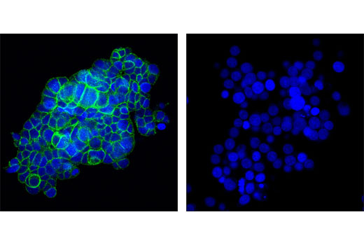
| c-Kit (D13A2) XP® Rabbit mAb | 3074 | 20 µl | 120 and 145 kDa | Rabbit |
| Anti-rabbit IgG, HRP-linked Antibody | 7074 | 100 µl | Goat |
Please visit cellsignal.com for individual component applications, species cross-reactivity, dilutions, protocols, and additional product information.
Description
The c-Kit Antibody Sampler Kit provides a fast and economical means of evaluating levels of c-Kit receptor protein phosphorylated at the specified sites, as well as total c-Kit receptor levels. The kit contains enough primary and secondary antibody to perform two Western blot experiments.
Storage
Background
c-Kit is a member of the subfamily of receptor tyrosine kinases that includes PDGF, CSF-1, and FLT3/flk-2 receptors (1,2). It plays a critical role in activation and growth in a number of cell types, including hematopoietic stem cells, mast cells, melanocytes, and germ cells (3). Upon binding with its stem cell factor (SCF) ligand, c-Kit undergoes dimerization/oligomerization and autophosphorylation. Activation of c-Kit results in the recruitment and tyrosine phosphorylation of downstream SH2-containing signaling components, including PLCγ, the p85 subunit of PI3 kinase, SHP2, and CrkL (4). Molecular lesions that impair the kinase activity of c-Kit are associated with a variety of developmental disorders (5), and mutations that constitutively activate c-Kit can lead to pathogenesis of mastocytosis and gastrointestinal stromal tumors (6). Tyr719 is located in the kinase insert region of the catalytic domain. c-Kit phosphorylated at Tyr719 binds to the p85 subunit of PI3 kinase in vitro and in vivo (7).
- Martin, F.H. et al. (1990) Cell 63, 203-11.
- Yarden, Y. et al. (1987) EMBO J 6, 3341-51.
- Gommerman, J.L. et al. (1997) J Biol Chem 272, 30519-25.
- Sattler, M. et al. (1997) J Biol Chem 272, 10248-53.
- Nocka, K. et al. (1990) EMBO J 9, 1805-13.
- Hirota, S. et al. (1998) Science 279, 577-80.
- Blume-Jensen, P. et al. (2000) Nat Genet 24, 157-62.
Background References
Trademarks and Patents
使用に関する制限
法的な権限を与えられたCSTの担当者が署名した書面によって別途明示的に合意された場合を除き、 CST、その関連会社または代理店が提供する製品には以下の条件が適用されます。お客様が定める条件でここに定められた条件に含まれるものを超えるもの、 または、ここに定められた条件と異なるものは、法的な権限を与えられたCSTの担当者が別途書面にて受諾した場合を除き、拒絶され、 いかなる効力も効果も有しません。
研究専用 (For Research Use Only) またはこれに類似する表示がされた製品は、 いかなる目的についても FDA または外国もしくは国内のその他の規制機関により承認、認可または許可を受けていません。 お客様は製品を診断もしくは治療目的で使用してはならず、また、製品に表示された内容に違反する方法で使用してはなりません。 CST が販売または使用許諾する製品は、エンドユーザーであるお客様に対し、使途を研究および開発のみに限定して提供されるものです。 診断、予防もしくは治療目的で製品を使用することまたは製品を再販売 (単独であるか他の製品等の一部であるかを問いません) もしくはその他の商業的利用の目的で購入することについては、CST から別途許諾を得る必要があります。 お客様は以下の事項を遵守しなければなりません。(a) CST の製品 (単独であるか他の資材と一緒であるかを問いません) を販売、使用許諾、貸与、寄付もしくはその他の態様で第三者に譲渡したり使用させたりしてはなりません。また、商用の製品を製造するために CST の製品を使用してはなりません。(b) 複製、改変、リバースエンジニアリング、逆コンパイル、 分解または他の方法により製品の構造または技術を解明しようとしてはなりません。また、 CST の製品またはサービスと競合する製品またはサービスを開発する目的で CST の製品を使用してはなりません。(c) CST の製品の商標、商号、ロゴ、特許または著作権に関する通知または表示を除去したり改変したりしてはなりません。(d) CST の製品をCST 製品販売条件(CST’s Product Terms of Sale) および該当する書面のみに従って使用しなければなりません。(e) CST の製品に関連してお客様が使用する第三者の製品またはサービスに関する使用許諾条件、 サービス提供条件またはこれに類する合意事項を遵守しなければなりません。