| Product Includes | Product # | Quantity | Mol. Wt | Isotype/Source |
|---|---|---|---|---|
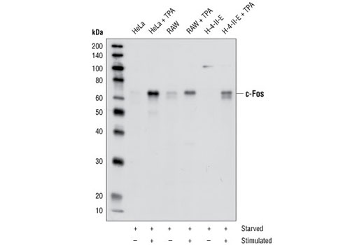
| c-Fos Antibody | 4384 | 20 µl | 62 kDa | Rabbit |
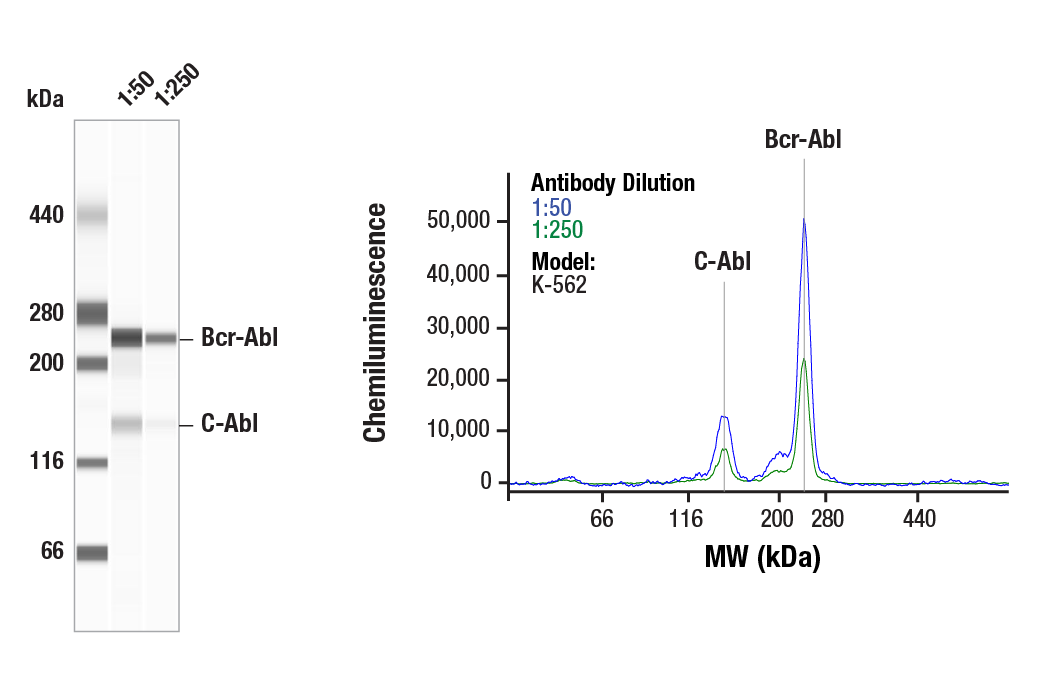
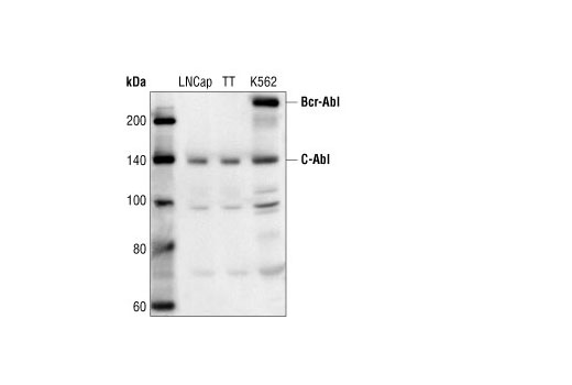
| c-Abl Antibody | 2862 | 20 µl | 135 (c-Abl); 210 (Bcr-Abl) kDa | Rabbit |
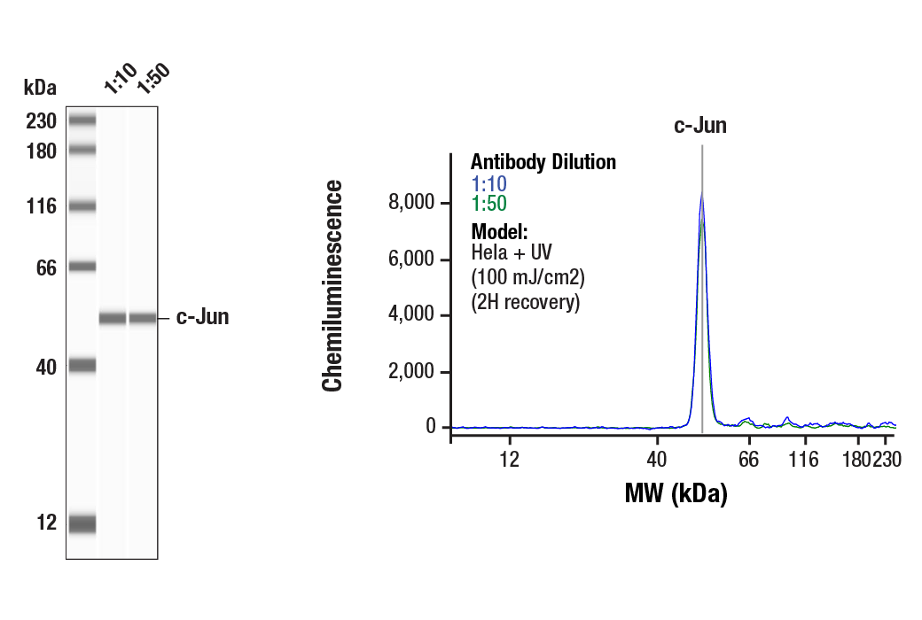
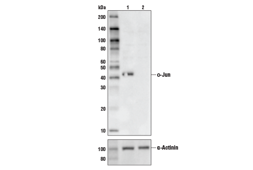
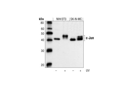
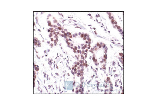
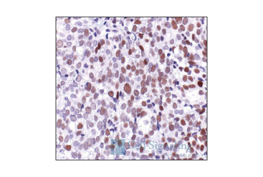
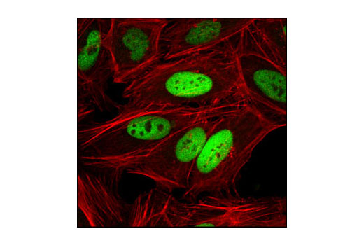
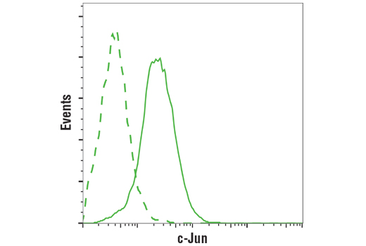
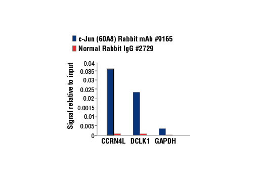
| c-Jun (60A8) Rabbit mAb | 9165 | 20 µl | 43, 48 kDa | Rabbit IgG |
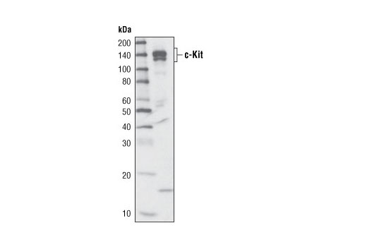
| c-Kit (D13A2) XP® Rabbit mAb | 3074 | 20 µl | 120 and 145 kDa | Rabbit |
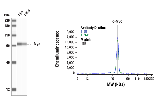
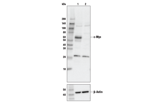
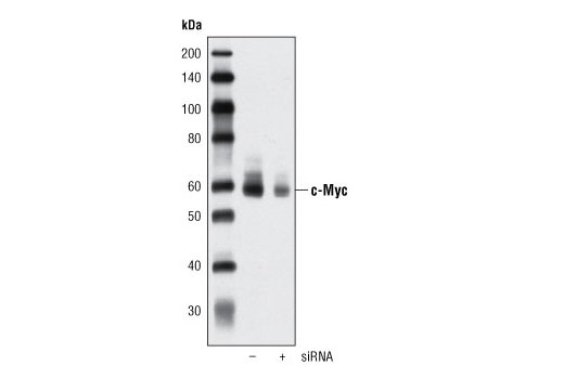
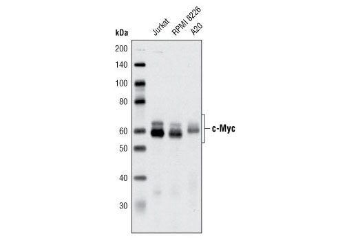
| c-Myc (D84C12) Rabbit mAb | 5605 | 20 µl | 57-65 kDa | Rabbit IgG |
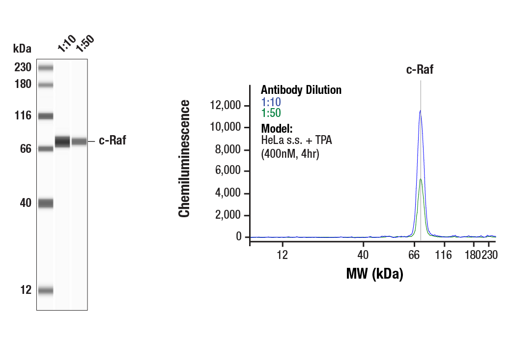
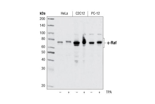
| c-Raf Antibody | 9422 | 20 µl | 65 to 75 kDa | Rabbit |
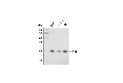
| Ras (27H5) Rabbit mAb | 3339 | 20 µl | 21 kDa | Rabbit IgG |
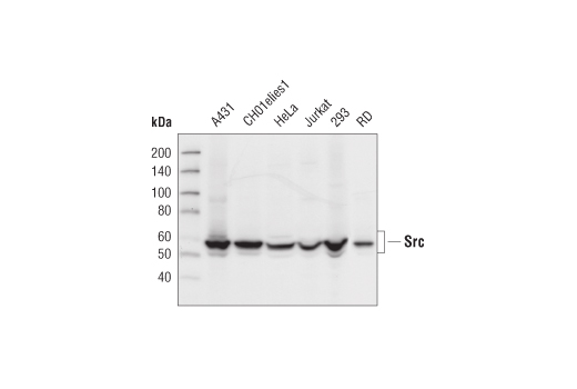
| Src (32G6) Rabbit mAb | 2123 | 20 µl | 60 kDa | Rabbit IgG |
| Anti-rabbit IgG, HRP-linked Antibody | 7074 | 100 µl | Goat | |
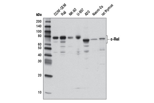
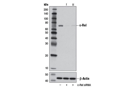
| c-Rel (D4Y6M) Rabbit mAb | 12707 | 20 µl | 68-78 kDa | Rabbit IgG |
Please visit cellsignal.com for individual component applications, species cross-reactivity, dilutions, protocols, and additional product information.
Description
The c-Oncogene Antibody Sampler Kit provides an economical means of evaluating total levels of various oncogenic proteins. The kit contains enough primary and secondary antibodies to perform two Western blot experiments.
Storage
Background
The regulation of cell growth, differentiation and programmed death is coordinated by several sets of proteins that comprise essential signal transduction pathways. Many of these key regulatory proteins are encoded by proto-oncogenes, which can be activated (altered) to change the typical cell program to one of abnormal cell growth and unregulated development. Proteins encoded by proto-oncogenes include growth factors and other ligands, receptor proteins, tyrosine kinases, various regulatory proteins (i.e. GTPases) and transcription factors. Together these proteins comprise the basic elements of cell signaling pathways; altered expression or mutation of one or more of these components can lead to oncogenic growth (reviewed in 1).
Non-receptor (i.e. cytoplasmic, nuclear) tyrosine kinases such as c-Abl and Src play key roles in the regulation of cell proliferation, differentiation, apoptosis, cell adhesion and stress responses (2,3). Alteration of the corresponding c-Abl and Src proto-oncogenes is associated with oncogenesis; Abl1-BCR gene translocations result in chronic myelogenous leukemia (CML) while constitutively active Src is seen in some patients with colon cancer and altered Src expression is seen in a wide array of cancers (2,4). Regulation of Raf tyrosine kinase by Ras GTPase controls downstream kinases in the MEK/MAPK signaling pathway (5). Activation of the Ras and Raf proto-oncogenes are common in human cancers and both proteins are seen as potential therapeutic targets (6). The receptor tyrosine kinase c-Kit plays a critical role in activation and growth of hematopoietic stem cells (7); mutations that inhibit c-Kit kinase activity are associated with a variety of developmental disorders while mutations producing constitutively active c-Kit can result in mastocytosis and gastrointestinal stromal tumors (8). The alteration of key transcription factors such as c-Fos, c-Jun, c-Myc and c-Rel that are normally responsible for regulating cell and tissue growth, differentiation and the inflammation/immune response, can also result in unregulated, oncogenic cell growth (9-12).
- Croce, C.M. (2008) N Engl J Med 358, 502-11.
- Wang, J.Y. (2000) Oncogene 19, 5643-50.
- Thomas, S.M. and Brugge, J.S. (1997) Annu Rev Cell Dev Biol 13, 513-609.
- Dehm, S.M. and Bonham, K. (2004) Biochem Cell Biol 82, 263-74.
- Avruch, J. et al. (1994) Trends Biochem Sci 19, 279-83.
- Stites, E.C. et al. (2007) Science 318, 463-7.
- Gommerman, J.L. et al. (1997) J Biol Chem 272, 30519-25.
- Nocka, K. et al. (1990) EMBO J 9, 1805-13.
- Milde-Langosch, K. (2005) Eur J Cancer 41, 2449-61.
- Shaulian, E. and Karin, M. (2002) Nat Cell Biol 4, E131-6.
- Yokota, J. et al. (1986) Science 231, 261-5.
- Rayet, B. and Gélinas, C. (1999) Oncogene 18, 6938-47.
Background References
Trademarks and Patents
使用に関する制限
法的な権限を与えられたCSTの担当者が署名した書面によって別途明示的に合意された場合を除き、 CST、その関連会社または代理店が提供する製品には以下の条件が適用されます。お客様が定める条件でここに定められた条件に含まれるものを超えるもの、 または、ここに定められた条件と異なるものは、法的な権限を与えられたCSTの担当者が別途書面にて受諾した場合を除き、拒絶され、 いかなる効力も効果も有しません。
研究専用 (For Research Use Only) またはこれに類似する表示がされた製品は、 いかなる目的についても FDA または外国もしくは国内のその他の規制機関により承認、認可または許可を受けていません。 お客様は製品を診断もしくは治療目的で使用してはならず、また、製品に表示された内容に違反する方法で使用してはなりません。 CST が販売または使用許諾する製品は、エンドユーザーであるお客様に対し、使途を研究および開発のみに限定して提供されるものです。 診断、予防もしくは治療目的で製品を使用することまたは製品を再販売 (単独であるか他の製品等の一部であるかを問いません) もしくはその他の商業的利用の目的で購入することについては、CST から別途許諾を得る必要があります。 お客様は以下の事項を遵守しなければなりません。(a) CST の製品 (単独であるか他の資材と一緒であるかを問いません) を販売、使用許諾、貸与、寄付もしくはその他の態様で第三者に譲渡したり使用させたりしてはなりません。また、商用の製品を製造するために CST の製品を使用してはなりません。(b) 複製、改変、リバースエンジニアリング、逆コンパイル、 分解または他の方法により製品の構造または技術を解明しようとしてはなりません。また、 CST の製品またはサービスと競合する製品またはサービスを開発する目的で CST の製品を使用してはなりません。(c) CST の製品の商標、商号、ロゴ、特許または著作権に関する通知または表示を除去したり改変したりしてはなりません。(d) CST の製品をCST 製品販売条件(CST’s Product Terms of Sale) および該当する書面のみに従って使用しなければなりません。(e) CST の製品に関連してお客様が使用する第三者の製品またはサービスに関する使用許諾条件、 サービス提供条件またはこれに類する合意事項を遵守しなければなりません。