Revision 1
Cell Signaling Technology

Orders: 877-616-CELL (2355) [email protected]

Support: 877-678-TECH (8324)

Web: [email protected] cellsignal.com

3 Trask LaneDanversMassachusetts01923USA
For Research Use Only. Not for Use in Diagnostic Procedures.
Applications:

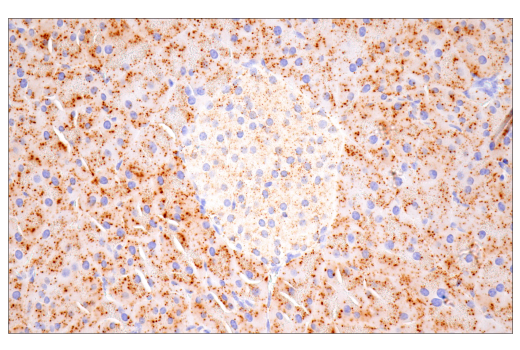
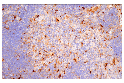
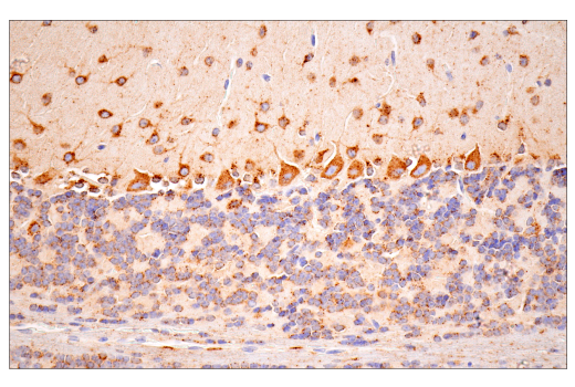
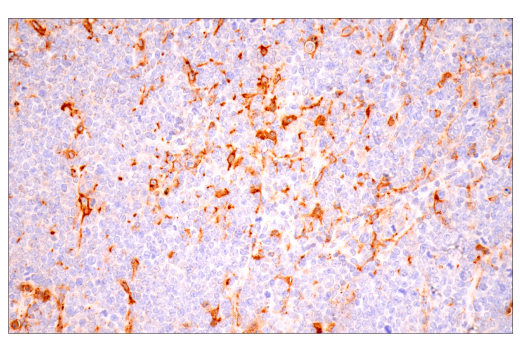
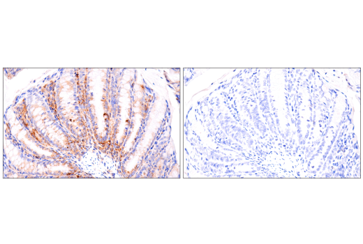
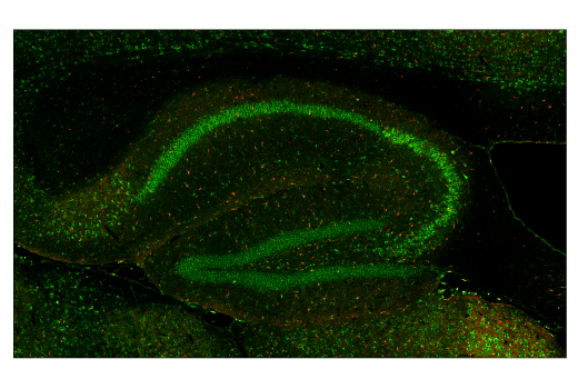
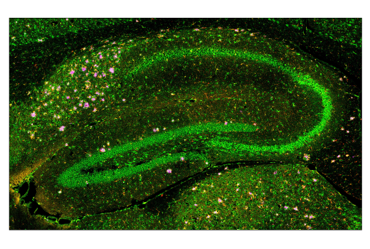
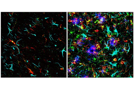
WB, IHC-P, IF-F, IF-IC

REACTIVITY:

M

SENSITIVITY:

Endogenous

MW (kDa):

46, 43, 28

Source/Isotype:

Rabbit IgG

UniProt ID:

#P18242

Entrez-Gene Id:

13033

Product Information

Product Usage Information

This product is the carrier free version of product #88239. All data were generated using the same antibody clone in the standard formulation which contains BSA and glycerol.

This formulation is ideal for use with technologies requiring specialized or custom antibody labeling, including fluorophores, metals, lanthanides, and oligonucleotides. It is not recommended for ChIP, ChIP-seq, CUT&RUN or CUT&Tag assays. If you require a carrier free formulation for chromatin profiling, please contact us. Optimal dilutions/concentrations should be determined by the end user.

Formulation

Supplied in 1X PBS, BSA and Azide Free.

For standard formulation of this product see product #88239

Storage

Store at -20°C. This product will freeze at -20°C so it is recommended to aliquot into single-use vials to avoid multiple freeze/thaw cycles. A slight precipitate may be present and can be dissolved by gently vortexing. This will not interfere with antibody performance.

Specificity / Sensitivity

Cathepsin D (E7Z4L) XP® Rabbit mAb (BSA and Azide Free) recognizes endogenous levels of preprocathepsin D, procathepsin D and the heavy and light chain subunit of mature cathepsin D protein.

Species Reactivity:

Mouse

Source / Purification

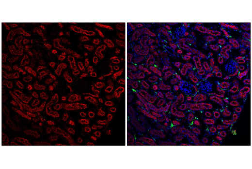
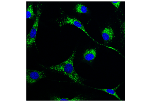
Monoclonal antibody is produced by immunizing animals with a synthetic peptide corresponding to residues near the carboxy terminus of mouse cathepsin D protein.

Background

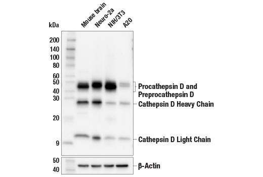
Cathepsin D is a ubiquitously expressed lysosomal aspartyl protease involved in the normal degradation of proteins (1). It is synthesized as an inactive 43 kDa preprocathepsin D that is cleaved and glycosylated to form a 46 kDa procathepsin D and then further cleaved to produce 28 kDa and 15 kDa subunits (heavy and light chains, respectively) (2). Cathepsin D may also be secreted into the cytosol during apoptosis and contribute to cleavage of substrates implicated in the apoptotic pathway (3). Numerous studies have suggested that cathepsin D plays a role in neuronal degradation and malignant transformation, particularly in breast cancer (4-9).

  1. Faust, P.L. et al. (1985) Proc. Natl. Acad. Sci. USA 82, 4910-4.
  2. Erickson, A.H. et al. (1981) J. Biol .Chem. 256, 11224-31.
  3. Liaudet-Coopman, E. et al. (2006) Cancer Lett. 237, 167-79.
  4. Berchem, G. et al. (2002) Oncogene 21, 5951-5.
  5. Nomura, T. and Katunuma, N. (2005) J. Med. Invest. 52, 1-9.
  6. Garcia, M. et al. (1996) Stem Cells 14, 642-50.
  7. Nogami, M. et al. (2000) Histochem. J. 32, 505-8.
  8. Nakanishi, H. (2003) Ageing Res. Rev. 2, 367-81.
  9. Callahan, L.M. et al. Neurobiol. Aging 19, S99-105.

Species Reactivity

Species reactivity is determined by testing in at least one approved application (e.g., western blot).

Applications Key

WB: Western Blotting IHC-P: Immunohistochemistry (Paraffin) IF-F: Immunofluorescence (Frozen) IF-IC: Immunofluorescence (Immunocytochemistry)

Cross-Reactivity Key

H: human M: mouse R: rat Hm: hamster Mk: monkey Vir: virus Mi: mink C: chicken Dm: D. melanogaster X: Xenopus Z: zebrafish B: bovine Dg: dog Pg: pig Sc: S. cerevisiae Ce: C. elegans Hr: horse GP: Guinea Pig Rab: rabbit All: all species expected

Trademarks and Patents

Cell Signaling Technology is a trademark of Cell Signaling Technology, Inc.
XP is a registered trademark of Cell Signaling Technology, Inc.
All other trademarks are the property of their respective owners. Visit cellsignal.com/trademarks for more information.

使用に関する制限

法的な権限を与えられたCSTの担当者が署名した書面によって別途明示的に合意された場合を除き、 CST、その関連会社または代理店が提供する製品には以下の条件が適用されます。お客様が定める条件でここに定められた条件に含まれるものを超えるもの、 または、ここに定められた条件と異なるものは、法的な権限を与えられたCSTの担当者が別途書面にて受諾した場合を除き、拒絶され、 いかなる効力も効果も有しません。

研究専用 (For Research Use Only) またはこれに類似する表示がされた製品は、 いかなる目的についても FDA または外国もしくは国内のその他の規制機関により承認、認可または許可を受けていません。 お客様は製品を診断もしくは治療目的で使用してはならず、また、製品に表示された内容に違反する方法で使用してはなりません。 CST が販売または使用許諾する製品は、エンドユーザーであるお客様に対し、使途を研究および開発のみに限定して提供されるものです。 診断、予防もしくは治療目的で製品を使用することまたは製品を再販売 (単独であるか他の製品等の一部であるかを問いません) もしくはその他の商業的利用の目的で購入することについては、CST から別途許諾を得る必要があります。 お客様は以下の事項を遵守しなければなりません。(a) CST の製品 (単独であるか他の資材と一緒であるかを問いません) を販売、使用許諾、貸与、寄付もしくはその他の態様で第三者に譲渡したり使用させたりしてはなりません。また、商用の製品を製造するために CST の製品を使用してはなりません。(b) 複製、改変、リバースエンジニアリング、逆コンパイル、 分解または他の方法により製品の構造または技術を解明しようとしてはなりません。また、 CST の製品またはサービスと競合する製品またはサービスを開発する目的で CST の製品を使用してはなりません。(c) CST の製品の商標、商号、ロゴ、特許または著作権に関する通知または表示を除去したり改変したりしてはなりません。(d) CST の製品をCST 製品販売条件(CST’s Product Terms of Sale) および該当する書面のみに従って使用しなければなりません。(e) CST の製品に関連してお客様が使用する第三者の製品またはサービスに関する使用許諾条件、 サービス提供条件またはこれに類する合意事項を遵守しなければなりません。

Revision 1
#52899

Cathepsin D (E7Z4L) XP® Rabbit mAb (BSA and Azide Free)

Western Blotting Image 1: Cathepsin D (E7Z4L) XP® Rabbit mAb (BSA and Azide Free) Expand Image
Western blot analysis of extracts from various cell lines and mouse brain tissue using Cathepsin D (E7Z4L) XP® Rabbit mAb (upper) or β-Actin (D6A8) Rabbit mAb #8457 (lower). Data were generated using the standard formulation of this product.
Immunohistochemistry Image 1: Cathepsin D (E7Z4L) XP® Rabbit mAb (BSA and Azide Free) Expand Image
Immunohistochemical analysis of paraffin-embedded mouse liver using Cathepsin D (E7Z4L) XP® Rabbit mAb. Data were generated using the standard formulation of this product.
Immunohistochemistry Image 2: Cathepsin D (E7Z4L) XP® Rabbit mAb (BSA and Azide Free) Expand Image
Immunohistochemical analysis of paraffin-embedded mouse kidney using Cathepsin D (E7Z4L) XP® Rabbit mAb. Data were generated using the standard formulation of this product.
Immunohistochemistry Image 3: Cathepsin D (E7Z4L) XP® Rabbit mAb (BSA and Azide Free) Expand Image
Immunohistochemical analysis of paraffin-embedded mouse lung using Cathepsin D (E7Z4L) XP® Rabbit mAb. Data were generated using the standard formulation of this product.
Immunohistochemistry Image 4: Cathepsin D (E7Z4L) XP® Rabbit mAb (BSA and Azide Free) Expand Image
Immunohistochemical analysis of paraffin-embedded mouse pancreas using Cathepsin D (E7Z4L) XP® Rabbit mAb. Data were generated using the standard formulation of this product.
Immunohistochemistry Image 5: Cathepsin D (E7Z4L) XP® Rabbit mAb (BSA and Azide Free) Expand Image
Immunohistochemical analysis of paraffin-embedded mouse thymus using Cathepsin D (E7Z4L) XP® Rabbit mAb. Data were generated using the standard formulation of this product.
Immunohistochemistry Image 6: Cathepsin D (E7Z4L) XP® Rabbit mAb (BSA and Azide Free) Expand Image
Immunohistochemical analysis of paraffin-embedded mouse cerebellum using Cathepsin D (E7Z4L) XP® Rabbit mAb. Data were generated using the standard formulation of this product.
Immunohistochemistry Image 7: Cathepsin D (E7Z4L) XP® Rabbit mAb (BSA and Azide Free) Expand Image
Immunohistochemical analysis of paraffin-embedded A20 syngeneic tumor using Cathepsin D (E7Z4L) XP® Rabbit mAb. Data were generated using the standard formulation of this product.
Immunohistochemistry Image 8: Cathepsin D (E7Z4L) XP® Rabbit mAb (BSA and Azide Free) Expand Image
Immunohistochemical analysis of paraffin-embedded mouse colon using Cathepsin D (E7Z4L) XP® Rabbit mAb (left) compared to concentration-matched Rabbit (DA1E) mAb IgG XP® Isotype Control #3900 (right). Data were generated using the standard formulation of this product.
Immunofluorescence Image 1: Cathepsin D (E7Z4L) XP® Rabbit mAb (BSA and Azide Free) Expand Image
Confocal immunofluorescent analysis of fixed frozen wild-type mouse brain using Cathepsin D (E7Z4L) XP® Rabbit mAb (green). After blocking free secondary antibody binding sites with Rabbit (DA1E) mAb IgG XP® Isotype Control #3900, the tissue was then labeled using Iba1/AIF-1 (E4O4W) XP® Rabbit mAb (Alexa Fluor® 555 Conjugate) #36618 (red) and β-Amyloid (D54D2) XP® Rabbit mAb (Alexa Fluor® 594 Conjugate) #35363 (magenta pseudocolor). Data were generated using the standard formulation of this product.
Immunofluorescence Image 2: Cathepsin D (E7Z4L) XP® Rabbit mAb (BSA and Azide Free) Expand Image
Confocal immunofluorescent analysis of fixed frozen brain from an amyloid mouse model of Alzheimer's disease using Cathepsin D (E7Z4L) XP® Rabbit mAb (green). After blocking free secondary antibody binding sites with Rabbit (DA1E) mAb IgG XP® Isotype Control #3900, the tissue was then labeled using Iba1/AIF-1 (E4O4W) XP® Rabbit mAb (Alexa Fluor® 555 Conjugate) #36618 (red) and β-Amyloid (D54D2) XP® Rabbit mAb (Alexa Fluor® 594 Conjugate) #35363 (magenta pseudocolor). Data were generated using the standard formulation of this product.
Immunofluorescence Image 3: Cathepsin D (E7Z4L) XP® Rabbit mAb (BSA and Azide Free) Expand Image
Confocal immunofluorescent analysis of fixed frozen wild-type mouse brain (left) or brain from an amyloid mouse model of Alzheimer's disease (right) using Cathepsin D (E7Z4L) XP® Rabbit mAb (green). After blocking free secondary antibody binding sites with Rabbit (DA1E) mAb IgG XP® Isotype Control #3900, the tissue was then labeled using Iba1/AIF-1 (E4O4W) XP® Rabbit mAb (Alexa Fluor® 555 Conjugate) #36618 (red), GFAP (GA5) Mouse mAb (Alexa Fluor® 647 Conjugate) #3657 (cyan pseudocolor), and β-Amyloid (D54D2) XP® Rabbit mAb (Alexa Fluor® 594 Conjugate) #35363 (blue pseudocolor). Data were generated using the standard formulation of this product.
Immunofluorescence Image 4: Cathepsin D (E7Z4L) XP® Rabbit mAb (BSA and Azide Free) Expand Image
Projected confocal z-stack of fixed frozen mouse kidney using Cathepsin D (E7Z4L) XP® Rabbit mAb (red). After blocking free secondary antibody binding sites with Rabbit (DA1E) mAb IgG XP® Isotype Control #3900, the tissue was then labeled using using Iba1/AIF-1 (E4O4W) XP® Rabbit mAb (Alexa Fluor® 488 Conjugate) #20825 (green) and DAPI #4083 (blue). Data were generated using the standard formulation of this product.
Immunofluorescence Image 1: Cathepsin D (E7Z4L) XP® Rabbit mAb (BSA and Azide Free) Expand Image
Confocal immunofluorescent analysis of NIH/3T3 cells using Cathepsin D (E7Z4L) XP® Rabbit mAb (green) and DAPI #4083 (blue). Data were generated using the standard formulation of this product.