WB, IHC-Bond, IHC-P
H
Endogenous
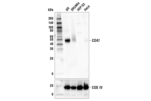
45-50
Rabbit IgG
#Q08722
961
Product Information
Product Usage Information
This product is the carrier free version of product #63000. All data were generated using the same antibody clone in the standard formulation which contains BSA and glycerol.
This formulation is ideal for use with technologies requiring specialized or custom antibody labeling, including fluorophores, metals, lanthanides, and oligonucleotides. It is not recommended for ChIP, ChIP-seq, CUT&RUN, or CUT&Tag assays. If you require a carrier-free formulation for chromatin profiling, please contact us. Optimal dilutions/concentrations should be determined by the end user.
Formulation
Storage
Specificity / Sensitivity
Species Reactivity:
Human
Source / Purification
Monoclonal antibody is produced by immunizing animals with a synthetic peptide corresponding to residues surrounding Leu72 of human CD47 protein.
Background
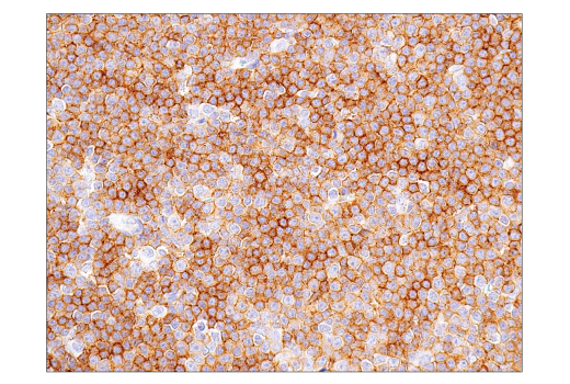
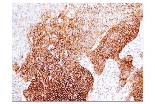
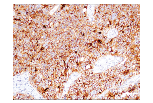
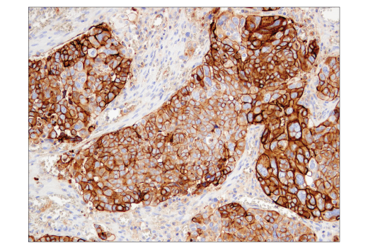
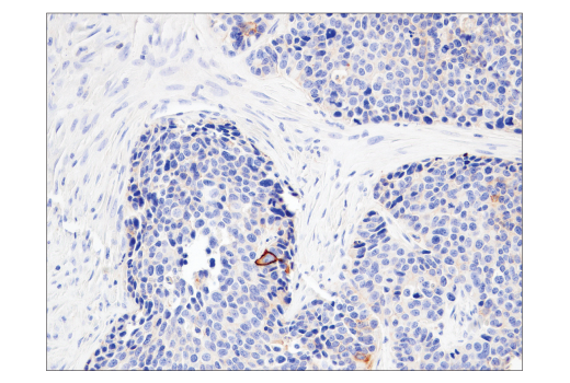
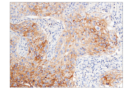
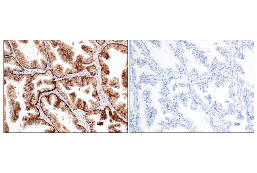
CD47 is a five-pass transmembrane protein expressed on all normal cells. It binds to the SIRPα that is expressed on myeloid cells, including macrophages, and neuronal cells in the central nervous system. Binding of CD47 to SIRPα promotes phosphorylation of tyrosine residues in the immunoreceptor tyrosine-based inhibitory motifs (ITIMs) within the SIRPα cytoplasmic tail, inhibiting macrophage phagocytosis toward CD47-expressing cells. In this way, CD47 serves as a "don't eat me" signal or a marker of "self", functioning as an innate immune checkpoint. Additionally, CD47 was reported to modulate lymphocyte cell activation and proliferation (1-3). CD47 is overexpressed in many types of cancer. The expression level of CD47 on cancer cells is negatively associated with the response to therapies, and low expression on tumor cells is associated with a better prognosis and survival. Reagents that can block CD47-SIRPα interaction are being actively pursued for therapeutic applications (4,5). In addition to SIRPα, other proteins have been reported to bind to CD47. Thrombospondin-1 (TSP1) competes with SIRPα to bind to CD47 in the extracellular region and activates signaling pathways downstream of CD47 (6). CD47 can laterally associate with VEGFR2, FAS, and certain integrins in different contexts, and influences their downstream signaling (7-9). CD47 can be shed from the cell surface by proteolytic cleavage. In addition, CD47 is present on extracellular vesicles including exosomes, suggesting additional extracellular signaling potential (10).
- Murata, Y. et al. (2014) J Biochem 155, 335-44.
- Legrand, N. et al. (2011) Proc Natl Acad Sci USA 108, 13224-9.
- Barclay, A.N. and Van den Berg, T.K. (2014) Annu Rev Immunol 32, 25-50.
- Weiskopf, K. (2017) Eur J Cancer 76, 100-109.
- Matlung, H.L. et al. (2017) Immunol Rev 276, 145-164.
- Roberts, D.D. et al. (2012) Matrix Biol 31, 162-9.
- Kaur, S. et al. (2010) J Biol Chem 285, 38923-32.
- Azcutia, V. et al. (2013) Mol Biol Cell 24, 3358-68.
- Quesada, A.J. et al. (2005) Cell Death Differ 12, 649-58.
- Soto-Pantoja, D.R. et al. (2015) Crit Rev Biochem Mol Biol 50, 212-30.
Species Reactivity
Species reactivity is determined by testing in at least one approved application (e.g., western blot).
Applications Key
WB: Western Blotting IHC-Bond: IHC Leica Bond IHC-P: Immunohistochemistry (Paraffin)
Cross-Reactivity Key
H: human M: mouse R: rat Hm: hamster Mk: monkey Vir: virus Mi: mink C: chicken Dm: D. melanogaster X: Xenopus Z: zebrafish B: bovine Dg: dog Pg: pig Sc: S. cerevisiae Ce: C. elegans Hr: horse GP: Guinea Pig Rab: rabbit All: all species expected
Trademarks and Patents
使用に関する制限
法的な権限を与えられたCSTの担当者が署名した書面によって別途明示的に合意された場合を除き、 CST、その関連会社または代理店が提供する製品には以下の条件が適用されます。お客様が定める条件でここに定められた条件に含まれるものを超えるもの、 または、ここに定められた条件と異なるものは、法的な権限を与えられたCSTの担当者が別途書面にて受諾した場合を除き、拒絶され、 いかなる効力も効果も有しません。
研究専用 (For Research Use Only) またはこれに類似する表示がされた製品は、 いかなる目的についても FDA または外国もしくは国内のその他の規制機関により承認、認可または許可を受けていません。 お客様は製品を診断もしくは治療目的で使用してはならず、また、製品に表示された内容に違反する方法で使用してはなりません。 CST が販売または使用許諾する製品は、エンドユーザーであるお客様に対し、使途を研究および開発のみに限定して提供されるものです。 診断、予防もしくは治療目的で製品を使用することまたは製品を再販売 (単独であるか他の製品等の一部であるかを問いません) もしくはその他の商業的利用の目的で購入することについては、CST から別途許諾を得る必要があります。 お客様は以下の事項を遵守しなければなりません。(a) CST の製品 (単独であるか他の資材と一緒であるかを問いません) を販売、使用許諾、貸与、寄付もしくはその他の態様で第三者に譲渡したり使用させたりしてはなりません。また、商用の製品を製造するために CST の製品を使用してはなりません。(b) 複製、改変、リバースエンジニアリング、逆コンパイル、 分解または他の方法により製品の構造または技術を解明しようとしてはなりません。また、 CST の製品またはサービスと競合する製品またはサービスを開発する目的で CST の製品を使用してはなりません。(c) CST の製品の商標、商号、ロゴ、特許または著作権に関する通知または表示を除去したり改変したりしてはなりません。(d) CST の製品をCST 製品販売条件(CST’s Product Terms of Sale) および該当する書面のみに従って使用しなければなりません。(e) CST の製品に関連してお客様が使用する第三者の製品またはサービスに関する使用許諾条件、 サービス提供条件またはこれに類する合意事項を遵守しなければなりません。