| Product Includes | Product # | Quantity | Mol. Wt | Isotype/Source |
|---|---|---|---|---|
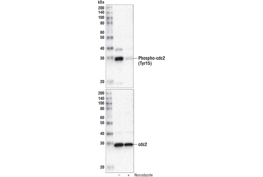
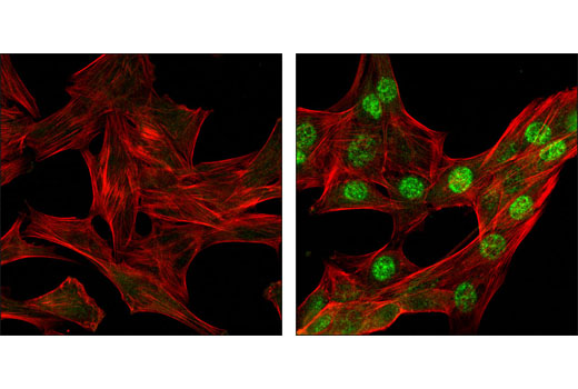
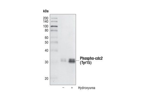
| Phospho-cdc2 (Tyr15) (10A11) Rabbit mAb | 4539 | 20 µl | 34 kDa | Rabbit |
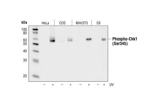
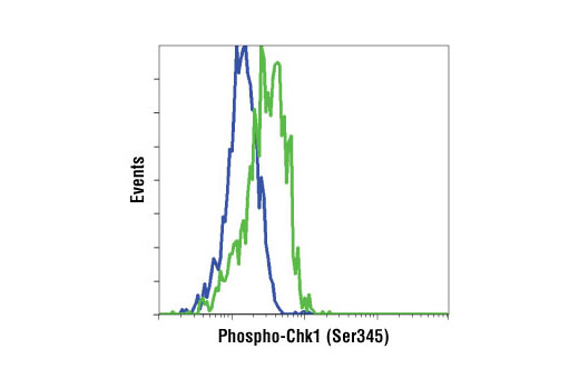
| Phospho-Chk1 (Ser345) (133D3) Rabbit mAb | 2348 | 20 µl | 56 kDa | Rabbit IgG |
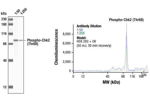
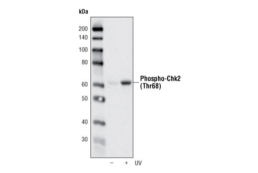
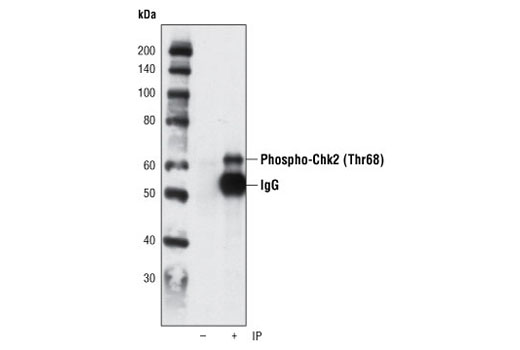
| Phospho-Chk2 (Thr68) (C13C1) Rabbit mAb | 2197 | 20 µl | 62 kDa | Rabbit IgG |
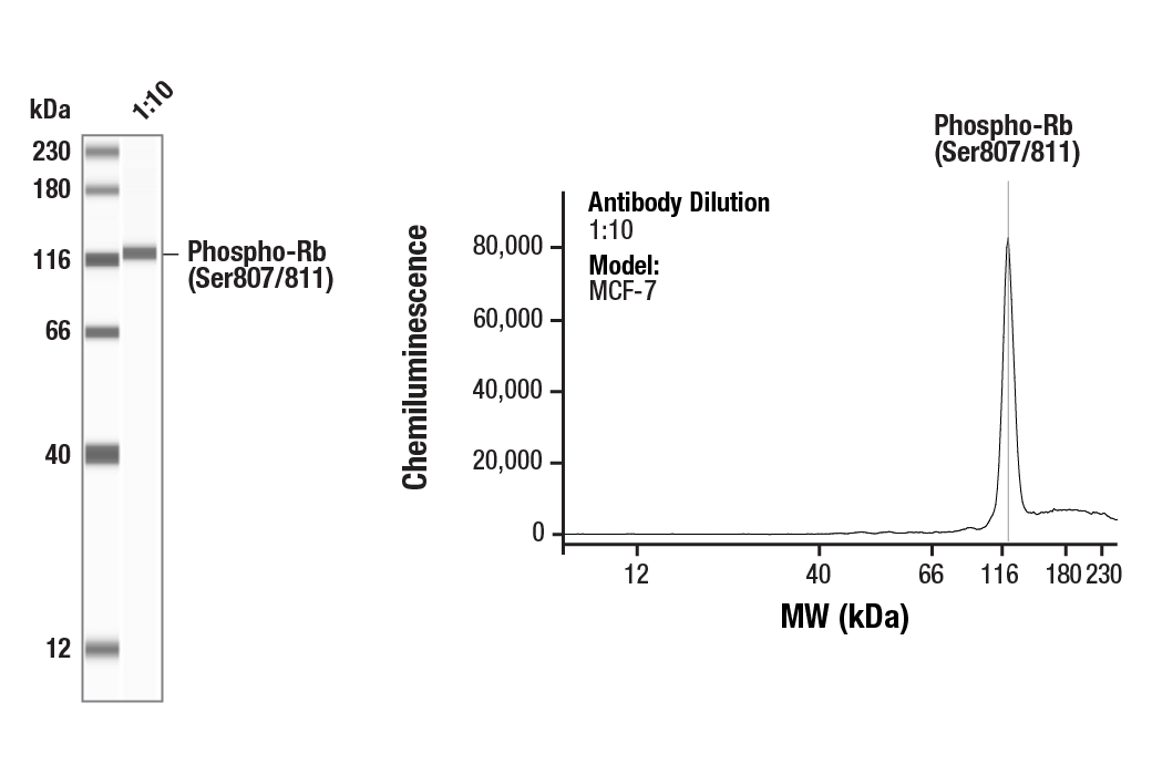
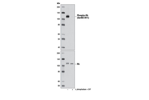
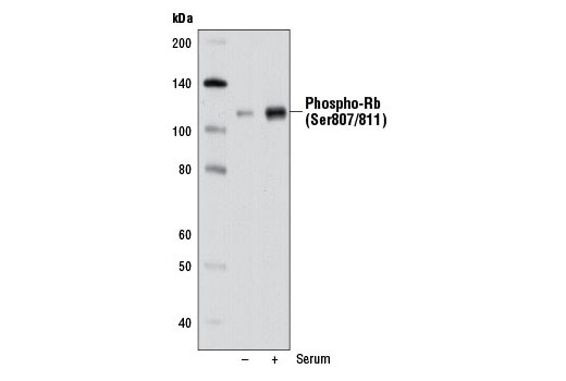
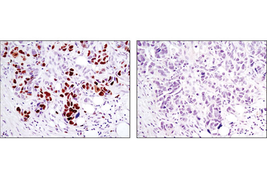
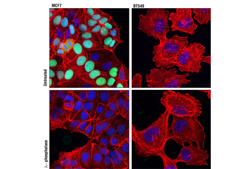
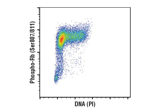
| Phospho-Rb (Ser807/811) (D20B12) XP® Rabbit mAb | 8516 | 20 µl | 110 kDa | Rabbit IgG |
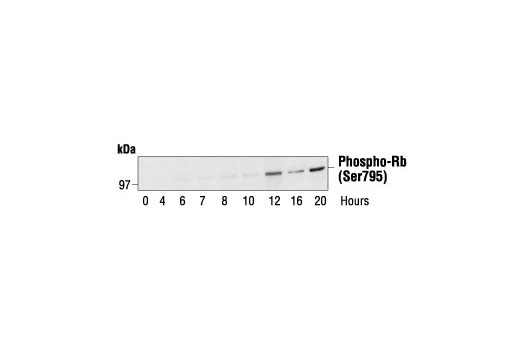
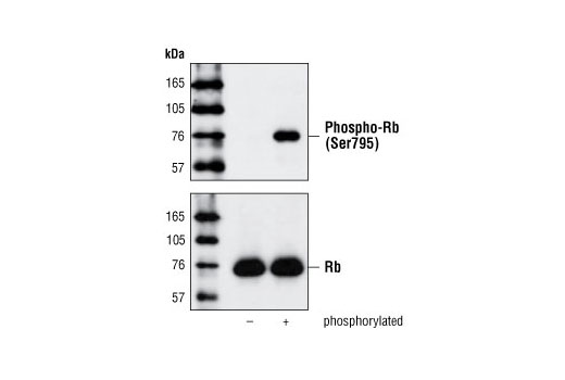
| Phospho-Rb (Ser795) Antibody | 9301 | 20 µl | 110 kDa | Rabbit |
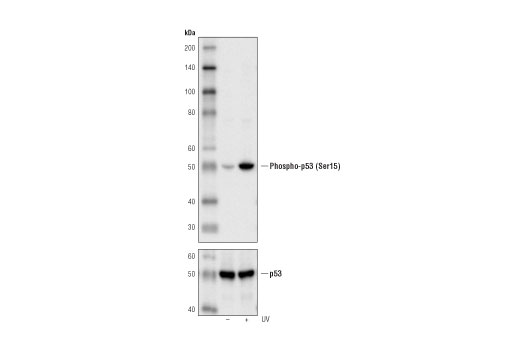
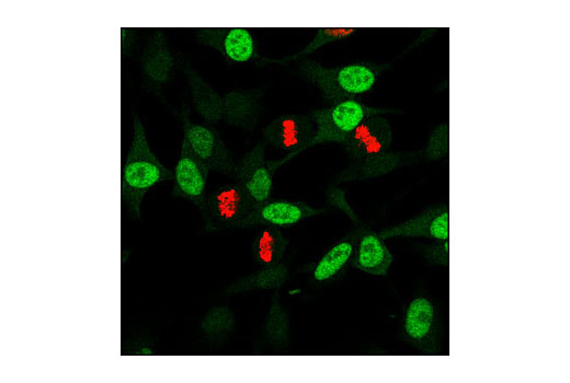
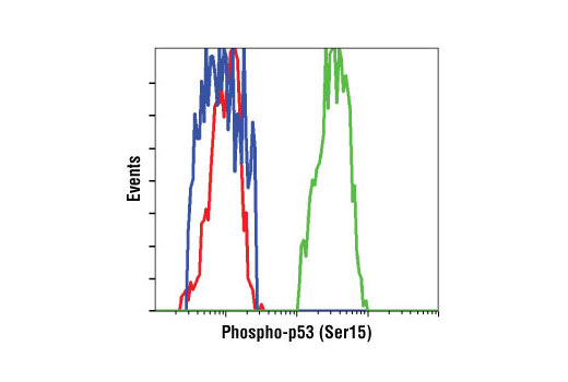
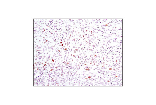
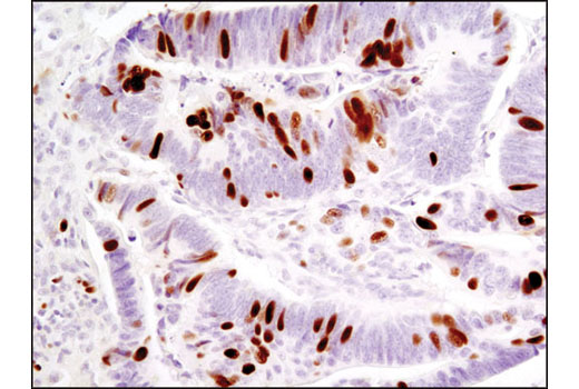
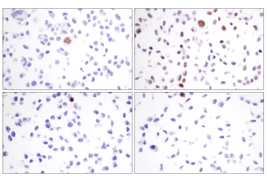
| Phospho-p53 (Ser15) (16G8) Mouse mAb | 9286 | 20 µl | 53 kDa | Mouse IgG1 |
| Anti-rabbit IgG, HRP-linked Antibody | 7074 | 100 µl | Goat | |
| Anti-mouse IgG, HRP-linked Antibody | 7076 | 100 µl | Horse |
Please visit cellsignal.com for individual component applications, species cross-reactivity, dilutions, protocols, and additional product information.
Description
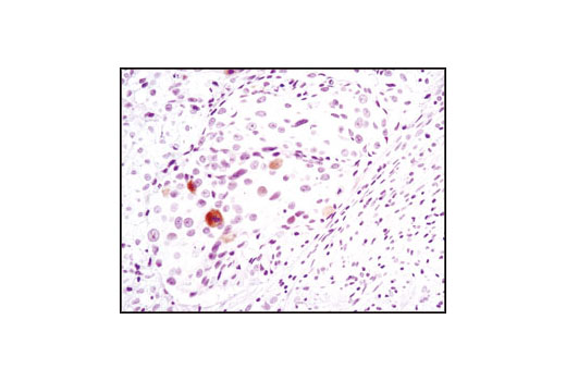
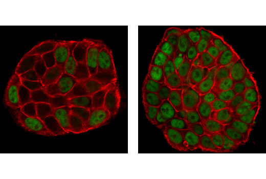
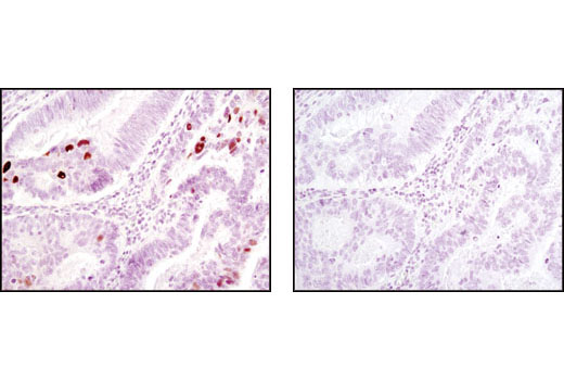
The Cell Cycle/Checkpoint Antibody Sampler Kit provides a fast and economical means of evaluating multiple proteins involved in the cell cyle and checkpoint control. The kit contains enough primary and secondary antibody to perform four Western blot experiments.
Storage
Background
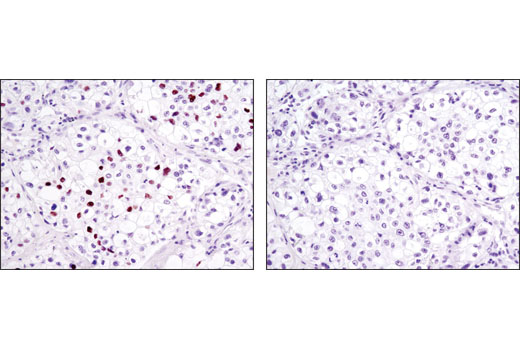
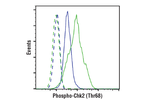
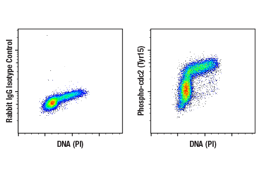
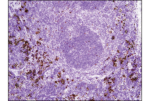
The cell division cycle demands accuracy to avoid the accumulation of genetic damage. This process is controlled by molecular circuits called "checkpoints" that are common to all eukaryotic cells (1). Checkpoints monitor DNA integrity and cell growth prior to replication and division at the G1/S and G2/M transitions, respectively. The cdc2-cyclin B kinase is pivotal in regulating the G2/M transition (2,3). Cdc2 is phosphorylated at Thr14 and Tyr15 during G2-phase by the kinases Wee1 and Myt1, rendering it inactive. The tumor suppressor protein retinoblastoma (Rb) controls progression through the late G1 restriction point (R) and is a major regulator of the G1/S transition (4). During early and mid G1-phase, Rb binds to and represses the transcription factor E2F (5). The phosphorylation of Rb late in G1-phase by CDKs induces Rb to dissociate from E2F, permitting the transcription of S-phase-promoting genes. In vitro, Rb can be phosphorylated at multiple sites by cdc2, cdk2, and cdk4/6 (6-8). DNA damage triggers both the G2/M and the G1/S checkpoints. DNA damage activates the DNA-PK/ATM/ATR kinases, which phosphorylate Chk at Ser345 (9), Chk2 at Thr68 (10) and p53 (11). The Chk kinases inactivate cdc25 via phosphorylation at Ser216, blocking the activation of cdc2.
- Nurse, P. (1997) Cell 91, 865-7.
- Norbury, C. and Nurse, P. (1992) Annu Rev Biochem 61, 441-70.
- Watanabe, N. et al. (1995) EMBO J 14, 1878-91.
- Sherr, C.J. (1996) Science 274, 1672-7.
- Dyson, N. (1998) Genes Dev 12, 2245-62.
- Kitagawa, M. et al. (1996) EMBO J 15, 7060-9.
- Lundberg, A.S. and Weinberg, R.A. (1998) Mol Cell Biol 18, 753-61.
- Harbour, J.W. et al. (1999) Cell 98, 859-69.
- Zhao, H. and Piwnica-Worms, H. (2001) Mol Cell Biol 21, 4129-39.
- Matsuoka, S. et al. (2000) Proc Natl Acad Sci USA 97, 10389-94.
- Tibbetts, R.S. et al. (1999) Genes Dev 13, 152-7.
Background References
Trademarks and Patents
使用に関する制限
法的な権限を与えられたCSTの担当者が署名した書面によって別途明示的に合意された場合を除き、 CST、その関連会社または代理店が提供する製品には以下の条件が適用されます。お客様が定める条件でここに定められた条件に含まれるものを超えるもの、 または、ここに定められた条件と異なるものは、法的な権限を与えられたCSTの担当者が別途書面にて受諾した場合を除き、拒絶され、 いかなる効力も効果も有しません。
研究専用 (For Research Use Only) またはこれに類似する表示がされた製品は、 いかなる目的についても FDA または外国もしくは国内のその他の規制機関により承認、認可または許可を受けていません。 お客様は製品を診断もしくは治療目的で使用してはならず、また、製品に表示された内容に違反する方法で使用してはなりません。 CST が販売または使用許諾する製品は、エンドユーザーであるお客様に対し、使途を研究および開発のみに限定して提供されるものです。 診断、予防もしくは治療目的で製品を使用することまたは製品を再販売 (単独であるか他の製品等の一部であるかを問いません) もしくはその他の商業的利用の目的で購入することについては、CST から別途許諾を得る必要があります。 お客様は以下の事項を遵守しなければなりません。(a) CST の製品 (単独であるか他の資材と一緒であるかを問いません) を販売、使用許諾、貸与、寄付もしくはその他の態様で第三者に譲渡したり使用させたりしてはなりません。また、商用の製品を製造するために CST の製品を使用してはなりません。(b) 複製、改変、リバースエンジニアリング、逆コンパイル、 分解または他の方法により製品の構造または技術を解明しようとしてはなりません。また、 CST の製品またはサービスと競合する製品またはサービスを開発する目的で CST の製品を使用してはなりません。(c) CST の製品の商標、商号、ロゴ、特許または著作権に関する通知または表示を除去したり改変したりしてはなりません。(d) CST の製品をCST 製品販売条件(CST’s Product Terms of Sale) および該当する書面のみに従って使用しなければなりません。(e) CST の製品に関連してお客様が使用する第三者の製品またはサービスに関する使用許諾条件、 サービス提供条件またはこれに類する合意事項を遵守しなければなりません。