| Product Includes | Product # | Quantity | Mol. Wt | Isotype/Source |
|---|---|---|---|---|
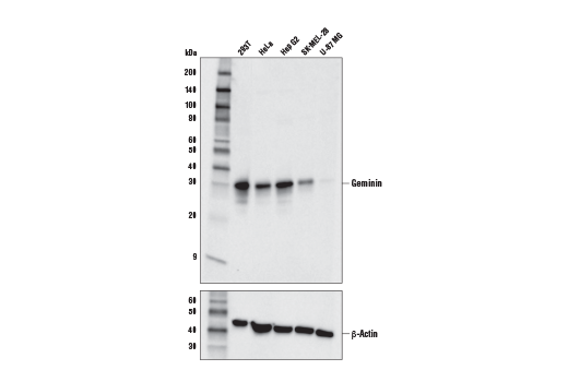
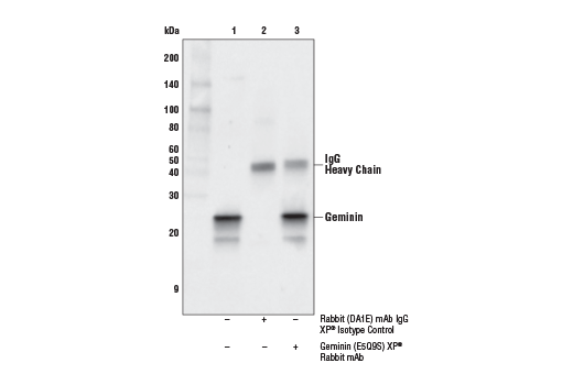
| Geminin (E5Q9S) XP® Rabbit mAb | 52508 | 20 µl | 25 kDa | Rabbit IgG |
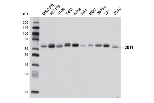
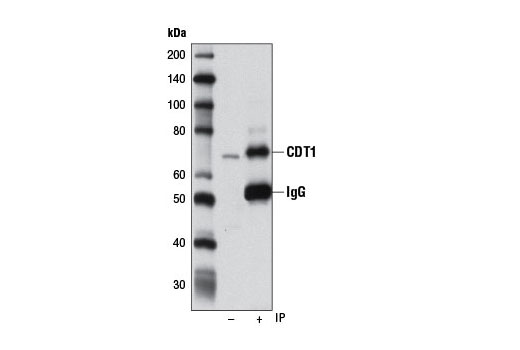
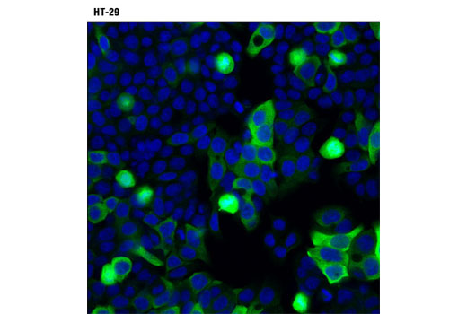
| CDT1 (D10F11) Rabbit mAb | 8064 | 20 µl | 65 kDa | Rabbit IgG |
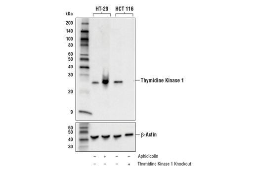
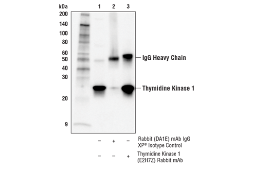
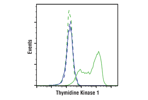
| Thymidine Kinase 1 (E2H7Z) Rabbit mAb | 28755 | 20 µl | 26 kDa | Rabbit IgG |
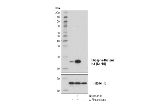
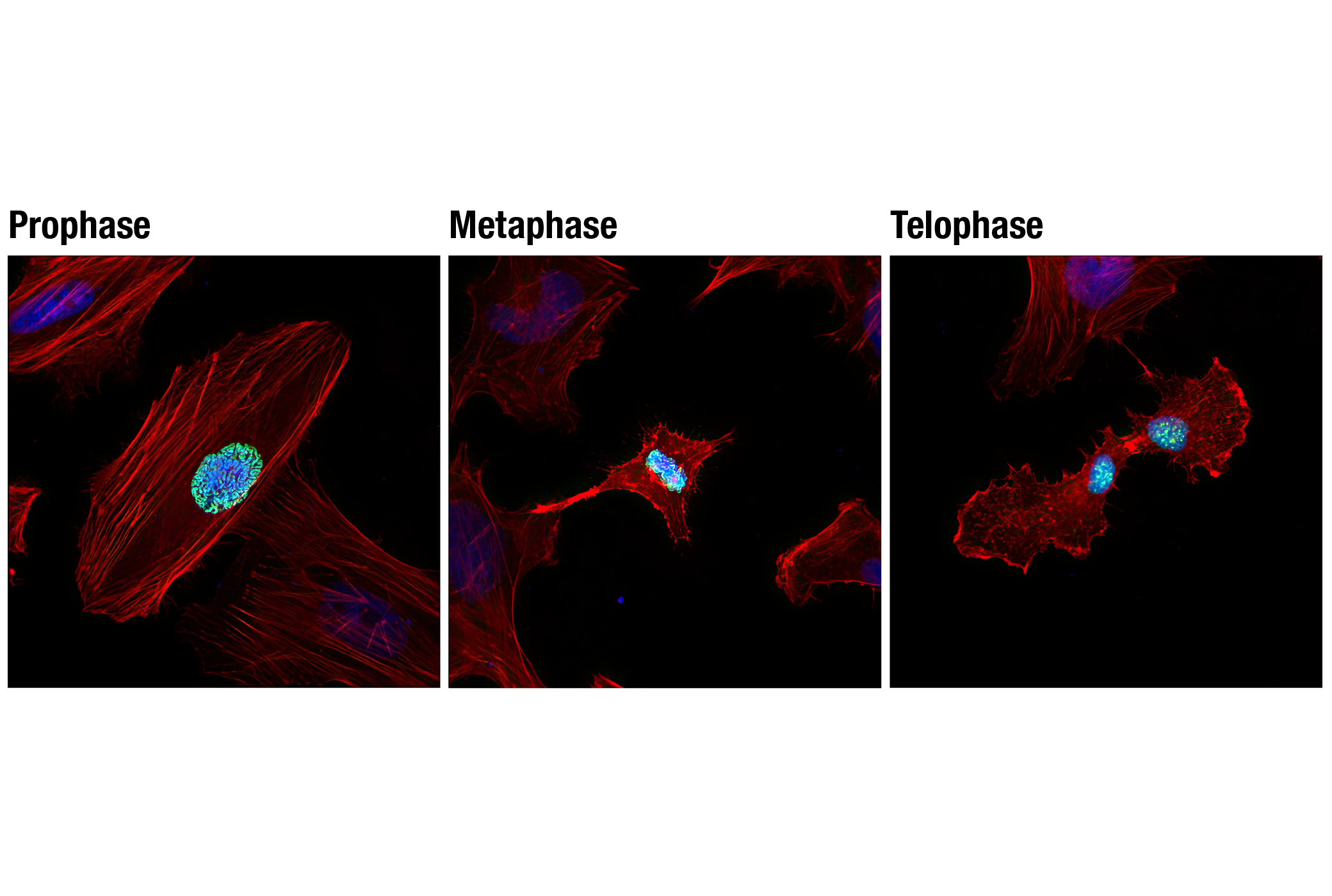
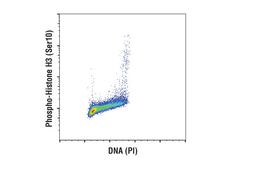
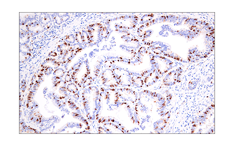
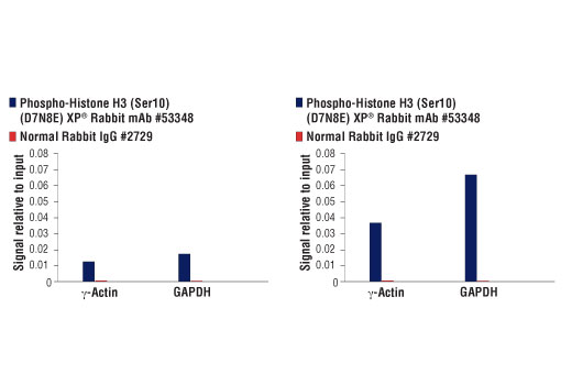
| Phospho-Histone H3 (Ser10) (D7N8E) XP® Rabbit mAb | 53348 | 20 µl | 17 kDa | Rabbit IgG |
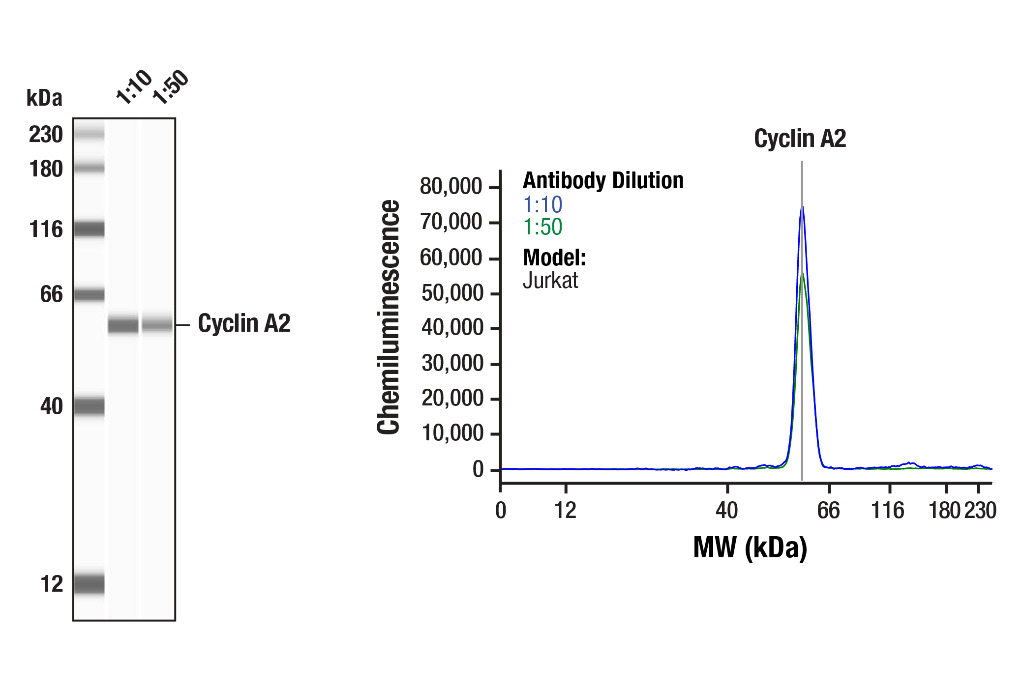
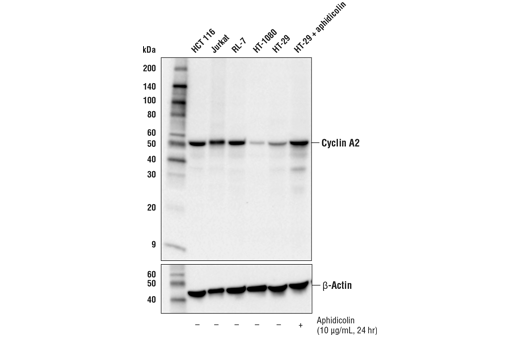
| Cyclin A2 (E1D9T) Rabbit mAb | 91500 | 20 µl | 55 kDa | Rabbit IgG |
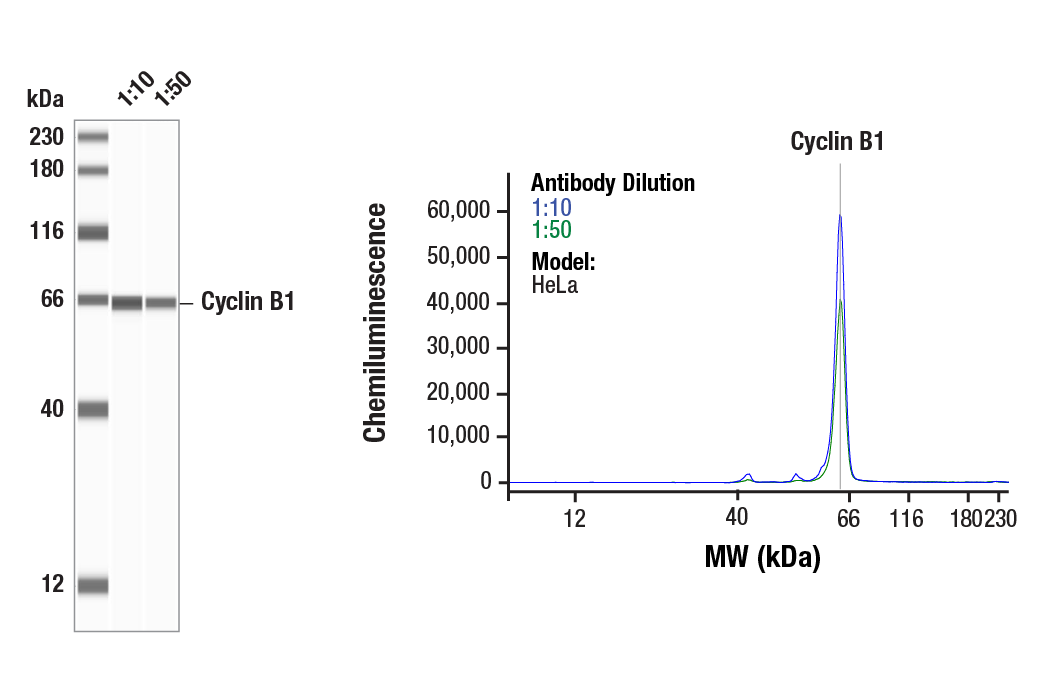
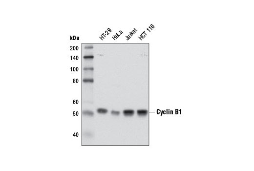
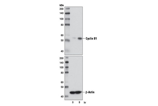
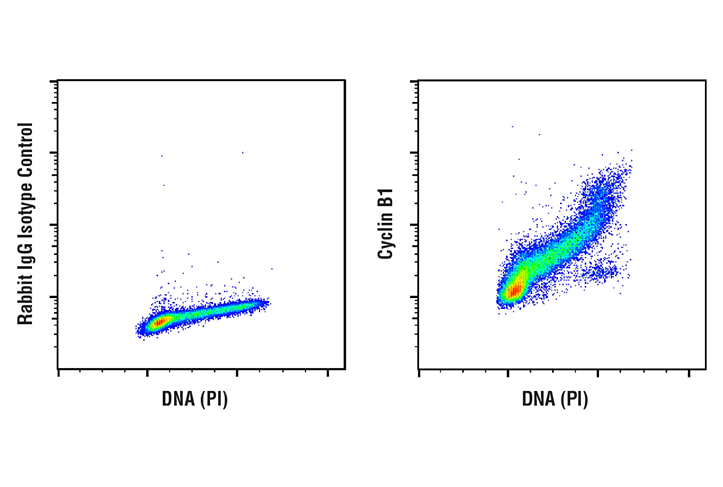
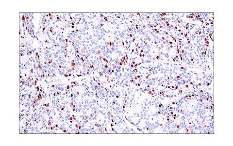
| Cyclin B1 (D5C10) XP® Rabbit mAb | 12231 | 20 µl | 55 kDa | Rabbit IgG |
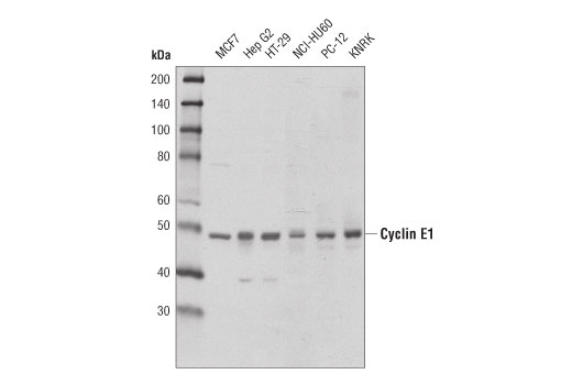
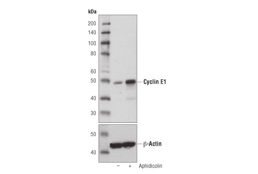
| Cyclin E1 (D7T3U) Rabbit mAb | 20808 | 20 µl | 48 kDa | Rabbit IgG |
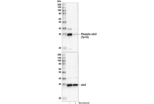
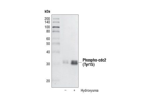
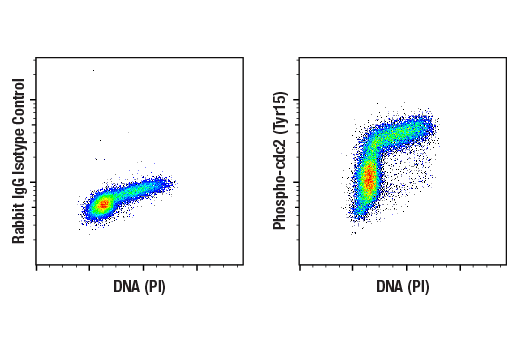
| Phospho-cdc2 (Tyr15) (10A11) Rabbit mAb | 4539 | 20 µl | 34 kDa | Rabbit |
| Anti-rabbit IgG, HRP-linked Antibody | 7074 | 100 µl | Goat |
Please visit cellsignal.com for individual component applications, species cross-reactivity, dilutions, protocols, and additional product information.
Description
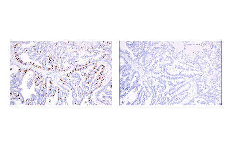
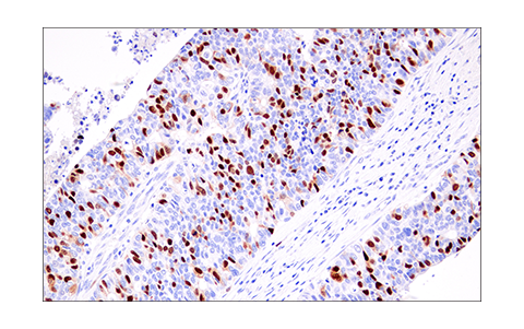
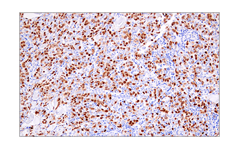
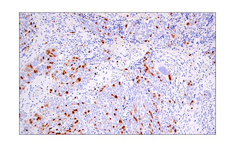
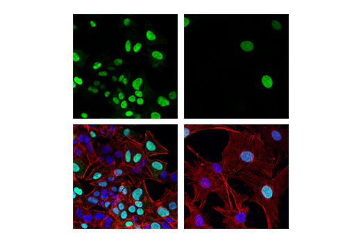
The Cell Cycle Phase Determination Antibody Sampler Kit provides an economical means of detecting total proteins or post-translational modifications present in cells at various phases of the cell cycle. Geminin is degraded in G1 phase, while CDT1 is degraded in S, G2, and M phases. Thymidine Kinase 1 accumulates in G1 phase, peaks in S phase, and is degraded before cell division. Phospho-Histone H3 (Ser10) is present only in M phase, while Phospho-cdc2 (Tyr15) is absent in M phase. Cyclins A2, B1, and E1 peak at G2 phase, late G2/M phase, and late G1/early S phase, respectively. The kit includes enough antibodies to perform two western blot experiments with each primary antibody.
Storage
Background
The entry of eukaryotic cells into mitosis is regulated by cdc2/CDK1 kinase activation, a process controlled at several steps including cyclin B1 nuclear accumulation and binding, and phosphorylation of cdc2/CDK1 at Thr161 (1). At the end of mitosis, cyclin B1 is targeted for degradation by the anaphase-promoting complex (APC), allowing for cell cycle progression (2). A critical regulatory step in activating cdc2 during progression into mitosis is dephosphorylation of cdc2/CDK1 at Thr14 and Tyr15 (3).
Phosphorylation of Histone H3 at Ser10 is tightly correlated with chromosome condensation during both mitosis and meiosis (4).
Overcoming the G1/S checkpoint to commence DNA replication requires cyclin E, traversing the G2/M checkpoint to initiate mitosis requires cyclin B, and cyclin A is required for both S-phase and M-phase (5). Cyclin A availability is apparently the rate-limiting step for entry into mitosis, and cyclin A is required for completion of prophase (6).
Thymidine kinases play a critical role in generating the DNA synthetic precursor deoxythymidine triphosphate (dTTP). Cytoplasmic thymidine kinase 1 (TK1) expression and activity are regulated in a cell cycle-dependent manner, accumulating during G1-phase to peak levels in S-phase before being degraded prior to cell division (7).
The initiation of S phase begins with the formation of the pre-replication complex (pre-RC) in late mitosis/early G1 phase. CDT1 and cdc6 bind to the origin of DNA replication, which allows binding of the MCM2-7 complex. In order to ensure that replication occurs only once per cell cycle, geminin inhibits and destabilizes CDT1 during the S, G2 and M phases. At the metaphase/anaphase transition, geminin is degraded by the anaphase-promoting complex (APC) allowing for the formation of new pre-RC (8).
- Atherton-Fessler, S. et al. (1994) Mol Biol Cell 5, 989-1001.
- Gong, D. and Ferrell, J.E. (2010) Mol Biol Cell 21, 3149-61.
- Norbury, C. et al. (1991) EMBO J 10, 3321-9.
- Hendzel, M.J. et al. (1997) Chromosoma 106, 348-60.
- Pagano, M. et al. (1992) EMBO J 11, 961-71.
- Furuno, N. et al. (1999) J Cell Biol 147, 295-306.
- Munch-Petersen, B. (2010) Nucleosides Nucleotides Nucleic Acids 29, 363-9.
- Caillat, C. and Perrakis, A. (2012) Subcell Biochem 62, 71-87.
Background References
Trademarks and Patents
使用に関する制限
法的な権限を与えられたCSTの担当者が署名した書面によって別途明示的に合意された場合を除き、 CST、その関連会社または代理店が提供する製品には以下の条件が適用されます。お客様が定める条件でここに定められた条件に含まれるものを超えるもの、 または、ここに定められた条件と異なるものは、法的な権限を与えられたCSTの担当者が別途書面にて受諾した場合を除き、拒絶され、 いかなる効力も効果も有しません。
研究専用 (For Research Use Only) またはこれに類似する表示がされた製品は、 いかなる目的についても FDA または外国もしくは国内のその他の規制機関により承認、認可または許可を受けていません。 お客様は製品を診断もしくは治療目的で使用してはならず、また、製品に表示された内容に違反する方法で使用してはなりません。 CST が販売または使用許諾する製品は、エンドユーザーであるお客様に対し、使途を研究および開発のみに限定して提供されるものです。 診断、予防もしくは治療目的で製品を使用することまたは製品を再販売 (単独であるか他の製品等の一部であるかを問いません) もしくはその他の商業的利用の目的で購入することについては、CST から別途許諾を得る必要があります。 お客様は以下の事項を遵守しなければなりません。(a) CST の製品 (単独であるか他の資材と一緒であるかを問いません) を販売、使用許諾、貸与、寄付もしくはその他の態様で第三者に譲渡したり使用させたりしてはなりません。また、商用の製品を製造するために CST の製品を使用してはなりません。(b) 複製、改変、リバースエンジニアリング、逆コンパイル、 分解または他の方法により製品の構造または技術を解明しようとしてはなりません。また、 CST の製品またはサービスと競合する製品またはサービスを開発する目的で CST の製品を使用してはなりません。(c) CST の製品の商標、商号、ロゴ、特許または著作権に関する通知または表示を除去したり改変したりしてはなりません。(d) CST の製品をCST 製品販売条件(CST’s Product Terms of Sale) および該当する書面のみに従って使用しなければなりません。(e) CST の製品に関連してお客様が使用する第三者の製品またはサービスに関する使用許諾条件、 サービス提供条件またはこれに類する合意事項を遵守しなければなりません。