Revision 5
Cell Signaling Technology

Orders: 877-616-CELL (2355) [email protected]

Support: 877-678-TECH (8324)

Web: [email protected] cellsignal.com

3 Trask LaneDanversMassachusetts01923USA
For Research Use Only. Not for Use in Diagnostic Procedures.
Applications:

WB, IP, IHC-P, IF-F, IF-IC

REACTIVITY:

H M R

SENSITIVITY:

Endogenous

MW (kDa):

39, 41, 43, 44

Source/Isotype:

Rabbit IgG

UniProt ID:

#P17302

Entrez-Gene Id:

2697

Product Information

Product Usage Information

Application Dilution
Western Blotting 1:1000
Immunoprecipitation 1:100
Immunohistochemistry (Paraffin) 1:200 - 1:800
Immunofluorescence (Frozen) 1:400 - 1:1600
Immunofluorescence (Immunocytochemistry) 1:400 - 1:1600

Storage

Supplied in 10 mM sodium HEPES (pH 7.5), 150 mM NaCl, 100 µg/ml BSA, 50% glycerol and less than 0.02% sodium azide. Store at –20°C. Do not aliquot the antibody.

For a carrier free (BSA and azide free) version of this product see product #89289.

Specificity / Sensitivity

Connexin 43 (E7N2R) XP® Rabbit mAb recognizes endogenous levels of total connexin 43 protein.

Species Reactivity:

Human, Mouse, Rat

Source / Purification

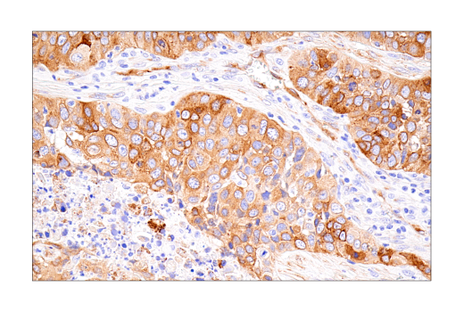
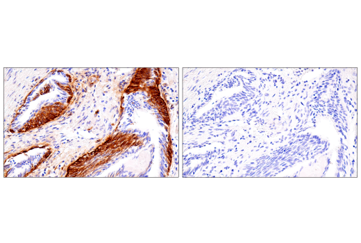
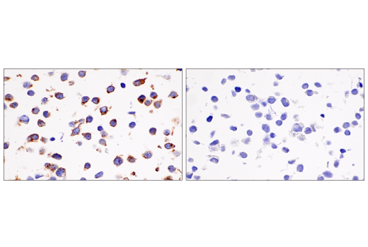
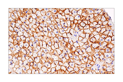
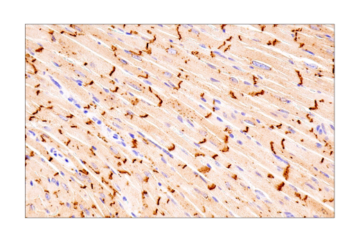
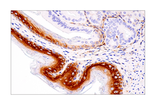
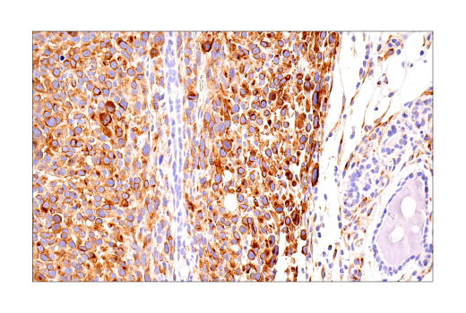
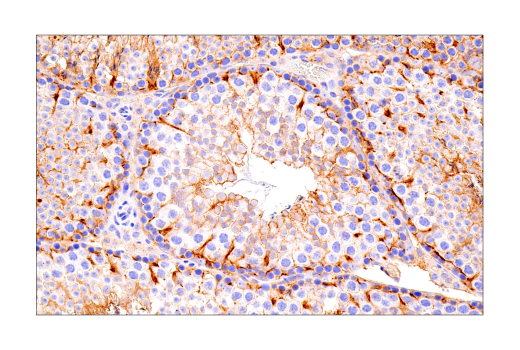
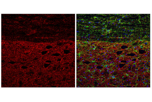
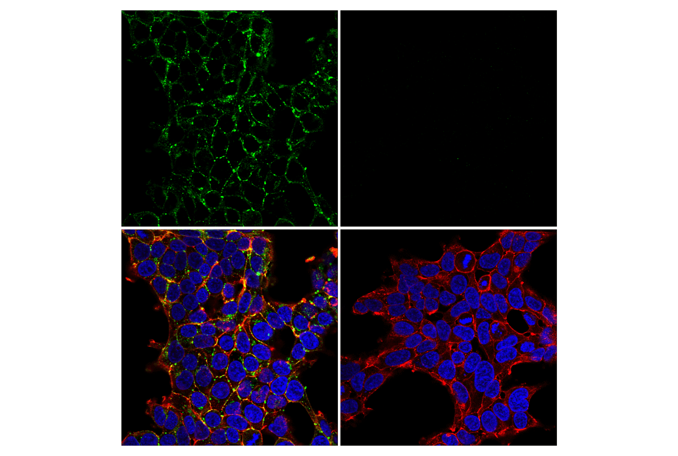
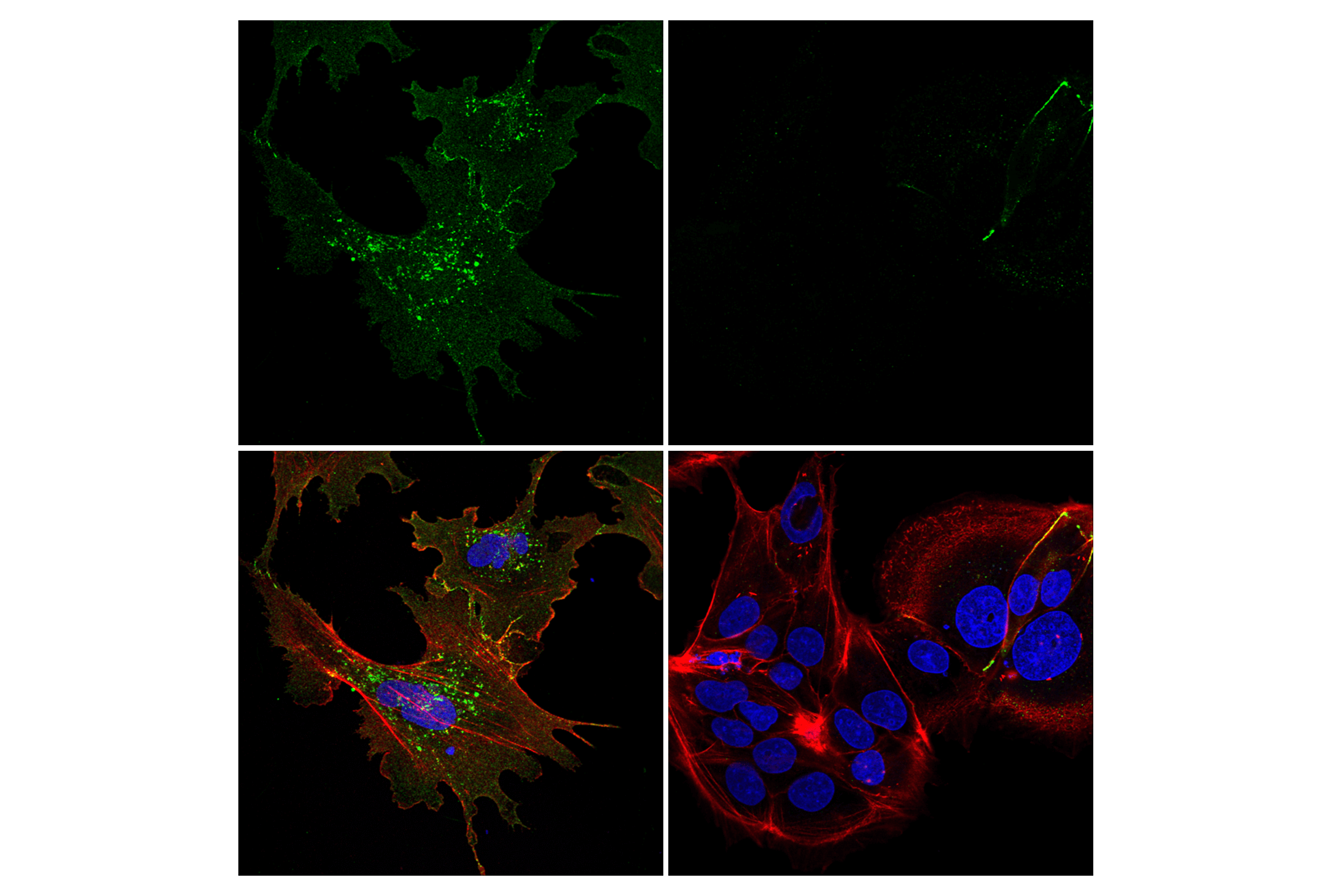
Monoclonal antibody is produced by immunizing animals with a synthetic peptide corresponding to residues surrounding Arg374 of human connexin 43 protein.

Background

Connexin 43 (Cx43) is a member of the large family of gap junction proteins. Connexins assemble as a hexamer and are transported to the plasma membrane to create a hemichannel that can associate with hemichannels on nearby cells to create cell-to-cell channels. Clusters of these channels assemble to make gap junctions. Gap junction communication is important in development and regulation of cell growth. Phosphorylation of Cx43 is important in regulating assembly and function of gap junctions (1,2). Ser368 of Cx43 is phosphorylated by protein kinase C (PKC) after activation by phorbol esters, which decreases cell-to-cell communication (3). Src can interact with and phosphorylate Cx43 to alter gap junction communication (4,5).

  1. Musil, L.S. et al. (1990) J Cell Biol 111, 2077-88.
  2. Musil, L.S. and Goodenough, D.A. (1991) J Cell Biol 115, 1357-74.
  3. Lampe, P.D. et al. (2000) J Cell Biol 149, 1503-12.
  4. Giepmans, B.N. et al. (2001) J Biol Chem 276, 8544-9.
  5. Lin, R. et al. (2001) J Cell Biol 154, 815-27.

Species Reactivity

Species reactivity is determined by testing in at least one approved application (e.g., western blot).

Western Blot Buffer

IMPORTANT: For western blots, incubate membrane with diluted primary antibody in 5% w/v BSA, 1X TBS, 0.1% Tween® 20 at 4°C with gentle shaking, overnight.

Applications Key

WB: Western Blotting IP: Immunoprecipitation IHC-P: Immunohistochemistry (Paraffin) IF-F: Immunofluorescence (Frozen) IF-IC: Immunofluorescence (Immunocytochemistry)

Cross-Reactivity Key

H: human M: mouse R: rat Hm: hamster Mk: monkey Vir: virus Mi: mink C: chicken Dm: D. melanogaster X: Xenopus Z: zebrafish B: bovine Dg: dog Pg: pig Sc: S. cerevisiae Ce: C. elegans Hr: horse GP: Guinea Pig Rab: rabbit All: all species expected

Trademarks and Patents

Cell Signaling Technology is a trademark of Cell Signaling Technology, Inc.
SignalStain is a registered trademark of Cell Signaling Technology, Inc.
XP is a registered trademark of Cell Signaling Technology, Inc.
All other trademarks are the property of their respective owners. Visit cellsignal.com/trademarks for more information.

使用に関する制限

法的な権限を与えられたCSTの担当者が署名した書面によって別途明示的に合意された場合を除き、 CST、その関連会社または代理店が提供する製品には以下の条件が適用されます。お客様が定める条件でここに定められた条件に含まれるものを超えるもの、 または、ここに定められた条件と異なるものは、法的な権限を与えられたCSTの担当者が別途書面にて受諾した場合を除き、拒絶され、 いかなる効力も効果も有しません。

研究専用 (For Research Use Only) またはこれに類似する表示がされた製品は、 いかなる目的についても FDA または外国もしくは国内のその他の規制機関により承認、認可または許可を受けていません。 お客様は製品を診断もしくは治療目的で使用してはならず、また、製品に表示された内容に違反する方法で使用してはなりません。 CST が販売または使用許諾する製品は、エンドユーザーであるお客様に対し、使途を研究および開発のみに限定して提供されるものです。 診断、予防もしくは治療目的で製品を使用することまたは製品を再販売 (単独であるか他の製品等の一部であるかを問いません) もしくはその他の商業的利用の目的で購入することについては、CST から別途許諾を得る必要があります。 お客様は以下の事項を遵守しなければなりません。(a) CST の製品 (単独であるか他の資材と一緒であるかを問いません) を販売、使用許諾、貸与、寄付もしくはその他の態様で第三者に譲渡したり使用させたりしてはなりません。また、商用の製品を製造するために CST の製品を使用してはなりません。(b) 複製、改変、リバースエンジニアリング、逆コンパイル、 分解または他の方法により製品の構造または技術を解明しようとしてはなりません。また、 CST の製品またはサービスと競合する製品またはサービスを開発する目的で CST の製品を使用してはなりません。(c) CST の製品の商標、商号、ロゴ、特許または著作権に関する通知または表示を除去したり改変したりしてはなりません。(d) CST の製品をCST 製品販売条件(CST’s Product Terms of Sale) および該当する書面のみに従って使用しなければなりません。(e) CST の製品に関連してお客様が使用する第三者の製品またはサービスに関する使用許諾条件、 サービス提供条件またはこれに類する合意事項を遵守しなければなりません。

Revision 5
#83649

Connexin 43 (E7N2R) XP® Rabbit mAb

Western Blotting Image 1: Connexin 43 (E7N2R) XP® Rabbit mAb Expand Image
Western blot analysis of extracts from various cell lines using Connexin 43 (E7N2R) XP® Rabbit mAb (upper) and GAPDH (D16H11) XP® Rabbit mAb #5174 (lower). Low expression of connexin 43 protein in BeWo cells is consistent with the predicted expression pattern.
Western Blotting Image 2: Connexin 43 (E7N2R) XP® Rabbit mAb Expand Image
Western blot analysis of extracts from control 293T cells (lane 1) or connexin 43 knockout 293T cells (lane 2) using Connexin 43 (E7N2R) XP® Rabbit mAb (upper) and GAPDH (D16H11) XP® Rabbit mAb #5174 (lower). The absence of signal in the connexin 43 knockout 293T cells confirms specificity of the antibody for connexin 43.
Immunoprecipitation Image 1: Connexin 43 (E7N2R) XP® Rabbit mAb Expand Image
Immunoprecipitation of connexin 43 protein from 293T cell extracts. Lane 1 is 10% input, lane 2 is Rabbit (DA1E) mAb IgG XP® Isotype Control #3900, and lane 3 is Connexin 43 (E7N2R) XP® Rabbit mAb. Western blot analysis was performed using Connexin 43 (E7N2R) XP® Rabbit mAb. Anti-rabbit IgG, HRP-linked Antibody #7074 was used as the secondary antibody. 
Immunohistochemistry Image 1: Connexin 43 (E7N2R) XP® Rabbit mAb Expand Image
Immunohistochemical analysis of paraffin-embedded human granulosa cell tumor of the ovary using Connexin 43 (E7N2R) XP® Rabbit mAb.
Immunohistochemistry Image 2: Connexin 43 (E7N2R) XP® Rabbit mAb Expand Image
Immunohistochemical analysis of paraffin-embedded normal human testis using Connexin 43 (E7N2R) XP® Rabbit mAb.
Immunohistochemistry Image 3: Connexin 43 (E7N2R) XP® Rabbit mAb Expand Image
Immunohistochemical analysis of paraffin-embedded human colon adenocarcinoma using Connexin 43 (E7N2R) XP® Rabbit mAb.
Immunohistochemistry Image 4: Connexin 43 (E7N2R) XP® Rabbit mAb Expand Image
Immunohistochemical analysis of paraffin-embedded human squamous cell carcinoma of the tonsil using Connexin 43 (E7N2R) XP® Rabbit mAb.
Immunohistochemistry Image 5: Connexin 43 (E7N2R) XP® Rabbit mAb Expand Image
Immunohistochemical analysis of paraffin-embedded human prostate adenocarcinoma using Connexin 43 (E7N2R) XP® Rabbit mAb.
Immunohistochemistry Image 6: Connexin 43 (E7N2R) XP® Rabbit mAb Expand Image
Immunohistochemical analysis of paraffin-embedded human non-small cell lung carcinoma using Connexin 43 (E7N2R) XP® Rabbit mAb.
Immunohistochemistry Image 7: Connexin 43 (E7N2R) XP® Rabbit mAb Expand Image
Immunohistochemical analysis of paraffin-embedded human prostate adenocarcinoma using Connexin 43 (E7N2R) XP® Rabbit mAb (left) compared to concentration-matched Rabbit (DA1E) mAb IgG XP® Isotype Control #3900 (right).
Immunohistochemistry Image 8: Connexin 43 (E7N2R) XP® Rabbit mAb Expand Image
Immunohistochemical analysis of paraffin-embedded 293T cell pellet, wild-type (left, positive) or connexin 43 knockout (right, negative), using Connexin 43 (E7N2R) XP® Rabbit mAb.
Immunohistochemistry Image 9: Connexin 43 (E7N2R) XP® Rabbit mAb Expand Image
Immunohistochemical analysis of paraffin-embedded normal mouse brown fat using Connexin 43 (E7N2R) XP® Rabbit mAb.
Immunohistochemistry Image 10: Connexin 43 (E7N2R) XP® Rabbit mAb Expand Image
Immunohistochemical analysis of paraffin-embedded normal mouse heart using Connexin 43 (E7N2R) XP® Rabbit mAb.
Immunohistochemistry Image 11: Connexin 43 (E7N2R) XP® Rabbit mAb Expand Image
Immunohistochemical analysis of paraffin-embedded normal mouse forestomach using Connexin 43 (E7N2R) XP® Rabbit mAb.
Immunohistochemistry Image 12: Connexin 43 (E7N2R) XP® Rabbit mAb Expand Image
Immunohistochemical analysis of paraffin-embedded 4T1 syngeneic mammary tumor using Connexin 43 (E7N2R) XP® Rabbit mAb.
Immunohistochemistry Image 13: Connexin 43 (E7N2R) XP® Rabbit mAb Expand Image
Immunohistochemical analysis of paraffin-embedded normal mouse testis using Connexin 43 (E7N2R) XP® Rabbit mAb.
Immunofluorescence Image 1: Connexin 43 (E7N2R) XP® Rabbit mAb Expand Image
Confocal immunofluorescent analysis of fixed frozen rat spinal cord labeled with Connexin 43 (E7N2R) XP® Rabbit mAb #83649 (left, red), and co-labeled with GFAP (GA5) Mouse mAb (Alexa Fluor® 488 Conjugate) #3655 (right, green) and DAPI #4083 (right, blue).
Immunofluorescence Image 1: Connexin 43 (E7N2R) XP® Rabbit mAb Expand Image
Confocal immunofluorescent analysis of control 293T cells (left, positive) or connexin 43 knockout 293T cells (right, negative) using Connexin 43 (E7N2R) XP® Rabbit mAb (green). Actin filaments were labeled with DyLight 650 Phalloidin #12956 (red). Samples were mounted in ProLong® Gold Antifade Reagent with DAPI #8961 (blue).
Immunofluorescence Image 2: Connexin 43 (E7N2R) XP® Rabbit mAb Expand Image
Confocal immunofluorescent analysis of NTERA-2 cl.D1 cells (left, moderate-expressing) or BeWo cells (right, low-expressing) using Connexin 43 (E7N2R) XP® Rabbit mAb (green). Actin filaments were labeled with DyLight 650 Phalloidin #12956 (red). Samples were mounted in ProLong® Gold Antifade Reagent with DAPI #8961 (blue).