| Product Includes | Product # | Quantity | Mol. Wt | Isotype/Source |
|---|---|---|---|---|
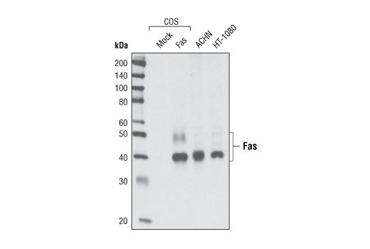
| Fas (C18C12) Rabbit mAb | 4233 | 20 µl | 40-50 kDa | Rabbit IgG |
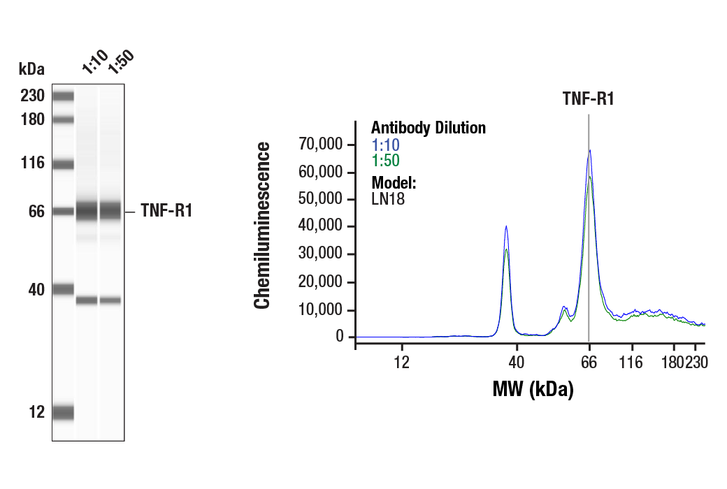
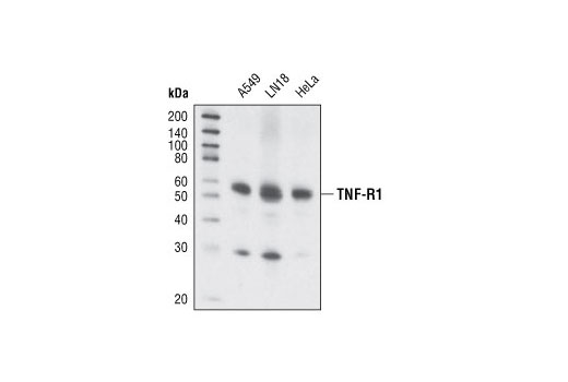
| TNF-R1 (C25C1) Rabbit mAb | 3736 | 20 µl | 55 kDa | Rabbit IgG |
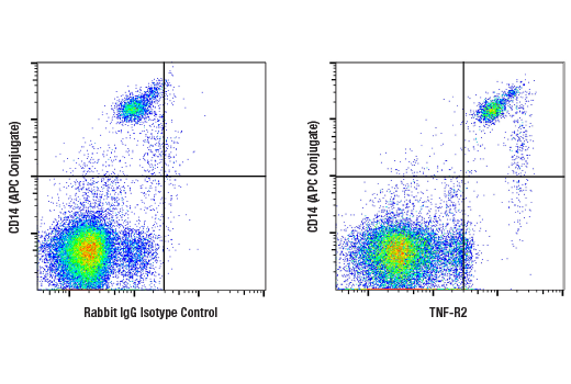
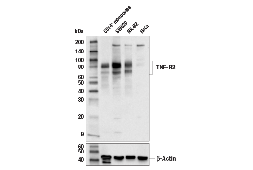
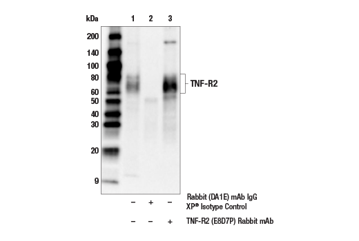
| TNF-R2 (E8D7P) Rabbit mAb | 72337 | 20 µl | 60-80 kDa | Rabbit IgG |
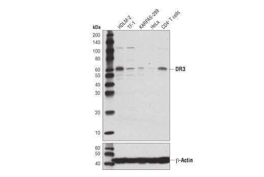
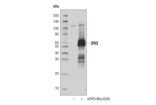
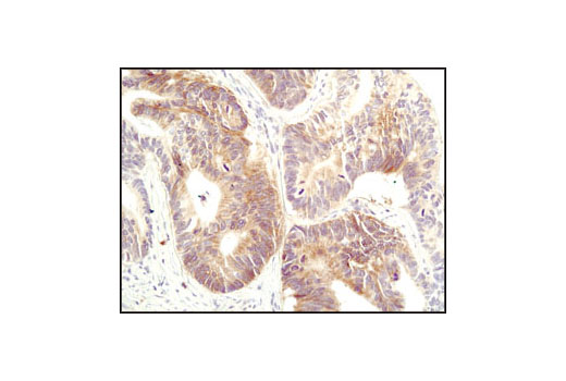
| DR3 (D4O3X) Rabbit mAb | 20772 | 20 µl | 55-60 kDa | Rabbit IgG |
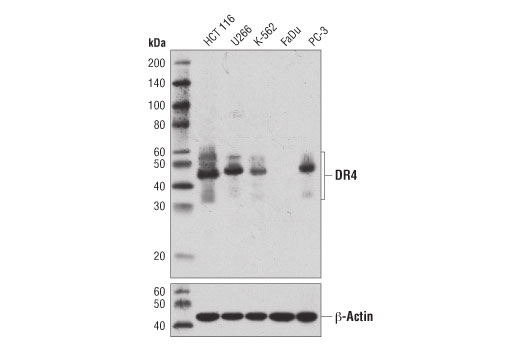
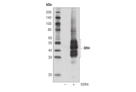
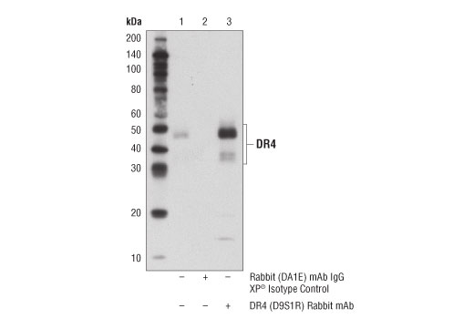
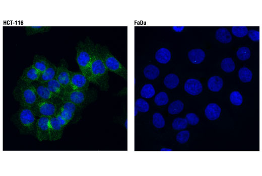
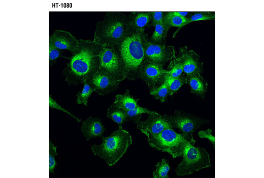
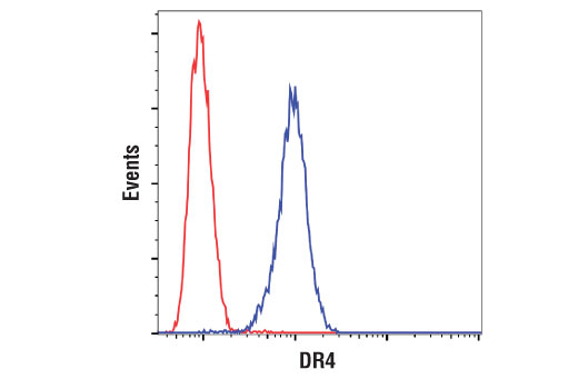
| DR4 (D9S1R) Rabbit mAb | 42533 | 20 µl | 35-55 kDa | Rabbit IgG |
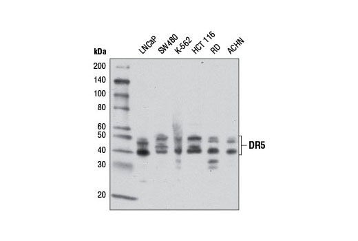
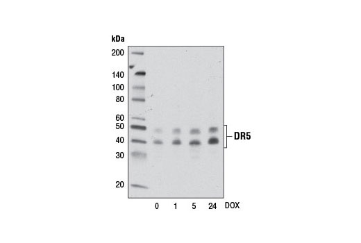
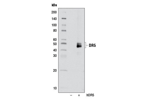
| DR5 (D4E9) XP® Rabbit mAb | 8074 | 20 µl | 40, 48 kDa | Rabbit IgG |
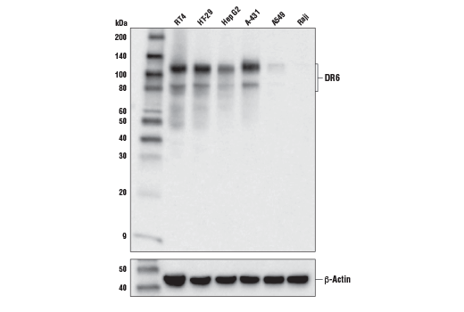
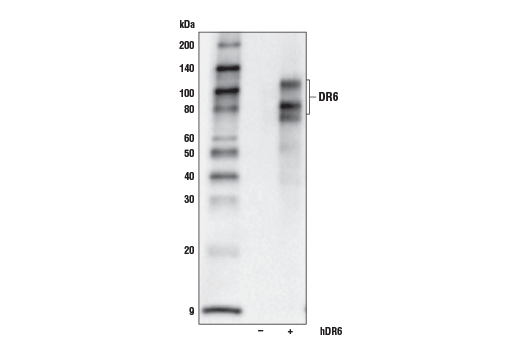
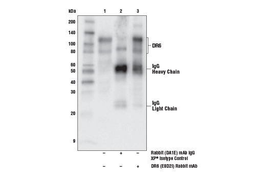
| DR6 (E8D2I) Rabbit mAb | 93026 | 20 µl | 80, 120 kDa | Rabbit IgG |
| Anti-rabbit IgG, HRP-linked Antibody | 7074 | 100 µl | Goat |
Please visit cellsignal.com for individual component applications, species cross-reactivity, dilutions, protocols, and additional product information.
Description
The Death Receptor Antibody Sampler Kit II provides an economical means to investigate members of the death receptor family. The kit includes enough antibody to perform two western blot experiments with each primary antibody.
Storage
Background
The tumor necrosis factor receptor family, which includes TNF-RI, TNF-R2, Fas, DR3, DR4, DR5, and DR6, plays an important role in the regulation of apoptosis in various physiological systems (1,2). The receptors are activated by a family of cytokines that include TNF, FasL, TWEAK, and TRAIL. They are characterized by a highly conserved extracellular region containing cysteine-rich repeats and a conserved intracellular region of about 80 amino acids termed the death domain (DD). The DD is important for transducing the death signal by recruiting other DD containing adaptor proteins (FADD, TRADD, RIP) to the death-inducing signaling complex (DISC) resulting in activation of caspases. The two receptors for TNF-α, TNF-R1 (55 kDa) and TNF-R2 (75 kDa) can mediate distinct cellular responses (3,4). In most cases cytotoxicity elicited by TNF has been reported to act through TNF-R1 (5,6). DR3/WSL-1/Apo-3/TRAMP/LARD is a TNFR family member containing the characteristic extracellular cysteine-repeats, transmembrane region, and an intracellular DD (7-11). DR3 is activated by its ligand Apo-3L/TWEAK to induce apoptosis and activation of NF-κB (12,13). Like TNF-R1, DR3 binds to the DD adaptor protein TRADD, which can then associate with other DD proteins like FADD and RIP as well as members of the TRAF family (7,8). Tissue expression of DR3 is very restricted, primarily seen on the surface of activated thymocytes and lymphocytes and plays an important role in thymocyte negative selection (7,8,14). Studies have also indicated an association with DR3 and rheumatoid arthritis (15,16). DR4 (TRAIL-RI, TNFRSF10A) and DR5 (TRAIL-R2, TNFRSF10B) are receptors for the cytokine TRAIL. Both receptors contain death domains that recruit DISC complexes triggering caspase activation and apoptosis (17-20). DR6, also known as TNFRSF21, is a TNFR family member able to induce apoptosis as well as activation of NF-κB and JNK (21). DR6 appears to play a critical role in the activation and differentiation of T and B lymphocytes (22,23). In the nervous system, β-amyloid precursor protein (APP) activates DR6 to trigger neuronal degeneration (24).
- Nagata, S. (1997) Cell 88, 355-65.
- Thorburn, A. (2004) Cell Signal 16, 139-44.
- Tartaglia, L.A. et al. (1991) Proc Natl Acad Sci U S A 88, 9292-6.
- Peschon, J.J. et al. (1998) J Immunol 160, 943-52.
- Tartaglia, L.A. et al. (1993) Cell 73, 213-6.
- Rothe, J. et al. (1993) Nature 364, 798-802.
- Chinnaiyan, A.M. et al. (1996) Science 274, 990-2.
- Kitson, J. et al. (1996) Nature 384, 372-5.
- Marsters, S.A. et al. (1996) Curr Biol 6, 1669-76.
- Bodmer, J.L. et al. (1997) Immunity 6, 79-88.
- Screaton, G.R. et al. (1997) Proc Natl Acad Sci U S A 94, 4615-9.
- Marsters, S.A. et al. (1998) Curr Biol 8, 525-8.
- Kaptein, A. et al. (2000) FEBS Lett 485, 135-41.
- Wang, E.C. et al. (2001) Mol Cell Biol 21, 3451-61.
- Osawa, K. et al. (2004) Genes Immun 5, 439-43.
- Borysenko, C.W. et al. (2005) Biochem Biophys Res Commun 328, 794-9.
- Pan, G. et al. (1997) Science 276, 111-3.
- Walczak, H. et al. (1997) EMBO J 16, 5386-97.
- Chaudhary, P.M. et al. (1997) Immunity 7, 821-30.
- Schneider, P. et al. (1997) Immunity 7, 831-6.
- Pan, G. et al. (1998) FEBS Lett 431, 351-6.
- Zhao, H. et al. (2001) J Exp Med 194, 1441-8.
- Schmidt, C.S. et al. (2003) J Exp Med 197, 51-62.
- Nikolaev, A. et al. (2009) Nature 457, 981-9.
Background References
Trademarks and Patents
使用に関する制限
法的な権限を与えられたCSTの担当者が署名した書面によって別途明示的に合意された場合を除き、 CST、その関連会社または代理店が提供する製品には以下の条件が適用されます。お客様が定める条件でここに定められた条件に含まれるものを超えるもの、 または、ここに定められた条件と異なるものは、法的な権限を与えられたCSTの担当者が別途書面にて受諾した場合を除き、拒絶され、 いかなる効力も効果も有しません。
研究専用 (For Research Use Only) またはこれに類似する表示がされた製品は、 いかなる目的についても FDA または外国もしくは国内のその他の規制機関により承認、認可または許可を受けていません。 お客様は製品を診断もしくは治療目的で使用してはならず、また、製品に表示された内容に違反する方法で使用してはなりません。 CST が販売または使用許諾する製品は、エンドユーザーであるお客様に対し、使途を研究および開発のみに限定して提供されるものです。 診断、予防もしくは治療目的で製品を使用することまたは製品を再販売 (単独であるか他の製品等の一部であるかを問いません) もしくはその他の商業的利用の目的で購入することについては、CST から別途許諾を得る必要があります。 お客様は以下の事項を遵守しなければなりません。(a) CST の製品 (単独であるか他の資材と一緒であるかを問いません) を販売、使用許諾、貸与、寄付もしくはその他の態様で第三者に譲渡したり使用させたりしてはなりません。また、商用の製品を製造するために CST の製品を使用してはなりません。(b) 複製、改変、リバースエンジニアリング、逆コンパイル、 分解または他の方法により製品の構造または技術を解明しようとしてはなりません。また、 CST の製品またはサービスと競合する製品またはサービスを開発する目的で CST の製品を使用してはなりません。(c) CST の製品の商標、商号、ロゴ、特許または著作権に関する通知または表示を除去したり改変したりしてはなりません。(d) CST の製品をCST 製品販売条件(CST’s Product Terms of Sale) および該当する書面のみに従って使用しなければなりません。(e) CST の製品に関連してお客様が使用する第三者の製品またはサービスに関する使用許諾条件、 サービス提供条件またはこれに類する合意事項を遵守しなければなりません。