| Product Includes | Product # | Quantity | Mol. Wt | Isotype/Source |
|---|---|---|---|---|
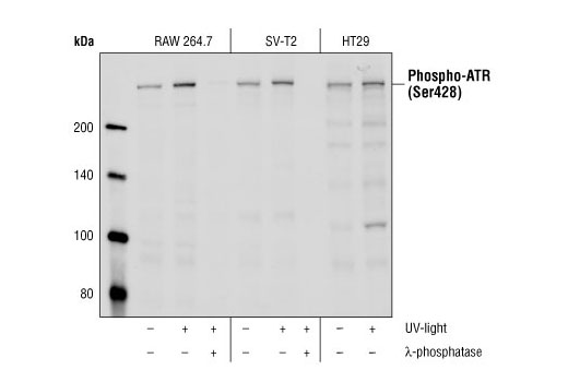
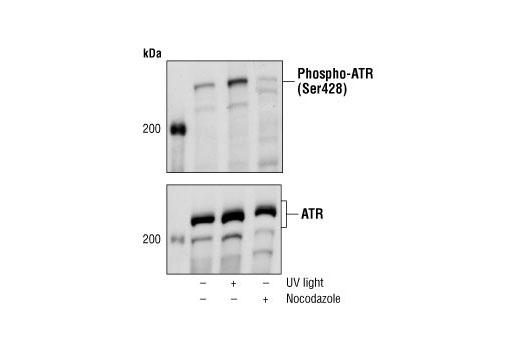
| Phospho-ATR (Ser428) Antibody | 2853 | 20 µl | 300 kDa | Rabbit |
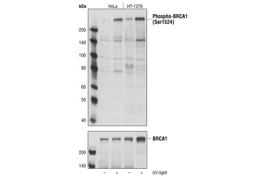
| Phospho-BRCA1 (Ser1524) Antibody | 9009 | 20 µl | 220 kDa | Rabbit |
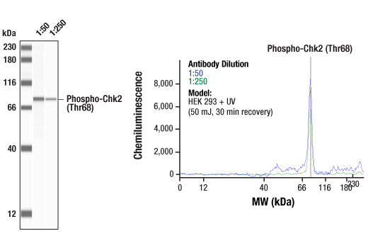
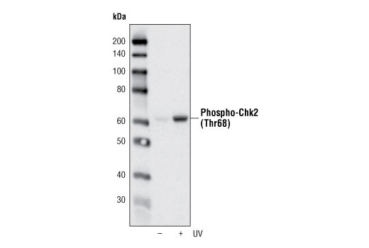
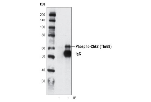
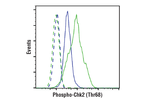
| Phospho-Chk2 (Thr68) (C13C1) Rabbit mAb | 2197 | 20 µl | 62 kDa | Rabbit IgG |
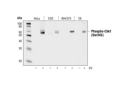
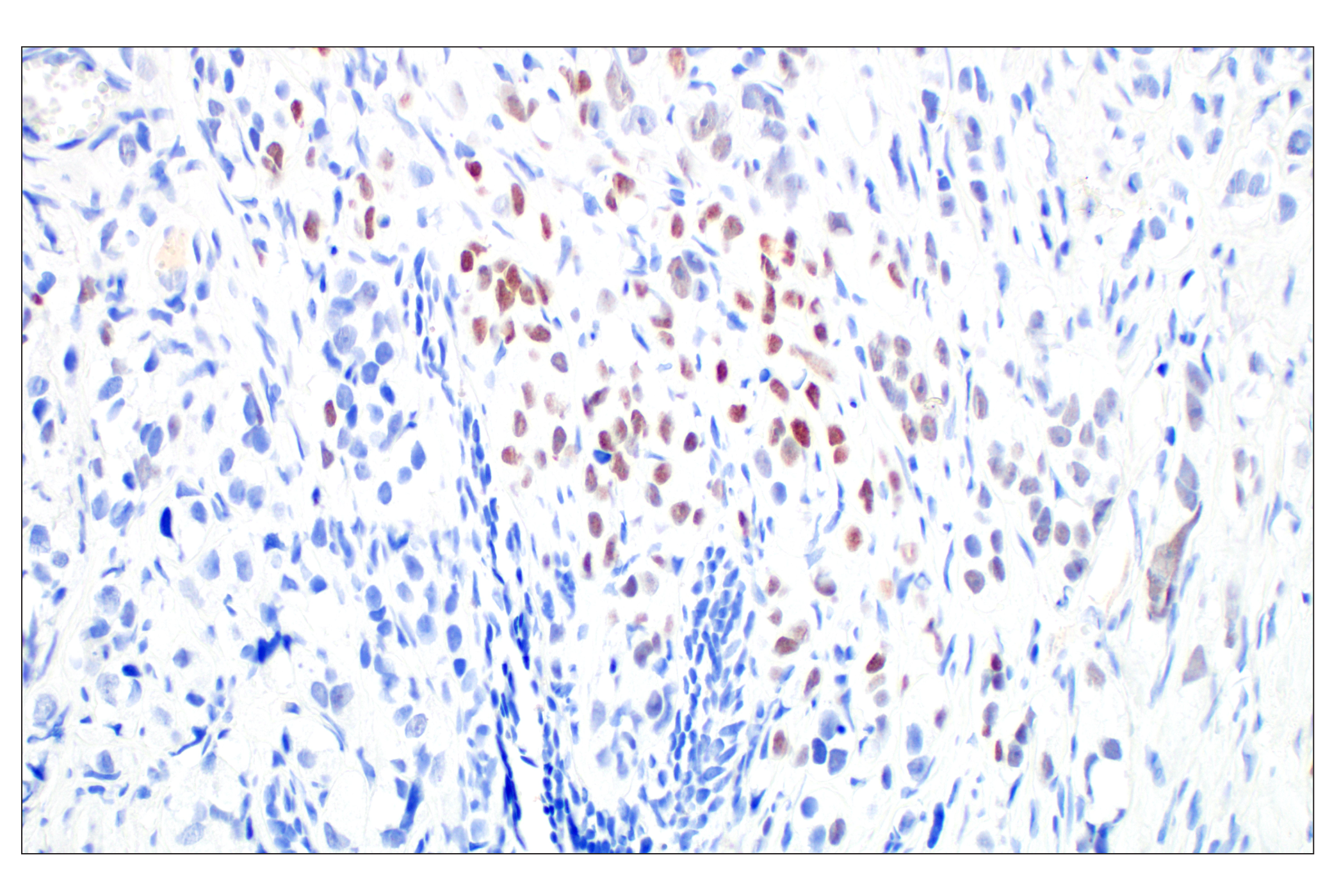
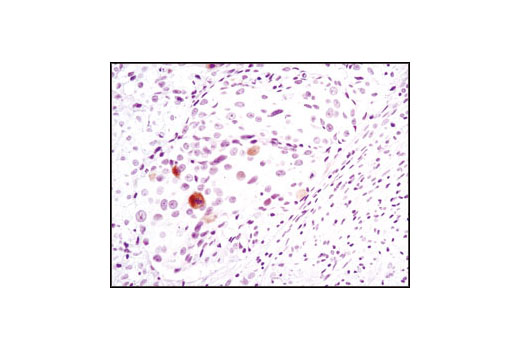
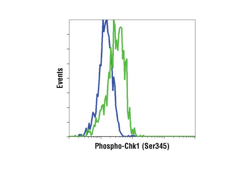
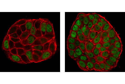
| Phospho-Chk1 (Ser345) (133D3) Rabbit mAb | 2348 | 20 µl | 56 kDa | Rabbit IgG |
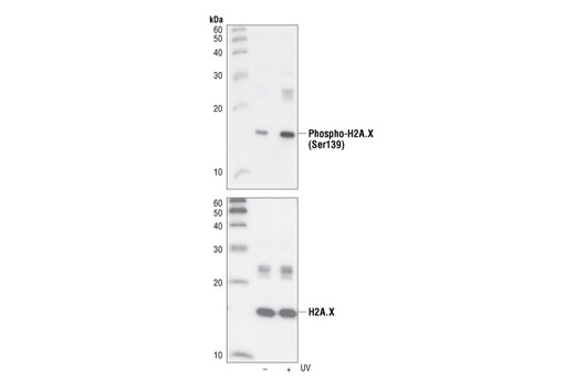
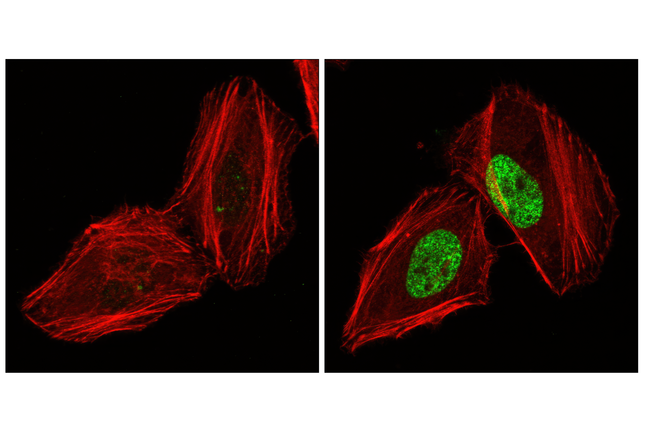
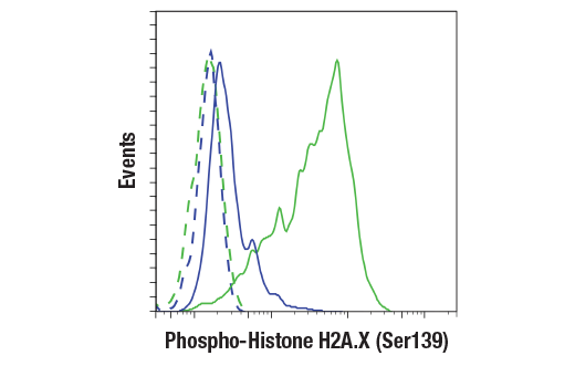
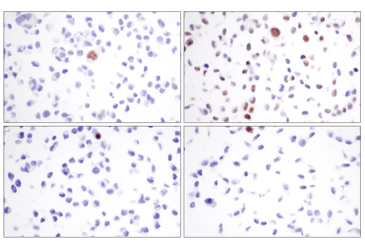
| Phospho-Histone H2A.X (Ser139) (20E3) Rabbit mAb | 9718 | 20 µl | 15 kDa | Rabbit IgG |
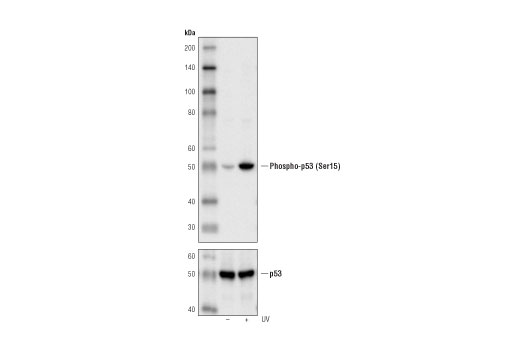
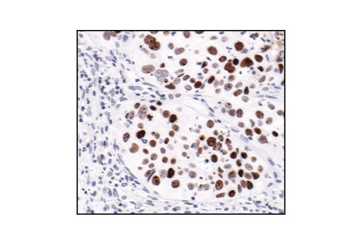
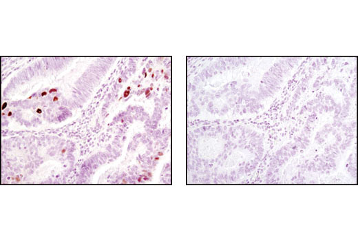
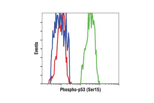
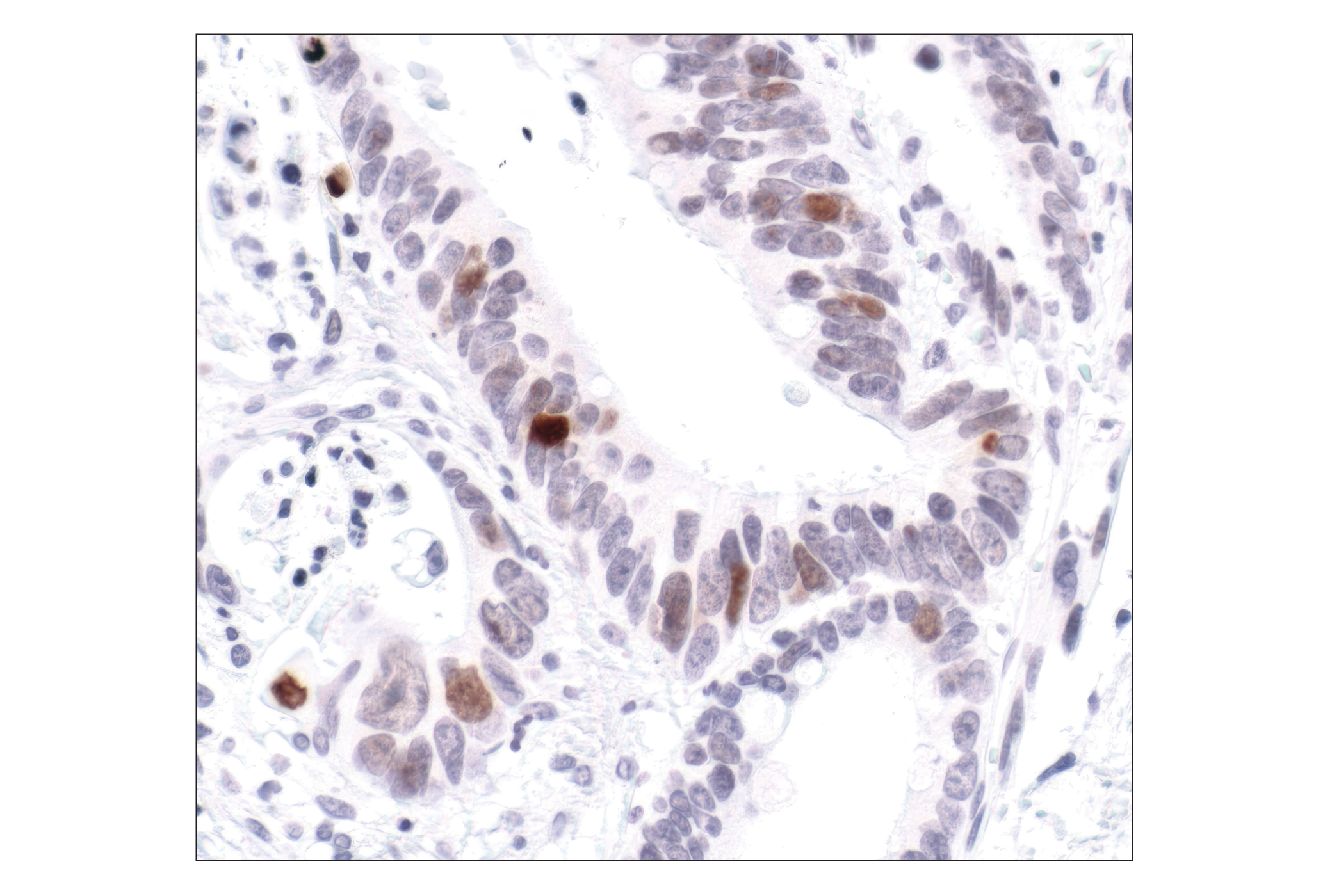
| Phospho-p53 (Ser15) (16G8) Mouse mAb | 9286 | 20 µl | 53 kDa | Mouse IgG1 |
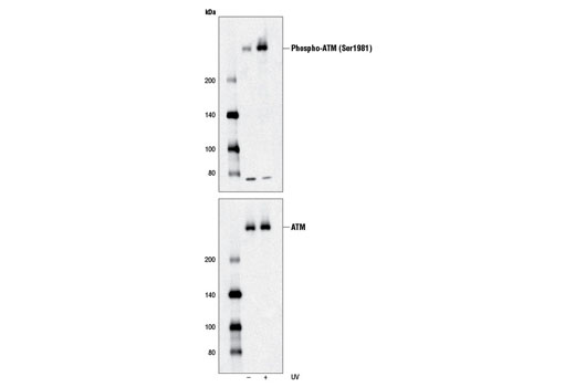
| Phospho-ATM (Ser1981) (D6H9) Rabbit mAb | 5883 | 20 µl | 350 kDa | Rabbit IgG |
| Anti-rabbit IgG, HRP-linked Antibody | 7074 | 100 µl | Goat | |
| Anti-mouse IgG, HRP-linked Antibody | 7076 | 100 µl | Horse |
Please visit cellsignal.com for individual component applications, species cross-reactivity, dilutions, protocols, and additional product information.
Description
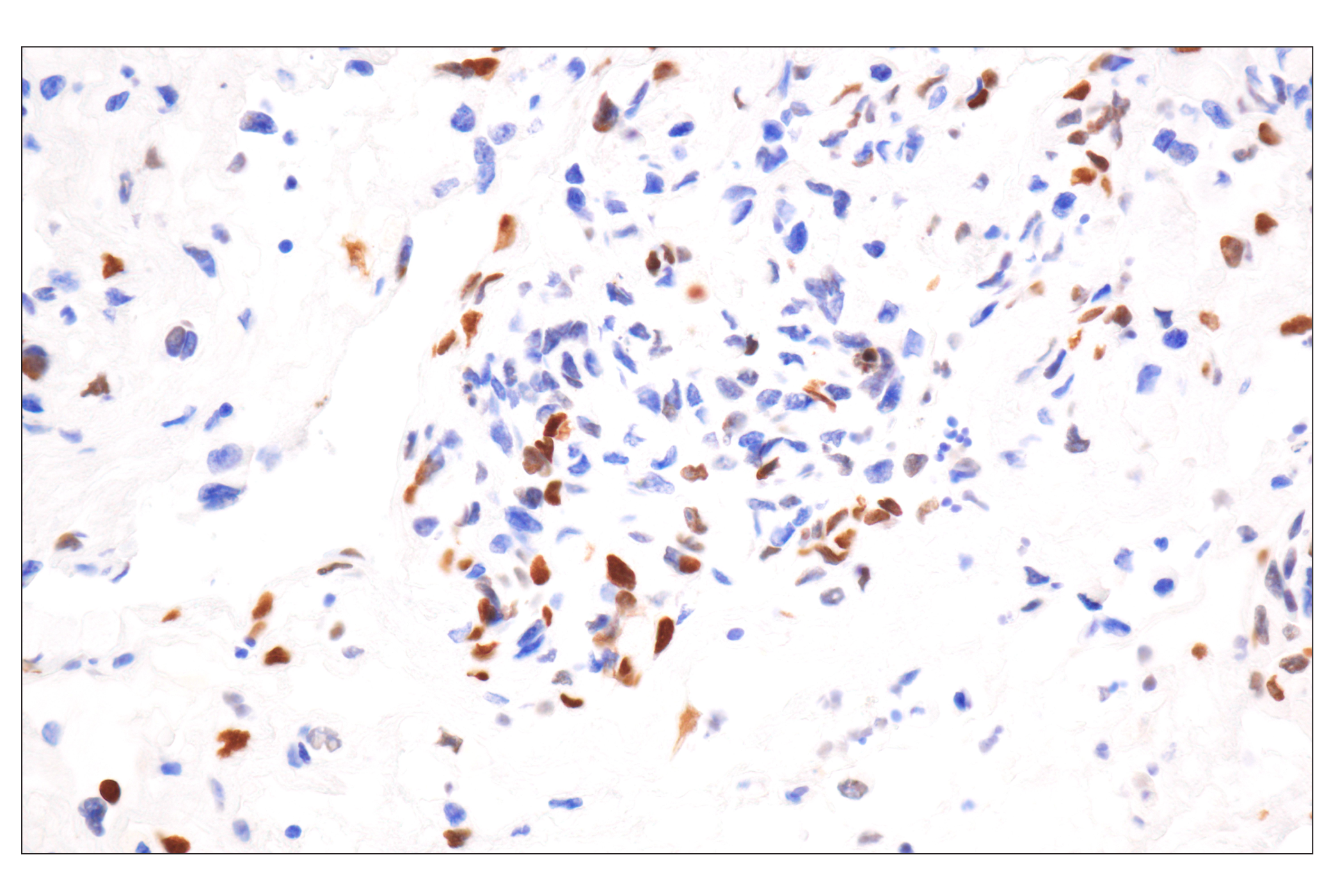
This kit provides an economical means to analyze major signaling checkpoints in response to DNA damage. The kit contains primary and secondary antibodies to perform two Western blots with each antibody.
Storage
Background
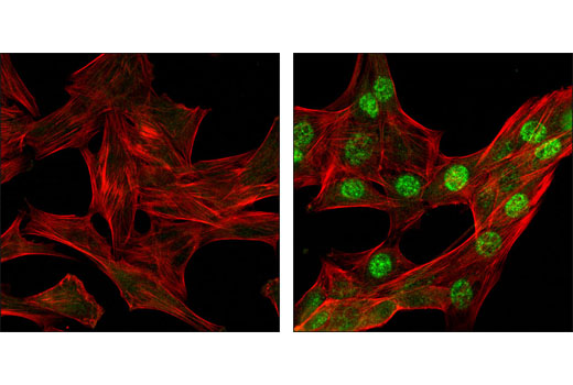
Ataxia telangiectasia mutated kinase (ATM) and ataxia telangiectasia and Rad3-related kinase (ATR) are PI3 Kinase-related kinase (PIKK) family members that phosphorylate multiple substrates on serine or threonine residues that are followed by a glutamine in response to DNA damage or replication blocks (1-3). p53 is phosphorylated by ATM, ATR and DNA-PK at Ser15. This phosphorylation impairs the ability of MDM2 to bind p53, promoting both the accumulation and activation of p53 in response to DNA damage (4,5). Chk1 and Chk2, downstream protein kinases of ATM/ATR, plays an important role in DNA damage checkpoint control, embryonic development and tumor suppression (6). Chk1 is phosphorylated at Ser280 and Ser296 following DNA damage. The amino-terminal domain of Chk2 contains a series of seven serine or threonine residues, including Thr68, each followed by glutamine (SQ or TQ motif). After DNA damage by ionizing radiation (IR), UV irradiation or hydroxyurea treatment, Thr68 and other sites in this region become phosphorylated by ATM/ATR (7-9). The breast cancer susceptibility proteins BRCA1 and BRCA2 are frequently mutated in cases of hereditary breast and ovarian cancers and have roles in multiple processes related to DNA damage, repair, cell cycle progression, transcription, ubiquitination and apoptosis. Numerous DNA-damage induced phosphorylation sites on BRCA1 have been identified, including serine 1524, and kinases activated in a cell cycle-dependent manner, including Aurora A and CDK2, can also phosphorylate BRCA1. IR, DNA and radiometric-induced DNA damage also results in rapid phosphorylation of the histone H2A family member H2A.X at Ser139 by ATM (10,11). Within minutes following DNA damage, Ser139-phosphorylated H2A.X localizes to sites of DNA damage at subnuclear foci (12).
- Kastan, M.B. and Lim, D.S. (2000) Nat. Rev. Mol. Cell Biol. 1, 179-186.
- Abraham, R.T. DNA Repair (Amst) 3, 883-887.
- Shechter, D. et al. DNA Repair (Amst) 3, 901-908.
- Shieh, S.Y. et al. (1997) Cell 91, 325-334.
- Tibbetts, R.S. et al. (1999) Genes Dev. 13, 152-157.
- Martinho, R.G. et al. (1998) EMBO J. 17, 7239-17249.
- Matsuoka, S. et al. (2000) Proc. Natl. Acad. Sci. USA 97, 10389-10394.
- Melchionna, R. et al. (2000) Nat. Cell Biol. 2, 762-765.
- Ahn, J.Y. et al. (2000) Cancer Res. 60, 5934-5936.
- Rogakou, E.P. et al. (1998) J. Biol. Chem. 273, 5858-5868.
- Burma, S. et al. (2001) J. Biol. Chem. 276, 42462-42467.
- Rogakou, E.P. et al. (1999) J. Cell Biol. 146, 905-916.
Background References
Trademarks and Patents
使用に関する制限
法的な権限を与えられたCSTの担当者が署名した書面によって別途明示的に合意された場合を除き、 CST、その関連会社または代理店が提供する製品には以下の条件が適用されます。お客様が定める条件でここに定められた条件に含まれるものを超えるもの、 または、ここに定められた条件と異なるものは、法的な権限を与えられたCSTの担当者が別途書面にて受諾した場合を除き、拒絶され、 いかなる効力も効果も有しません。
研究専用 (For Research Use Only) またはこれに類似する表示がされた製品は、 いかなる目的についても FDA または外国もしくは国内のその他の規制機関により承認、認可または許可を受けていません。 お客様は製品を診断もしくは治療目的で使用してはならず、また、製品に表示された内容に違反する方法で使用してはなりません。 CST が販売または使用許諾する製品は、エンドユーザーであるお客様に対し、使途を研究および開発のみに限定して提供されるものです。 診断、予防もしくは治療目的で製品を使用することまたは製品を再販売 (単独であるか他の製品等の一部であるかを問いません) もしくはその他の商業的利用の目的で購入することについては、CST から別途許諾を得る必要があります。 お客様は以下の事項を遵守しなければなりません。(a) CST の製品 (単独であるか他の資材と一緒であるかを問いません) を販売、使用許諾、貸与、寄付もしくはその他の態様で第三者に譲渡したり使用させたりしてはなりません。また、商用の製品を製造するために CST の製品を使用してはなりません。(b) 複製、改変、リバースエンジニアリング、逆コンパイル、 分解または他の方法により製品の構造または技術を解明しようとしてはなりません。また、 CST の製品またはサービスと競合する製品またはサービスを開発する目的で CST の製品を使用してはなりません。(c) CST の製品の商標、商号、ロゴ、特許または著作権に関する通知または表示を除去したり改変したりしてはなりません。(d) CST の製品をCST 製品販売条件(CST’s Product Terms of Sale) および該当する書面のみに従って使用しなければなりません。(e) CST の製品に関連してお客様が使用する第三者の製品またはサービスに関する使用許諾条件、 サービス提供条件またはこれに類する合意事項を遵守しなければなりません。