| Product Includes | Product # | Quantity | Mol. Wt | Isotype/Source |
|---|---|---|---|---|
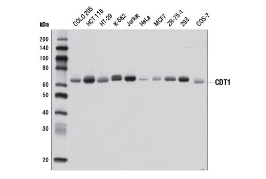
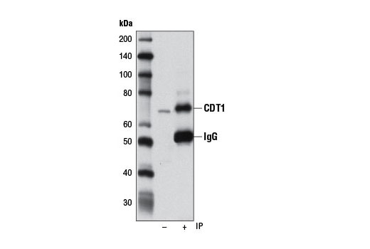
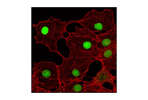
| CDT1 (D10F11) Rabbit mAb | 8064 | 40 µl | 65 kDa | Rabbit IgG |
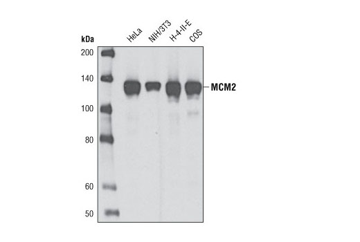
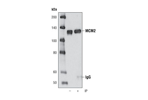
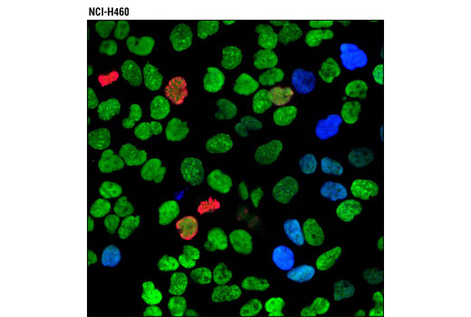
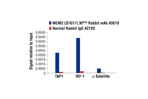
| MCM2 (D7G11) XP® Rabbit mAb | 3619 | 40 µl | 125 kDa | Rabbit IgG |
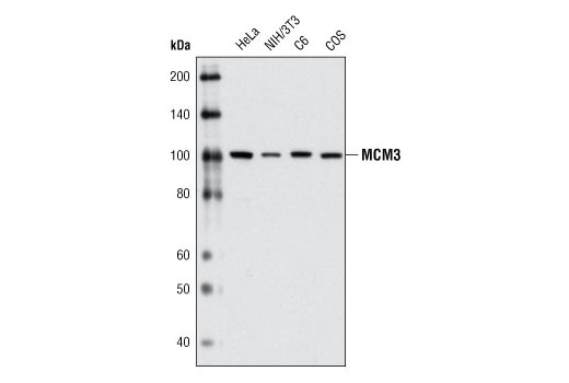
| MCM3 (D47B6) Rabbit mAb | 4003 | 40 µl | 100 kDa | Rabbit IgG |
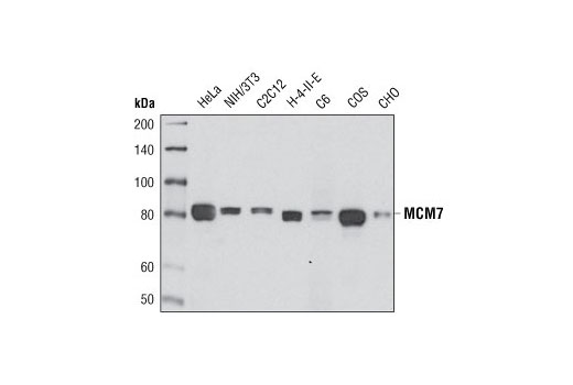
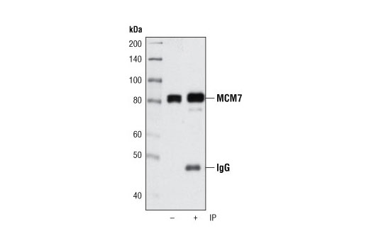
| MCM7 (D10A11) XP® Rabbit mAb | 3735 | 40 µl | 80 kDa | Rabbit IgG |
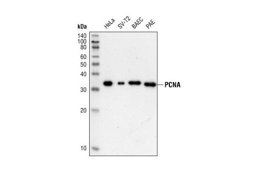
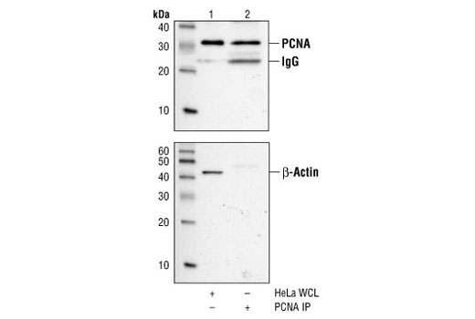
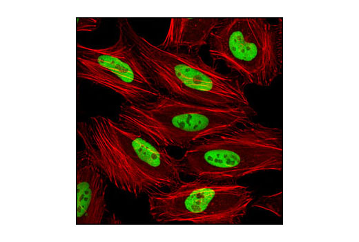
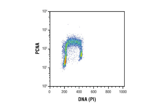
| PCNA (PC10) Mouse mAb | 2586 | 40 µl | 36 kDa | Mouse IgG2a |
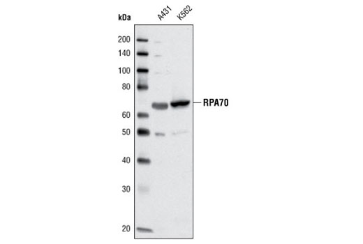
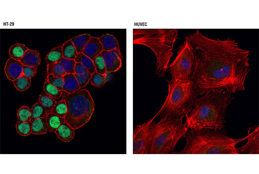
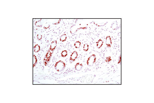
| RPA70/RPA1 (C24F2) Rabbit mAb | 2193 | 40 µl | 70 kDa | Rabbit IgG |
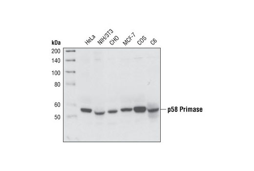
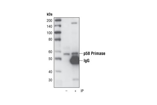
| p58 Primase (8D3) Rat mAb | 4726 | 40 µl | 58 kDa | Rat IgG2a |
| Anti-rabbit IgG, HRP-linked Antibody | 7074 | 100 µl | Goat | |
| Anti-mouse IgG, HRP-linked Antibody | 7076 | 100 µl | Horse | |
| Anti-rat IgG, HRP-linked Antibody | 7077 | 100 µl | Goat |
Please visit cellsignal.com for individual component applications, species cross-reactivity, dilutions, protocols, and additional product information.
Description
The DNA Replication Antibody Sampler Kit provides a fast and economical means of evaluating multiple targets regulating DNA replication. The kit contains enough primary antibodies to perform four western blots with each antibody.
Storage
Background
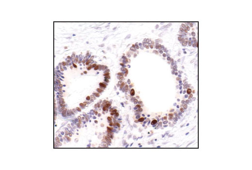
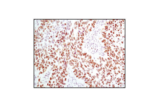
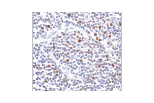
The initiation of DNA replication in mammalian cells is a highly coordinated process that is regulated by several protein complexes. Origins of replication (ORCs), at which replication is initiated, are dispersed throughout the genome. Their activities are regulated via the sequential binding of pre-replication and replication factors that initiate formation of replication forks, the active structures at which DNA is synthesized. The origin recognition complex is thought to be bound to chromatin throughout the cell cycle (1,2). The pre-replication complex (Pre-RC) forms in late mitosis/early G1 phase beginning with the binding of CDT1 and CDC6 to the origin. Together CDT1 and CDC6 promote the loading of the heterohexameric minichromosome maintenance (MCM) complex. This process is referred to as chromatin licensing. Licensing of the chromatin permits the DNA to replicate only once per cell cycle, helping to ensure that genetic alterations and malignant cell growth do not occur (reviewed in 3). The canonical MCM complex proteins (MCM2-7) are a family of six related phospho-proteins that function, in part, as the eukaryotic replicative DNA helicase (3,4). Phosphorylation and ubiquitination of the MCM2, MCM3, MCM4, and MCM6 subunits appears to regulate MCM complex activity and the initiation of DNA synthesis (5-7). MCM proteins are removed during DNA replication, causing chromatin to become unlicensed, inhibiting Pre-RC reformation. In addition to DNA polymerase, initiation of DNA replication requires a pair of primase subunits. DNA Primase activity catalyzes de novo synthesis of an RNA/DNA primer (initiator DNA) on the leading and lagging strands, while polymerase activity extends the initiator DNA (8). The 48 and 58 kDa primase subunits cooperate in the synthesis of small RNA primers. p48 is the catalytically active subunit (9), while p58 couples p48 to the polymerase to allow the transfer of primers to the active site. The p58 subunit may also play a role in regulation of primer length (10,11). Once replication is initiated, Proliferating Cell Nuclear Antigen (PCNA) serves as an accessory factor for DNA polymerases delta and epsilon, acting to tether these polymerases to template DNA during replication. Interactions of PCNA with DNA polymerases increase the processivity of leading strand synthesis. PCNA, a member of DNA sliding clamp family, is a homotrimeric ring complex that encircles and slides along the DNA double helix as the replication fork progresses (12). Multiple proteins involved in DNA replication, DNA repair, and cell cycle control bind to PCNA and regulate DNA synthesis. PCNA is loaded onto the DNA in an ATP-dependent manner by a multiprotein clamp loader, Replication Factor C (RFC) (13). RFC, in turn, associates with DNA via interactions with the single-stranded DNA binding protein complex, Replication Protein A (RPA). The canonical RPA complex is heterotrimeric and composed of RPA1 (RPA70), RPA2 (RPA32), and RPA3 (RPA14) subunits. RPA recognizes and stabilizes single stranded DNA in repair processes and DNA recombination, and plays a role in replication (14-17).
- Okuno, Y. et al. (2001) EMBO J 20, 4263-77.
- McNairn, A.J. et al. (2005) Exp Cell Res 308, 345-56.
- Forsburg, S.L. (2004) Microbiol Mol Biol Rev 68, 109-31.
- Johnson, A. and O'Donnell, M. (2005) Annu Rev Biochem 74, 283-315.
- Charych, D.H. et al. (2008) J Cell Biochem 104, 1075-86.
- Masai, H. et al. (2006) J Biol Chem 281, 39249-61.
- Lin, D.I. et al. (2008) Proc Natl Acad Sci U S A 105, 8079-84.
- Shiratori, A. et al. (1995) Genomics 28, 350-3.
- Copeland, W.C. (1997) Protein Expr Purif 9, 1-9.
- Copeland, W.C. and Wang, T.S. (1993) J Biol Chem 268, 26179-89.
- Arezi, B. and Kuchta, R.D. (2000) Trends Biochem Sci 25, 572-6.
- Bowman, G.D. et al. (2004) Nature 429, 724-30.
- Zhang, G. et al. (1999) Proc Natl Acad Sci U S A 96, 1869-74.
- Sakaguchi, K. et al. (2009) FEBS J 276, 943-63.
- Zou, Y. et al. (2006) J Cell Physiol 208, 267-73.
- Wold, M.S. (1997) Annu Rev Biochem 66, 61-92.
- Binz, S.K. et al. DNA Repair (Amst) 3, 1015-24.
Background References
Trademarks and Patents
使用に関する制限
法的な権限を与えられたCSTの担当者が署名した書面によって別途明示的に合意された場合を除き、 CST、その関連会社または代理店が提供する製品には以下の条件が適用されます。お客様が定める条件でここに定められた条件に含まれるものを超えるもの、 または、ここに定められた条件と異なるものは、法的な権限を与えられたCSTの担当者が別途書面にて受諾した場合を除き、拒絶され、 いかなる効力も効果も有しません。
研究専用 (For Research Use Only) またはこれに類似する表示がされた製品は、 いかなる目的についても FDA または外国もしくは国内のその他の規制機関により承認、認可または許可を受けていません。 お客様は製品を診断もしくは治療目的で使用してはならず、また、製品に表示された内容に違反する方法で使用してはなりません。 CST が販売または使用許諾する製品は、エンドユーザーであるお客様に対し、使途を研究および開発のみに限定して提供されるものです。 診断、予防もしくは治療目的で製品を使用することまたは製品を再販売 (単独であるか他の製品等の一部であるかを問いません) もしくはその他の商業的利用の目的で購入することについては、CST から別途許諾を得る必要があります。 お客様は以下の事項を遵守しなければなりません。(a) CST の製品 (単独であるか他の資材と一緒であるかを問いません) を販売、使用許諾、貸与、寄付もしくはその他の態様で第三者に譲渡したり使用させたりしてはなりません。また、商用の製品を製造するために CST の製品を使用してはなりません。(b) 複製、改変、リバースエンジニアリング、逆コンパイル、 分解または他の方法により製品の構造または技術を解明しようとしてはなりません。また、 CST の製品またはサービスと競合する製品またはサービスを開発する目的で CST の製品を使用してはなりません。(c) CST の製品の商標、商号、ロゴ、特許または著作権に関する通知または表示を除去したり改変したりしてはなりません。(d) CST の製品をCST 製品販売条件(CST’s Product Terms of Sale) および該当する書面のみに従って使用しなければなりません。(e) CST の製品に関連してお客様が使用する第三者の製品またはサービスに関する使用許諾条件、 サービス提供条件またはこれに類する合意事項を遵守しなければなりません。