WB, IP, IF-IC
H
Endogenous
95
Rabbit IgG
#Q9Y6K1-2
1788
Product Information
Product Usage Information
| Application | Dilution |
|---|---|
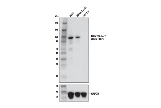
| Western Blotting | 1:1000 |
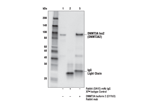
| Immunoprecipitation | 1:50 |
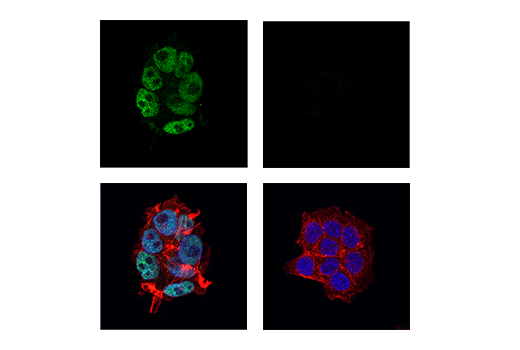
| Immunofluorescence (Immunocytochemistry) | 1:400 - 1:1600 |
Storage
Specificity / Sensitivity
Species Reactivity:
Human
Source / Purification
Monoclonal antibody is produced by immunizing animals with a synthetic peptide corresponding to residues near the amino terminus of human DNMT3A isoform 2 (DNMT3A2) protein.
Background
Methylation of DNA at cytosine residues in mammalian cells is a heritable, epigenetic modification that is critical for proper regulation of gene expression, genomic imprinting and development (1,2). Three families of mammalian DNA methyltransferases have been identified: DNMT1, DNMT2, and DNMT3 (1,2). DNMT1 is constitutively expressed in proliferating cells and functions as a maintenance methyltransferase, transferring proper methylation patterns to newly synthesized DNA during replication. DNMT3A and DNMT3B are strongly expressed in embryonic stem cells with reduced expression in adult somatic tissues. DNMT3A and DNMT3B function as de novo methyltransferases that methylate previously unmethylated regions of DNA. DNMT2 is expressed at low levels in adult somatic tissues and its inactivation affects neither de novo nor maintenance DNA methylation. DNMT1, DNMT3A, and DNMT3B together form a protein complex that interacts with histone deacetylases (HDAC1, HDAC2, Sin3A), transcriptional repressor proteins (RB, TAZ-1), and heterochromatin proteins (HP1, SUV39H1) to maintain proper levels of DNA methylation and facilitate gene silencing (3-8). Improper DNA methylation contributes to diseased states such as cancer (1,2). Hypermethylation of promoter CpG islands within tumor suppressor genes correlates with gene silencing and the development of cancer. In addition, hypomethylation of bulk genomic DNA correlates with and may contribute to the onset of cancer. DNMT1, DNMT3A, and DNMT3B are overexpressed in many cancers, including acute and chronic myelogenous leukemias, in addition to colon, breast, and stomach carcinomas (9-12).~There are at least two protein isoforms expressed from the DNMT3A locus, DNMT3A isoform 1 (DNMT3A1) and DNMT3A isoform 2 (DNMT3A2). DNMT3A2 is expressed from an intronic promoter that is downstream from the DNMT3A1 promoter (13,14). As a result, the N-terminal 223 amino acids found in DNMT3A1 are replaced by a different 24 amino acid found in DNMT3A2. Although they have distinct N-termini, both isoforms contain the PWWP domain required for binding to tri-methylated histone H3 lysine 36 (H3K36me3) and the ADD domain required for histone binding and transcriptional regulation. DNMT3A1 is lowly expressed in most cell and tissue types and is localized to heterochromatic regions. DNMT3A2 expression appears to be developmentally regulated and limited to embryonic stem cells, where it is localized to euchromatic regions of the genome, suggesting distinct functions for DNMT3A1 and DNMT3A2. DNMT3A2 is the predominant isoform responsible for de novo DNA methylation in embryonic stem cells. In addition, DNMT3A2 is mutated and/or highly expressed in a number of different cancers, including acute myeloid leukemia (AML), teratocarcinoma, neuroblastoma, and lung, testicular, gastric, and breast cancer (13-17).
- Hermann, A. et al. (2004) Cell. Mol. Life Sci. 61, 2571-87.
- Turek-Plewa, J. and Jagodziński, P.P. (2005) Cell. Mol. Biol. Lett. 10, 631-47.
- Kim, G.D. et al. (2002) EMBO J. 21, 4183-95.
- Fuks, F. et al. (2001) EMBO J. 20, 2536-44.
- Geiman, T.M. et al. (2004) Biochem. Biophys. Res. Commun. 318, 544-55.
- Rountree, M.R. et al. (2000) Nat. Genet. 25, 269-77.
- Pradhan, S. and Kim, G.D. (2002) EMBO J. 21, 779-88.
- Fuks, F. et al. (2003) Nucleic Acids Res. 31, 2305-12.
- Mizuno, S. et al. (2001) Blood 97, 1172-9.
- Robertson, K.D. et al. (1999) Nucleic Acids Res. 27, 2291-8.
- Xie, S. et al. (1999) Gene 236, 87-95.
- Kanai, Y. et al. (2001) Int. J. Cancer 91, 205-12.
- Gujar, H. et al. (2019) Genes (Basel) 10, pii: E172. doi: 10.3390/genes10020172.
- Chen, B.F. and Chan, W.Y. (2014) Epigenetics 9, 669-77.
- Chen, B.F. et al. (2014) Epigenetics 9, 119-28.
- Gao, J. et al. (2013) J Exp Clin Cancer Res 32, 86.
- Yu, Z. et al. (2015) Mol Carcinog 54, 707-19.
Species Reactivity
Species reactivity is determined by testing in at least one approved application (e.g., western blot).
Western Blot Buffer
IMPORTANT: For western blots, incubate membrane with diluted primary antibody in 5% w/v BSA, 1X TBS, 0.1% Tween® 20 at 4°C with gentle shaking, overnight.
Applications Key
WB: Western Blotting IP: Immunoprecipitation IF-IC: Immunofluorescence (Immunocytochemistry)
Cross-Reactivity Key
H: human M: mouse R: rat Hm: hamster Mk: monkey Vir: virus Mi: mink C: chicken Dm: D. melanogaster X: Xenopus Z: zebrafish B: bovine Dg: dog Pg: pig Sc: S. cerevisiae Ce: C. elegans Hr: horse GP: Guinea Pig Rab: rabbit All: all species expected
Trademarks and Patents
使用に関する制限
法的な権限を与えられたCSTの担当者が署名した書面によって別途明示的に合意された場合を除き、 CST、その関連会社または代理店が提供する製品には以下の条件が適用されます。お客様が定める条件でここに定められた条件に含まれるものを超えるもの、 または、ここに定められた条件と異なるものは、法的な権限を与えられたCSTの担当者が別途書面にて受諾した場合を除き、拒絶され、 いかなる効力も効果も有しません。
研究専用 (For Research Use Only) またはこれに類似する表示がされた製品は、 いかなる目的についても FDA または外国もしくは国内のその他の規制機関により承認、認可または許可を受けていません。 お客様は製品を診断もしくは治療目的で使用してはならず、また、製品に表示された内容に違反する方法で使用してはなりません。 CST が販売または使用許諾する製品は、エンドユーザーであるお客様に対し、使途を研究および開発のみに限定して提供されるものです。 診断、予防もしくは治療目的で製品を使用することまたは製品を再販売 (単独であるか他の製品等の一部であるかを問いません) もしくはその他の商業的利用の目的で購入することについては、CST から別途許諾を得る必要があります。 お客様は以下の事項を遵守しなければなりません。(a) CST の製品 (単独であるか他の資材と一緒であるかを問いません) を販売、使用許諾、貸与、寄付もしくはその他の態様で第三者に譲渡したり使用させたりしてはなりません。また、商用の製品を製造するために CST の製品を使用してはなりません。(b) 複製、改変、リバースエンジニアリング、逆コンパイル、 分解または他の方法により製品の構造または技術を解明しようとしてはなりません。また、 CST の製品またはサービスと競合する製品またはサービスを開発する目的で CST の製品を使用してはなりません。(c) CST の製品の商標、商号、ロゴ、特許または著作権に関する通知または表示を除去したり改変したりしてはなりません。(d) CST の製品をCST 製品販売条件(CST’s Product Terms of Sale) および該当する書面のみに従って使用しなければなりません。(e) CST の製品に関連してお客様が使用する第三者の製品またはサービスに関する使用許諾条件、 サービス提供条件またはこれに類する合意事項を遵守しなければなりません。