| Product Includes | Product # | Quantity | Mol. Wt | Isotype/Source |
|---|---|---|---|---|
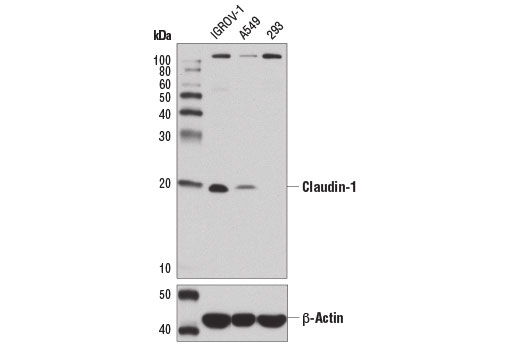
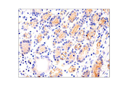
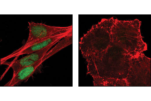
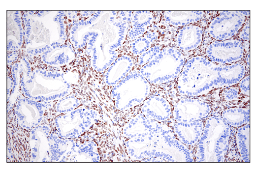
| Claudin-1 (D3H7C) Rabbit mAb | 13995 | 20 µl | 20 kDa | Rabbit IgG |
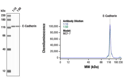
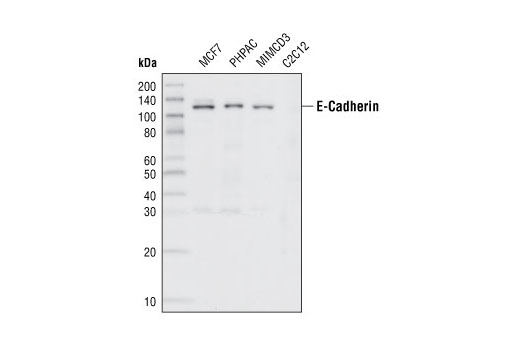
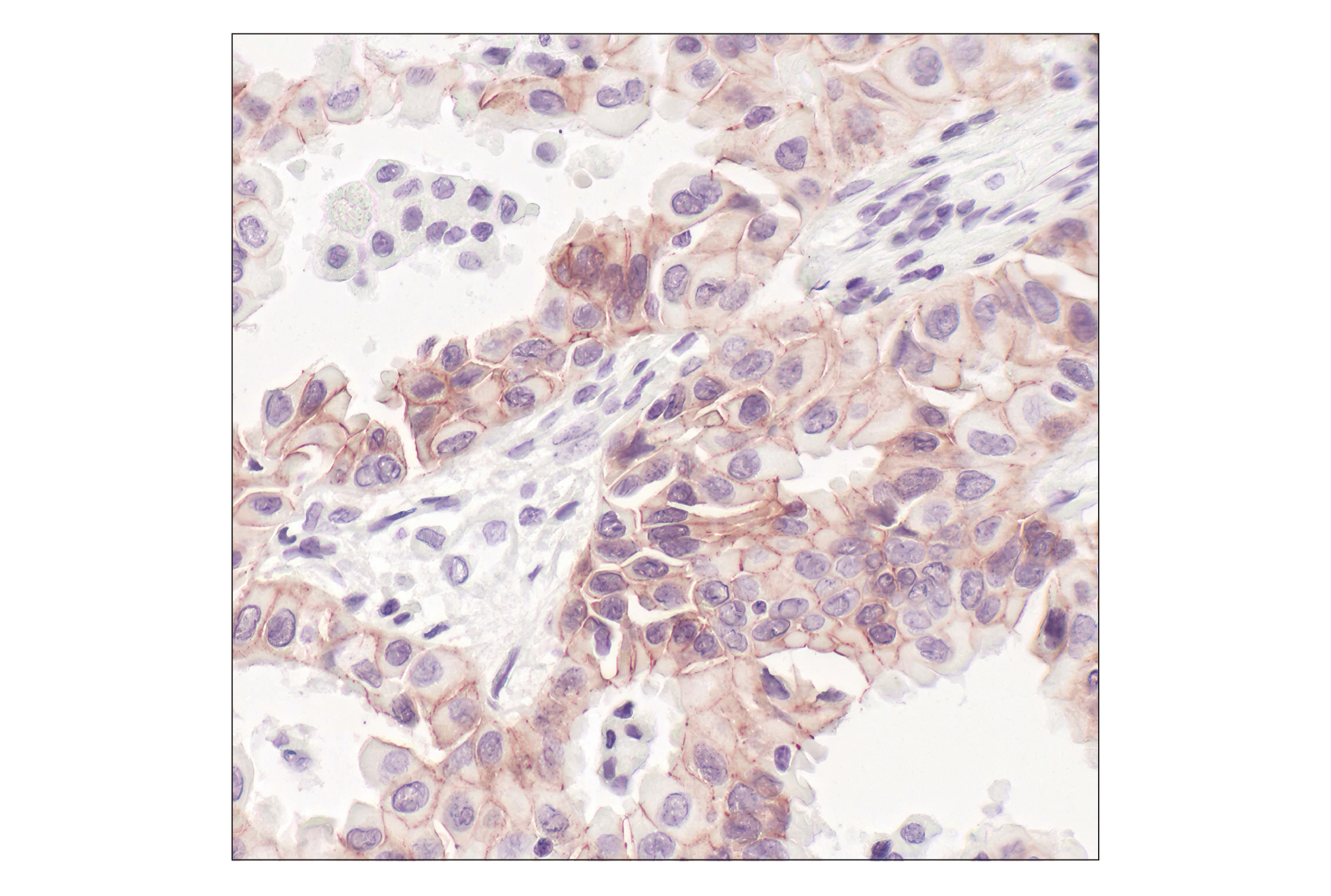
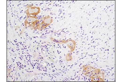
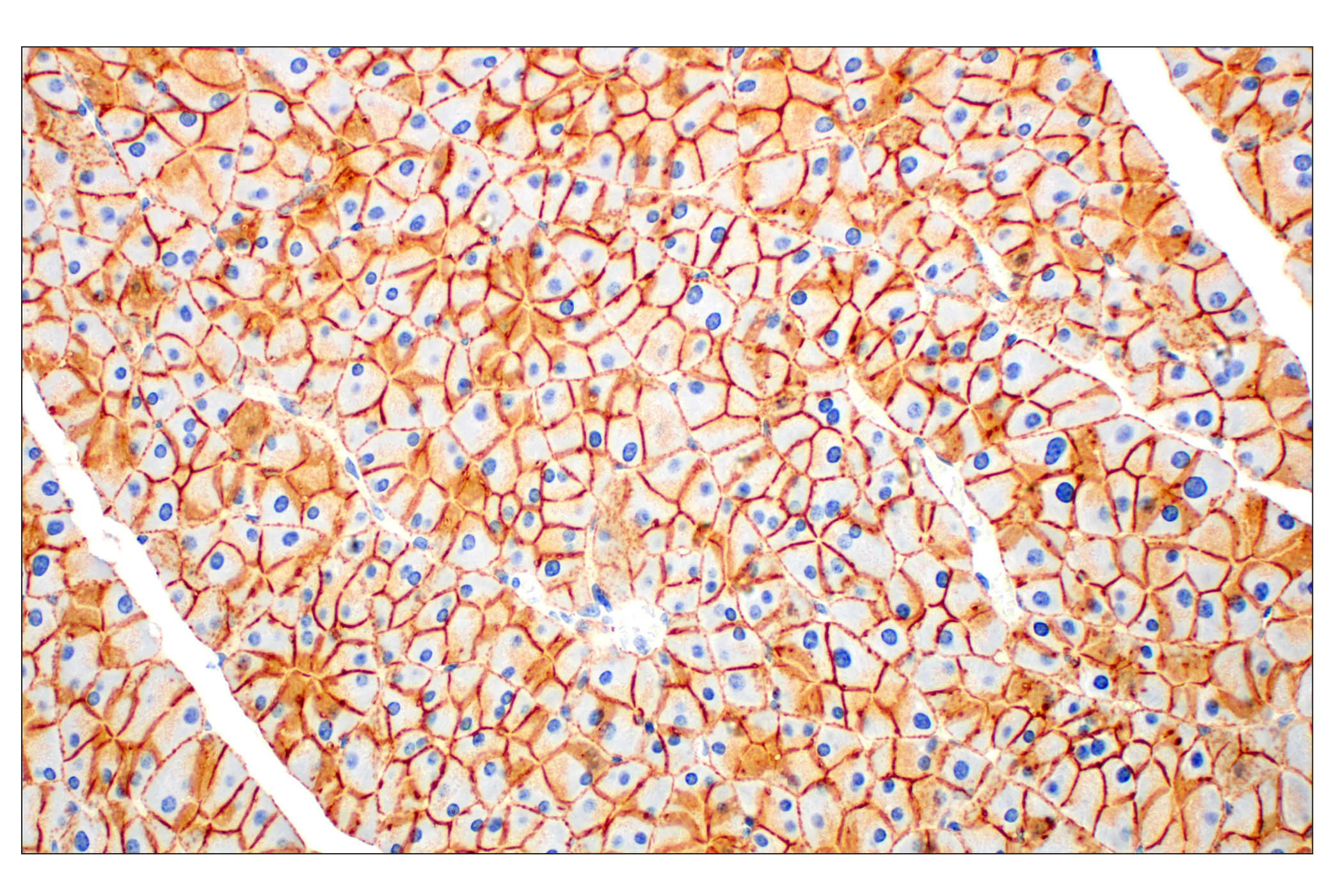
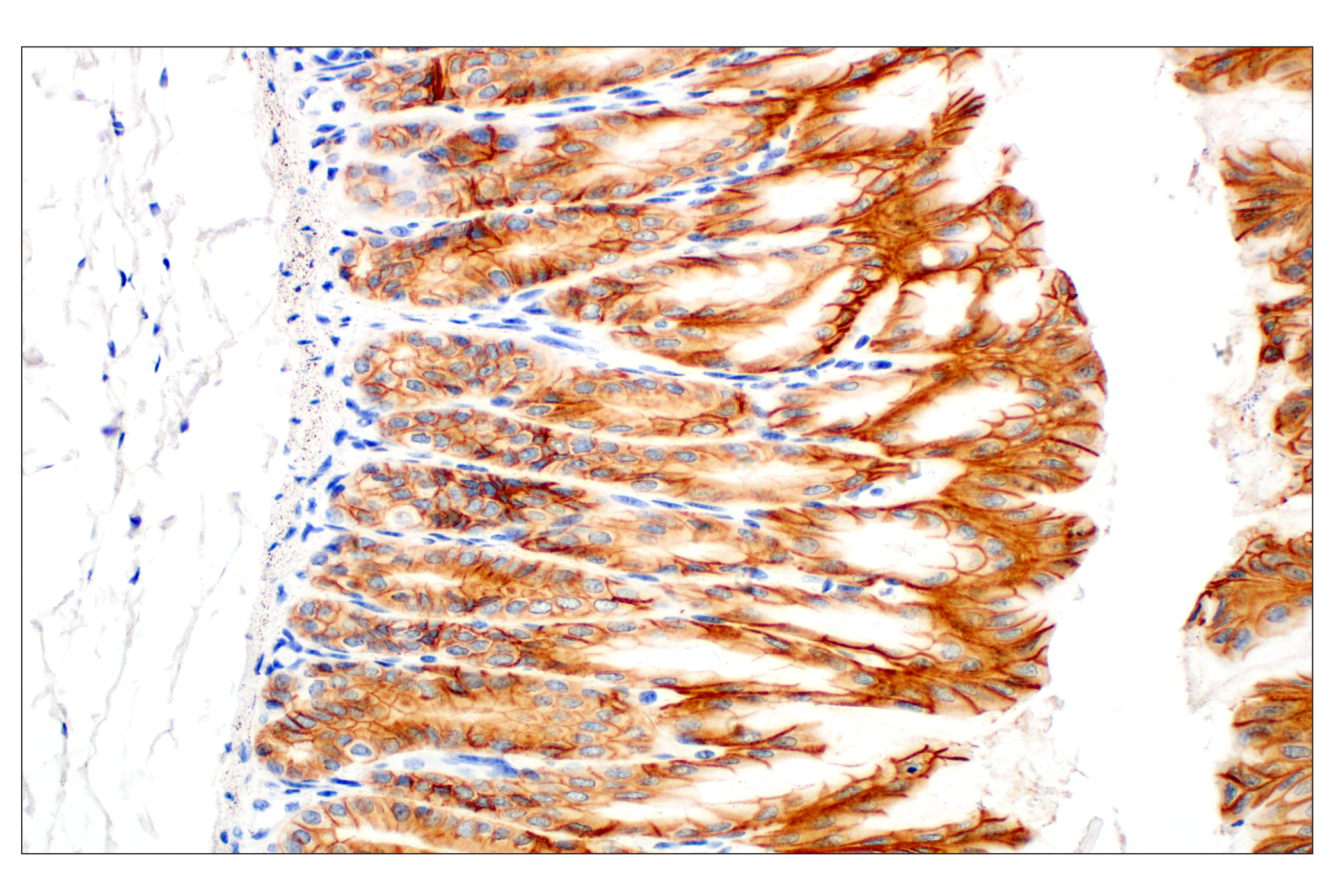
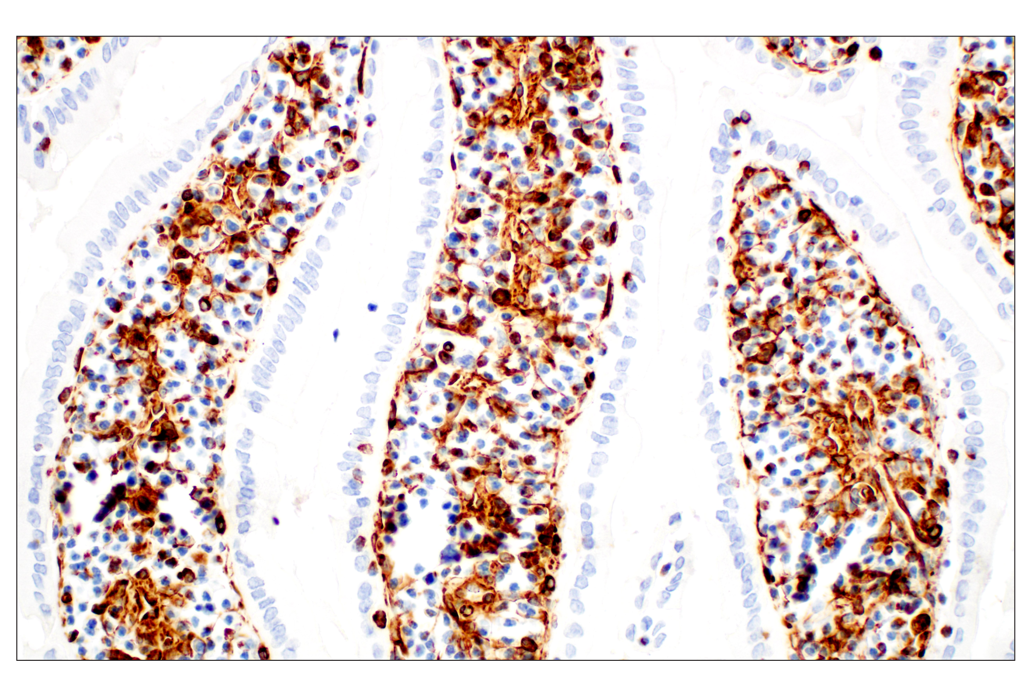
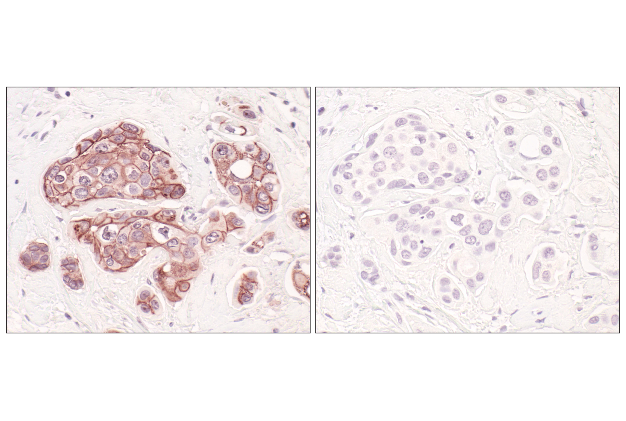
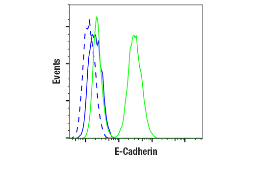
| E-Cadherin (24E10) Rabbit mAb | 3195 | 20 µl | 135 kDa | Rabbit IgG |
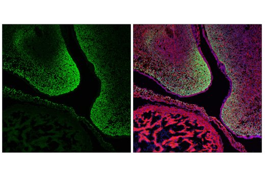
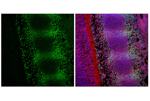
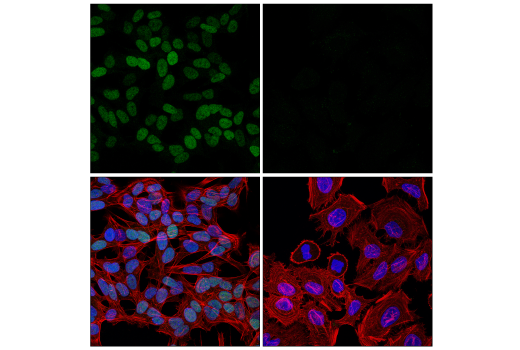
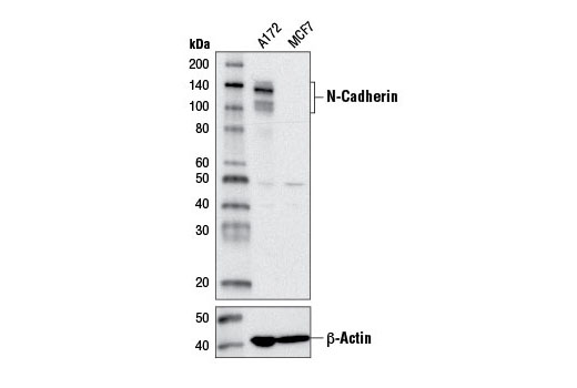
| N-Cadherin (D4R1H) XP® Rabbit mAb | 13116 | 20 µl | 140 kDa | Rabbit IgG |
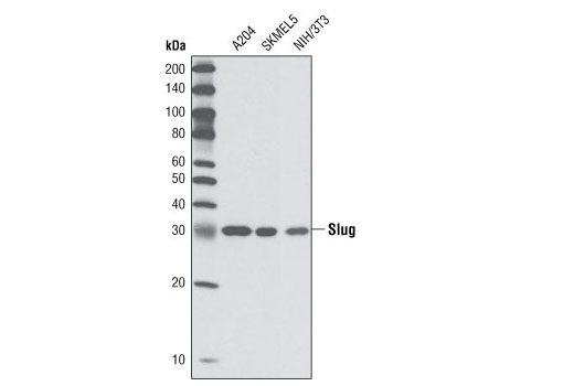
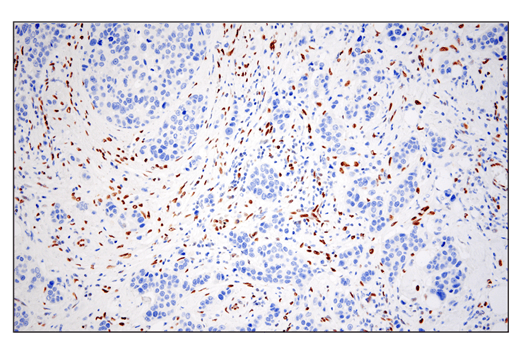
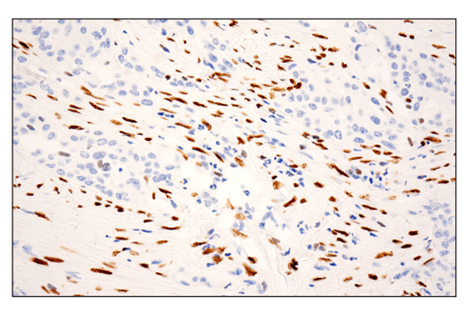
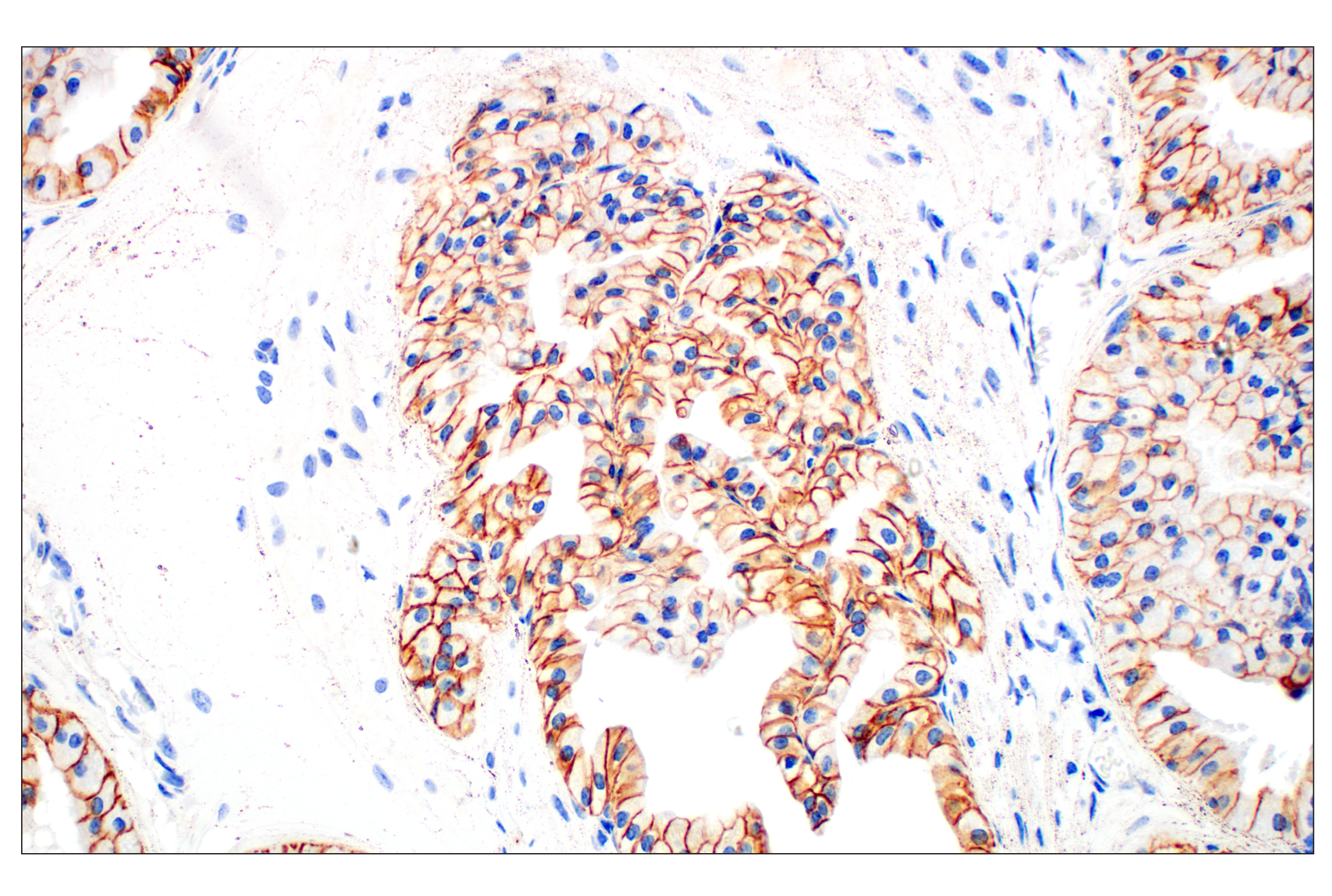
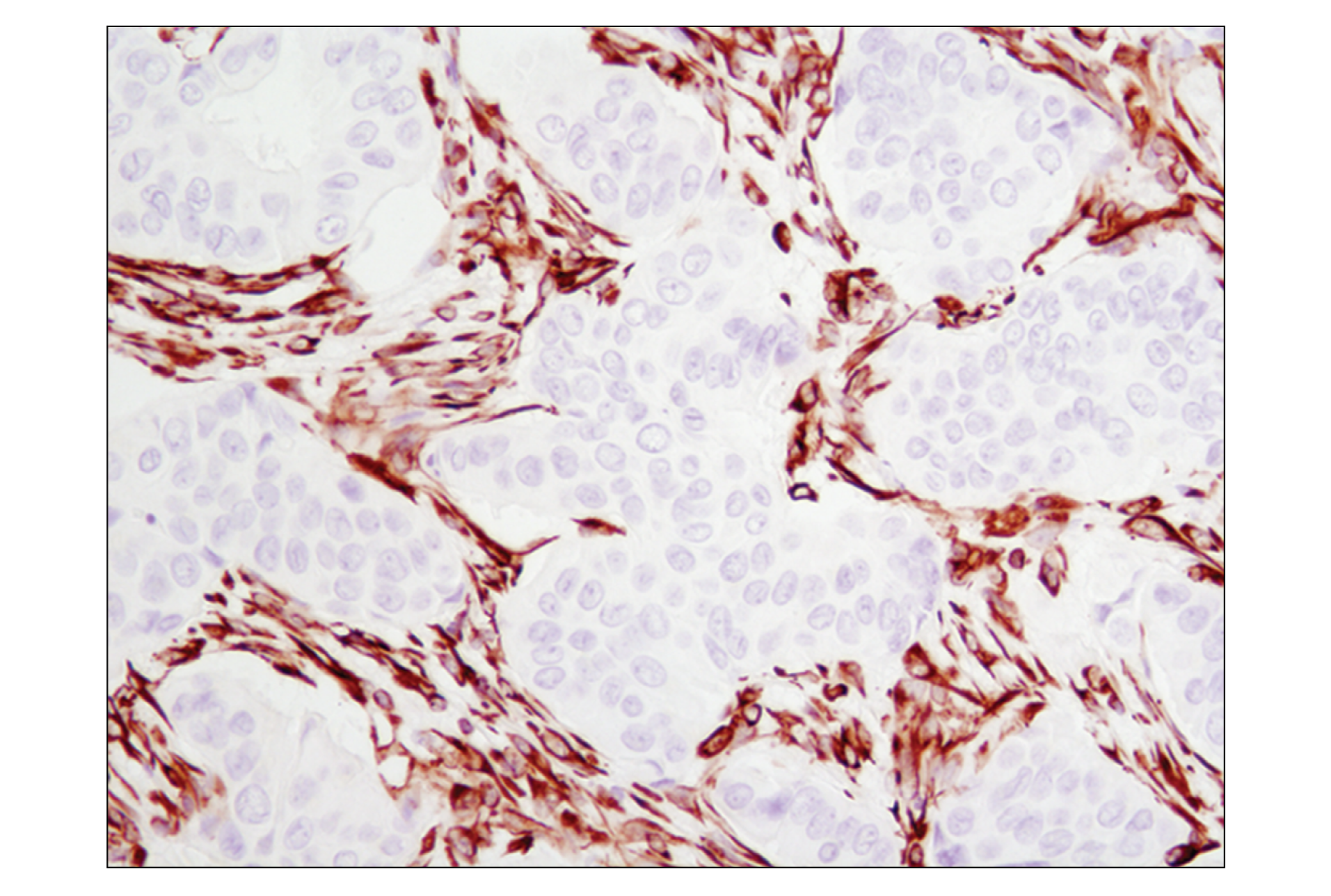
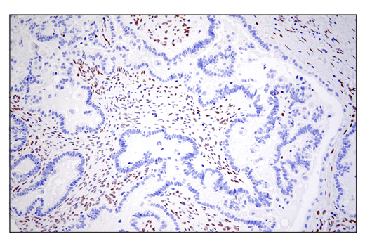
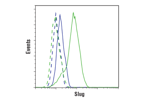
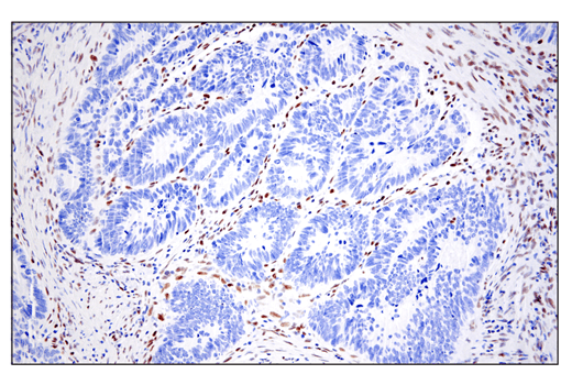
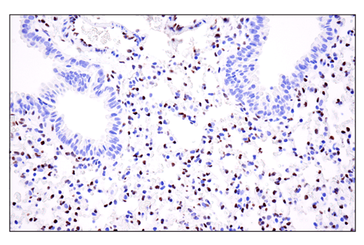
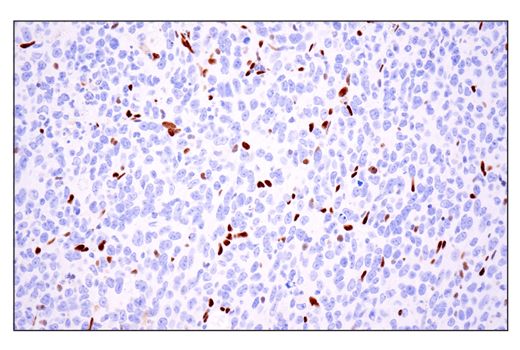
| Slug (C19G7) Rabbit mAb | 9585 | 20 µl | 30 kDa | Rabbit IgG |
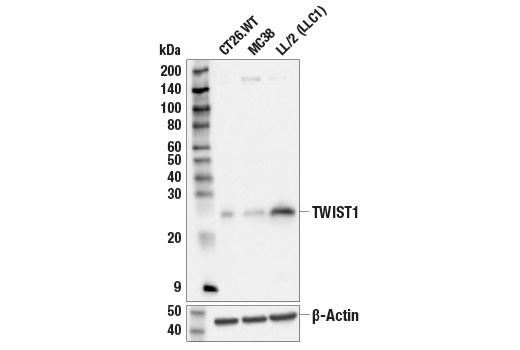
| TWIST1 (E5G9Y) Rabbit mAb | 90445 | 20 µl | 26 kDa | Rabbit IgG |
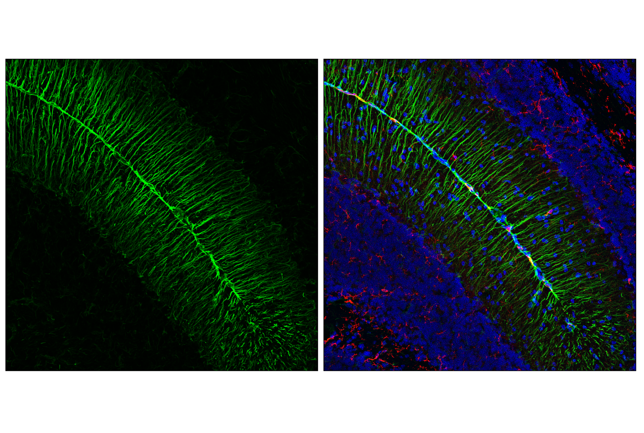
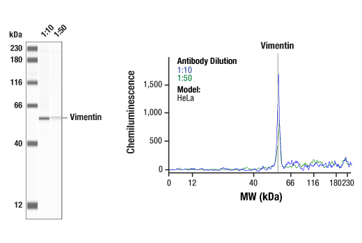
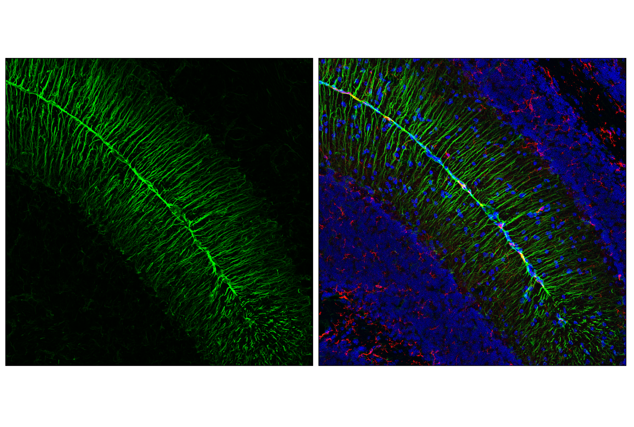
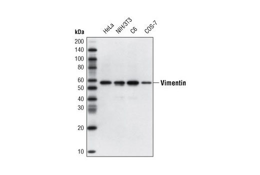
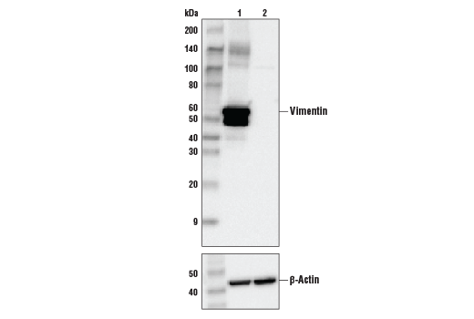
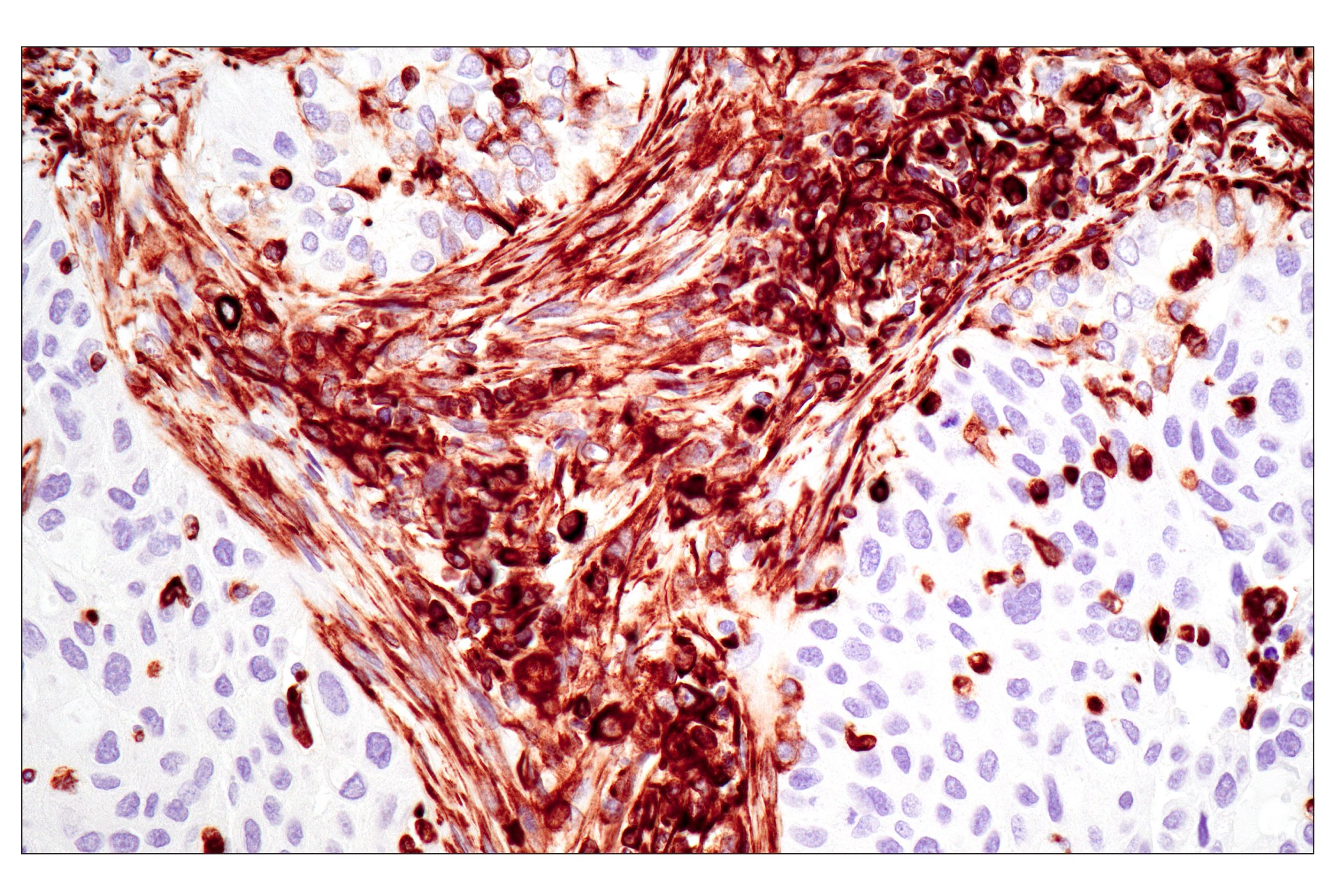
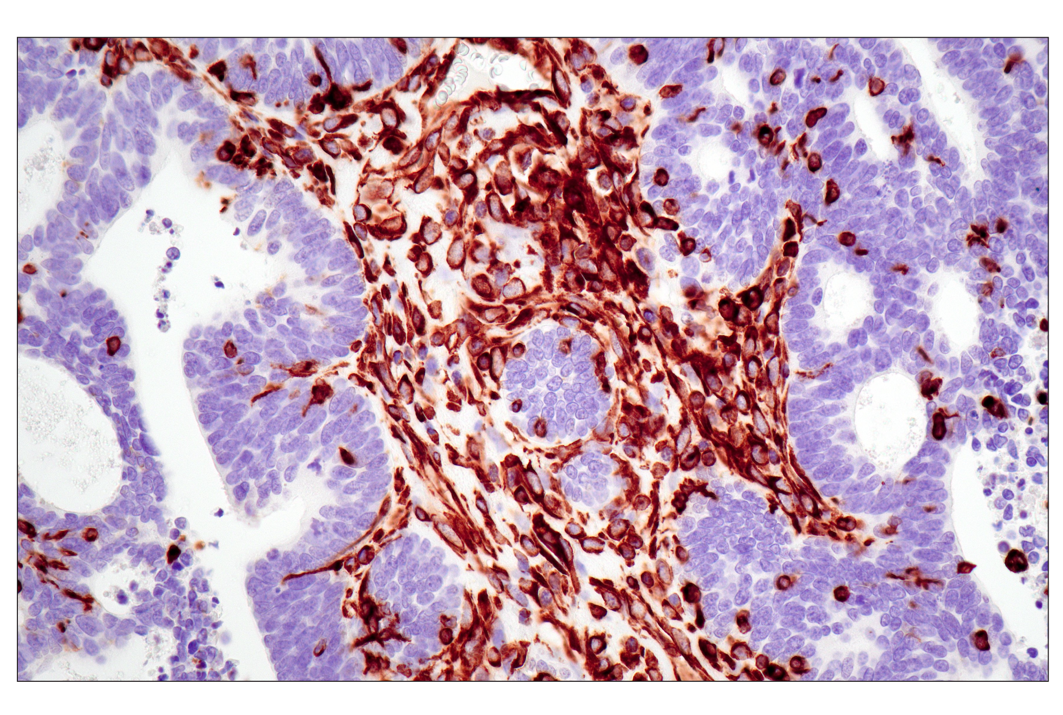
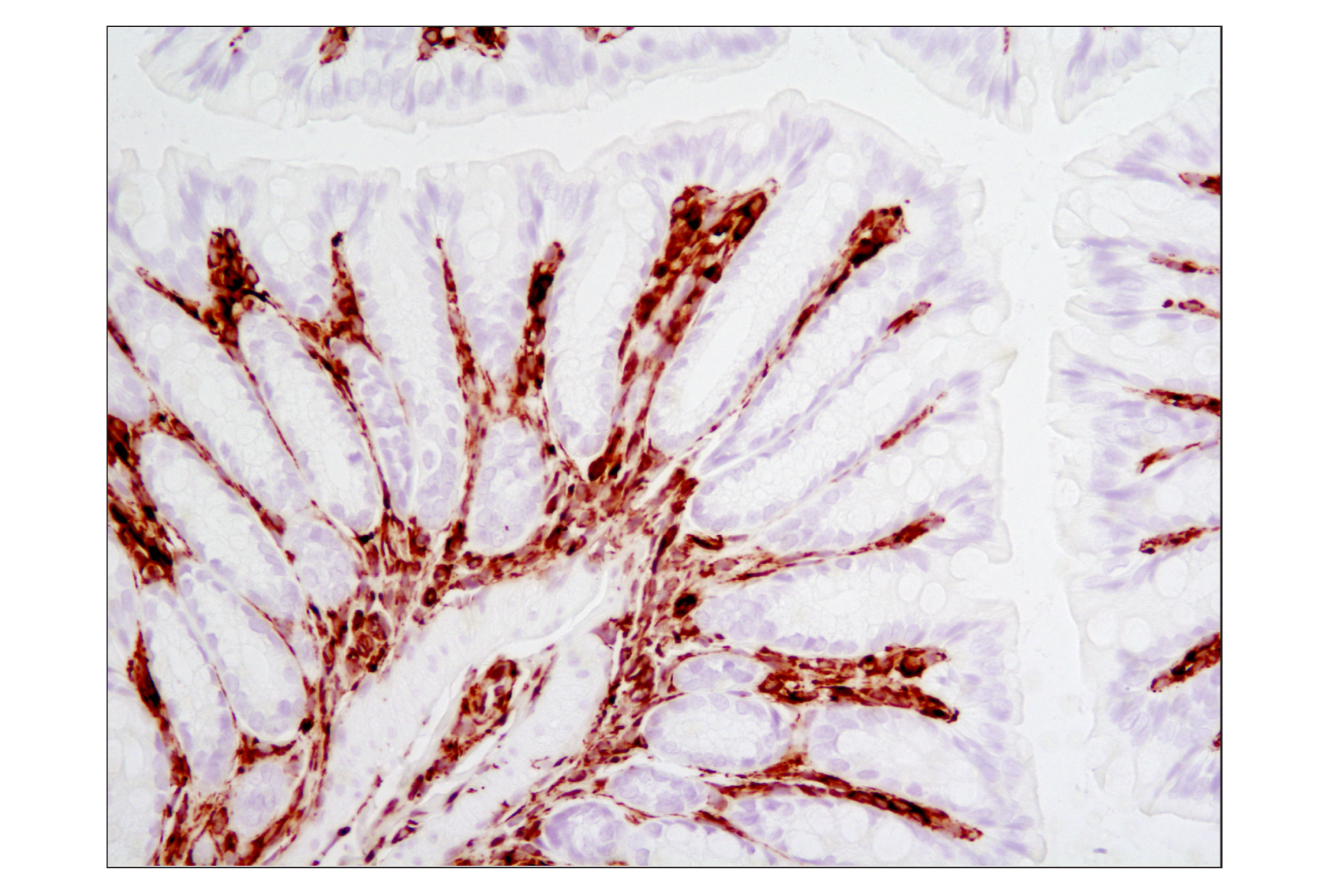
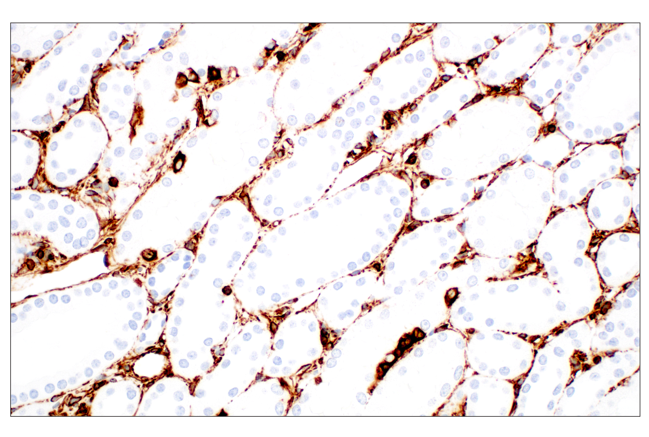
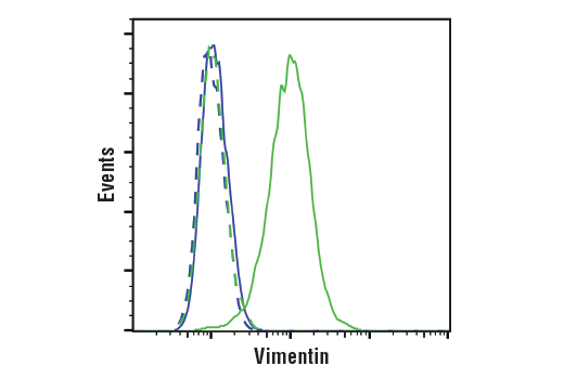
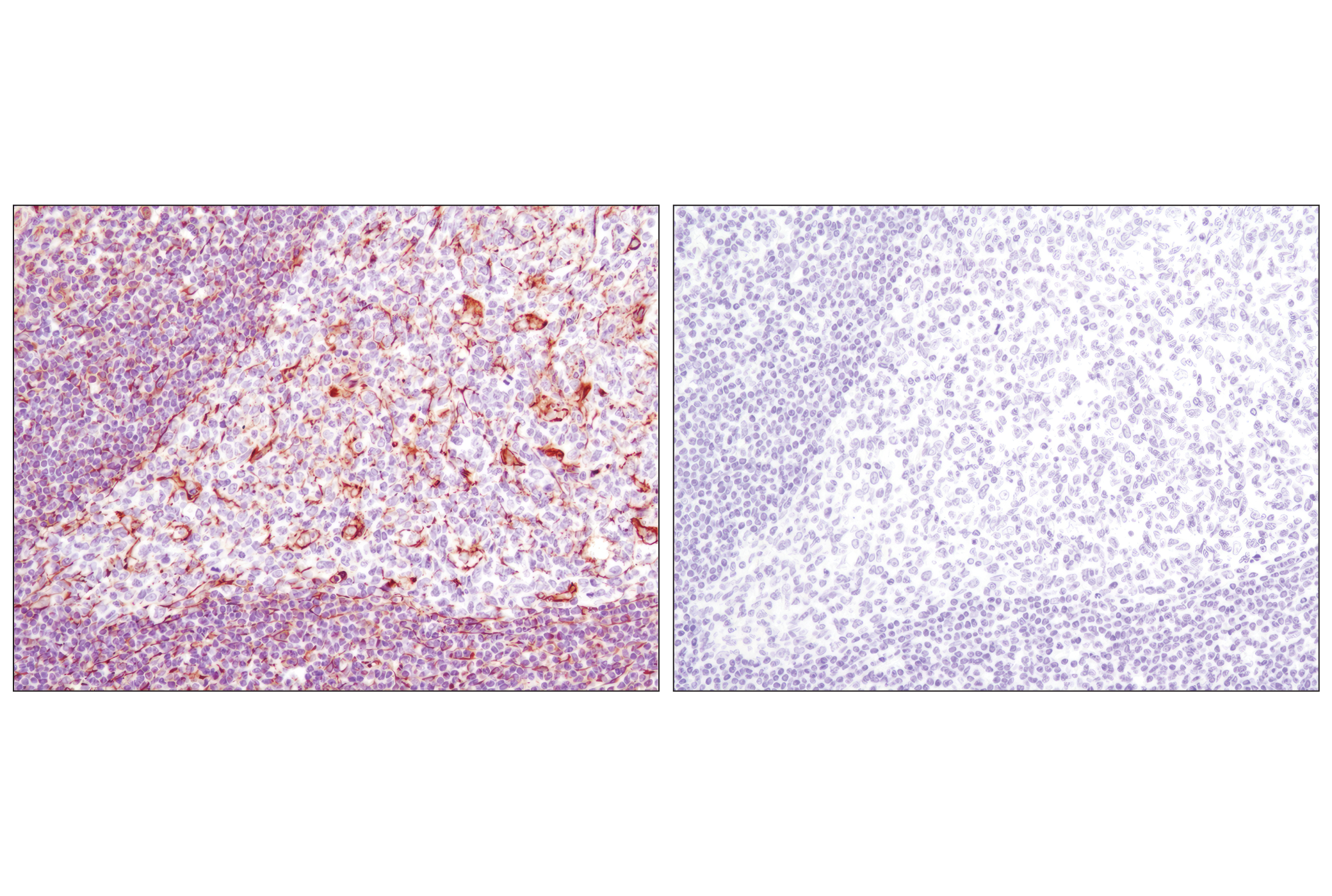
| Vimentin (D21H3) XP® Rabbit mAb | 5741 | 20 µl | 57 kDa | Rabbit IgG |
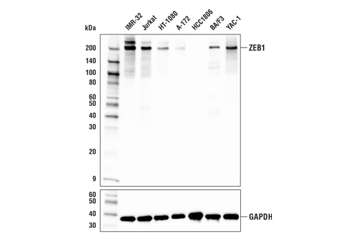
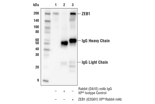
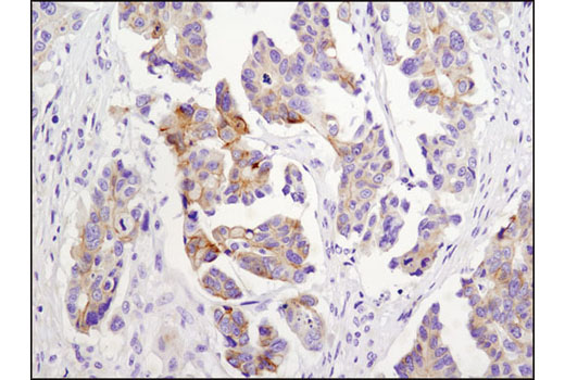
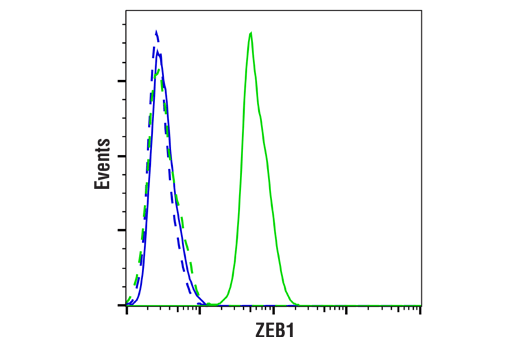
| ZEB1 (E2G6Y) XP® Rabbit mAb | 70512 | 20 µl | 200 kDa | Rabbit IgG |
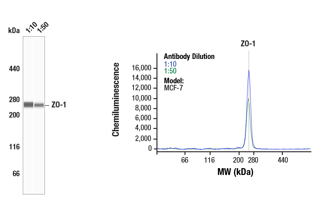
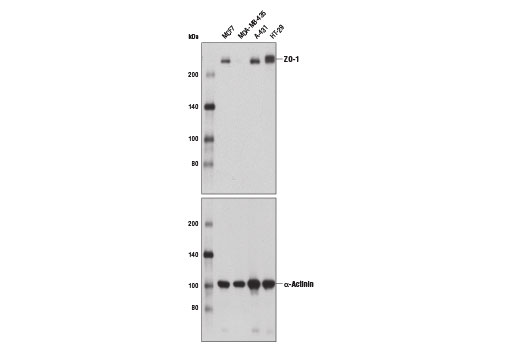
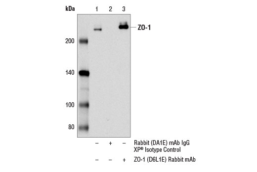
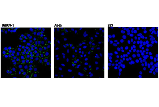
| ZO-1 (D6L1E) Rabbit mAb | 13663 | 20 µl | 220 kDa | Rabbit IgG |
Please visit cellsignal.com for individual component applications, species cross-reactivity, dilutions, protocols, and additional product information.
Description
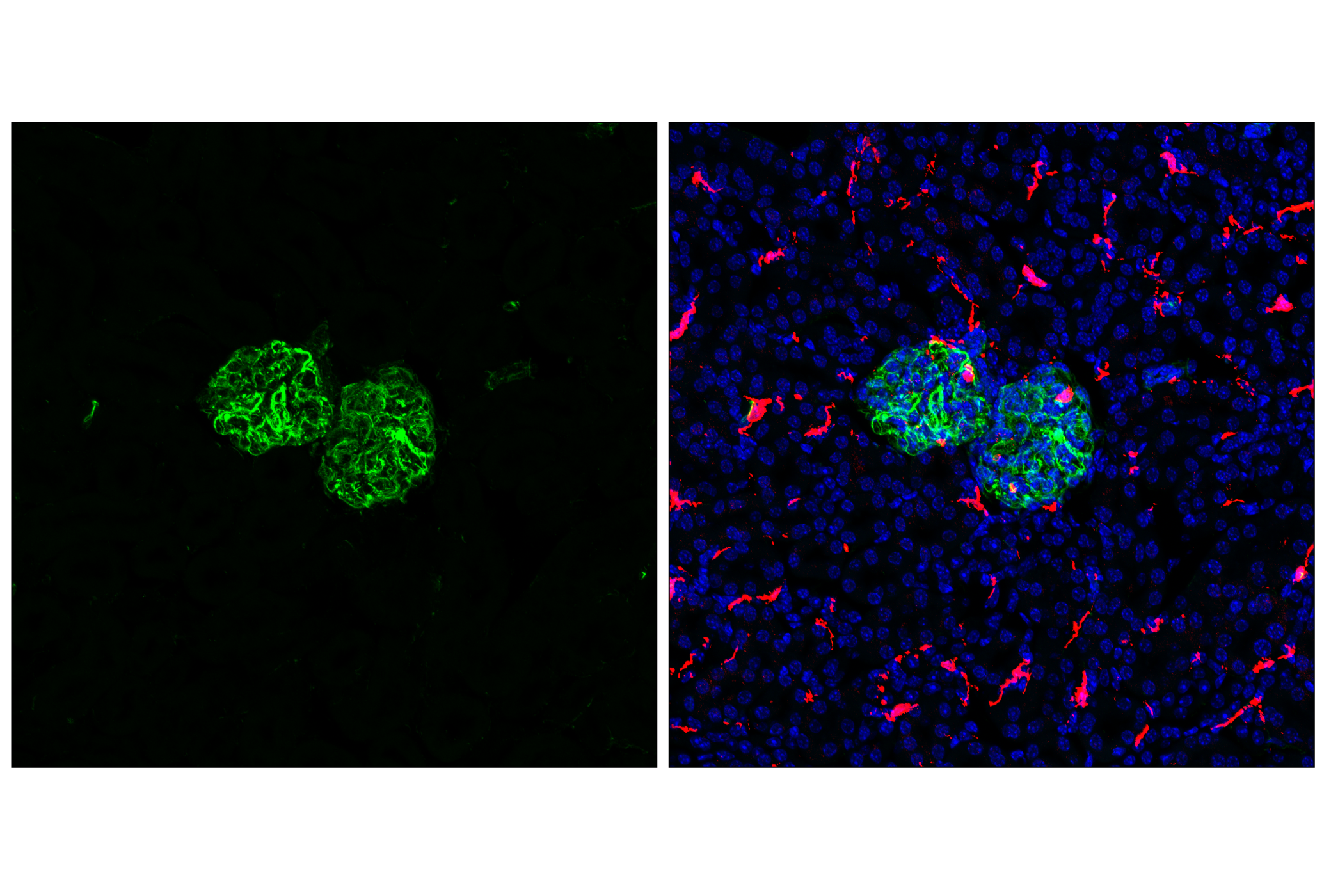
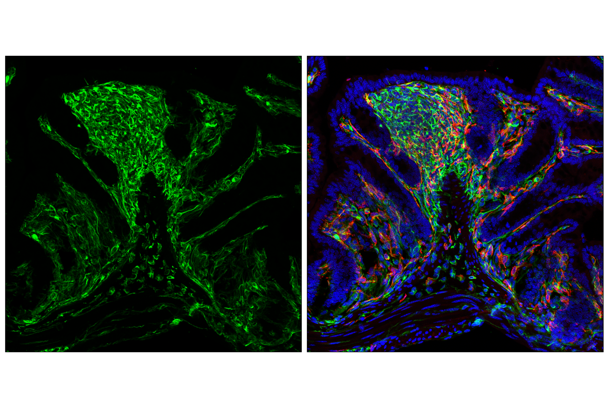
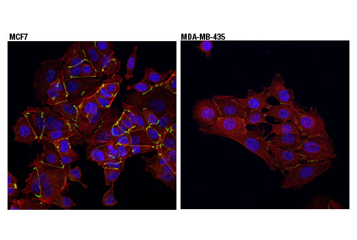
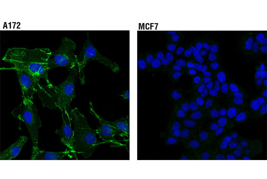
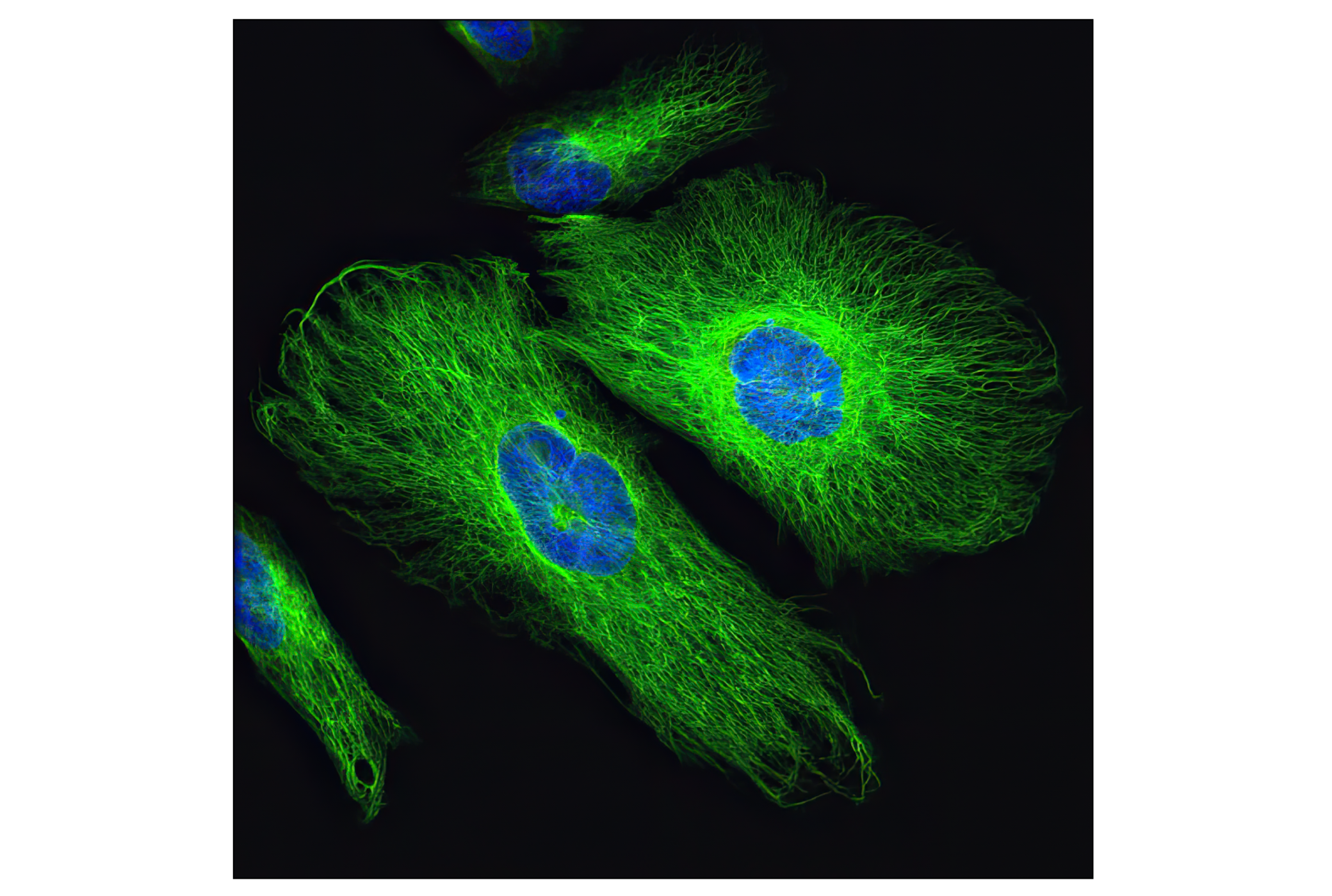
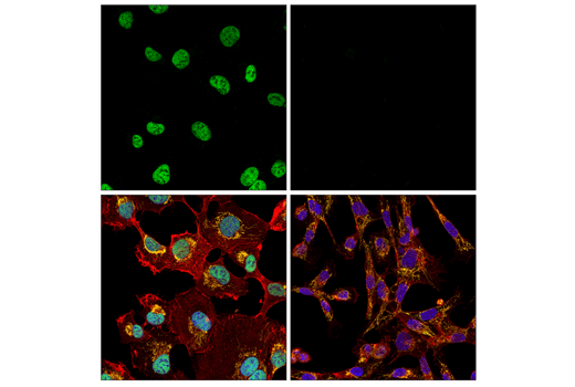
The Epithelial-Mesenchymal Transition (EMT) IF Antibody Sampler Kit provides an economical means to evaluate the expression of established markers of EMT by immunofluorescence (IF).
Storage
Background
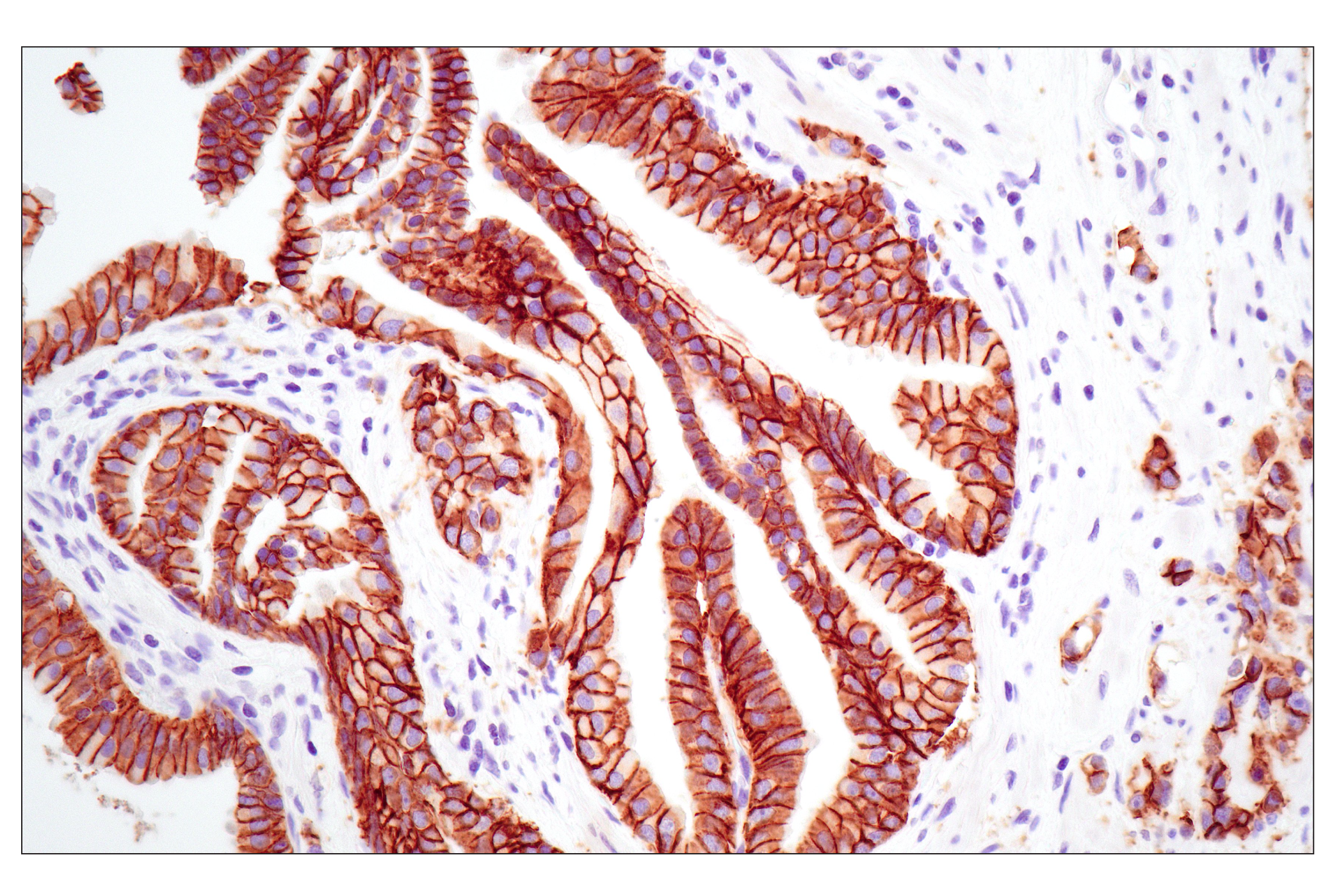
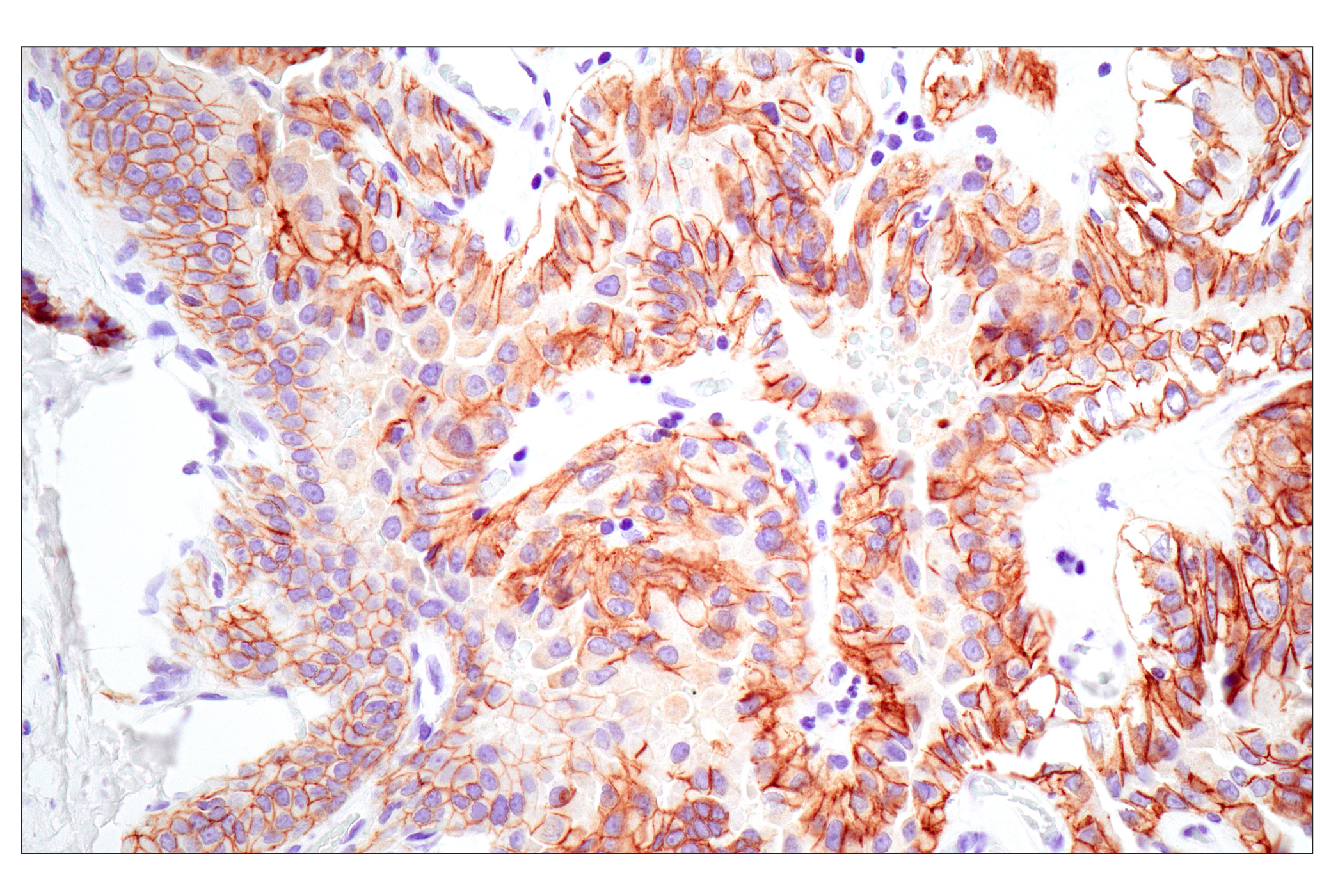
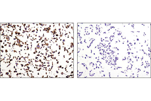
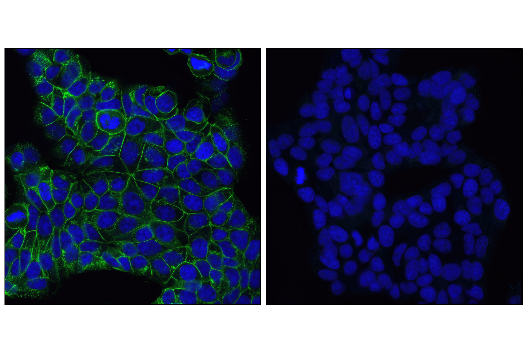
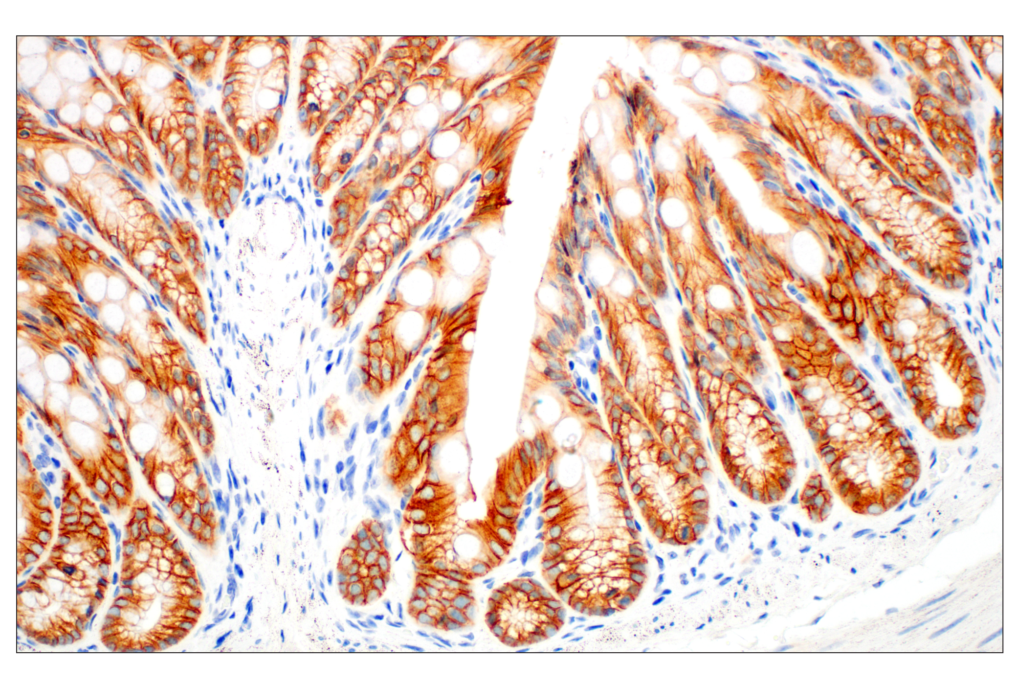
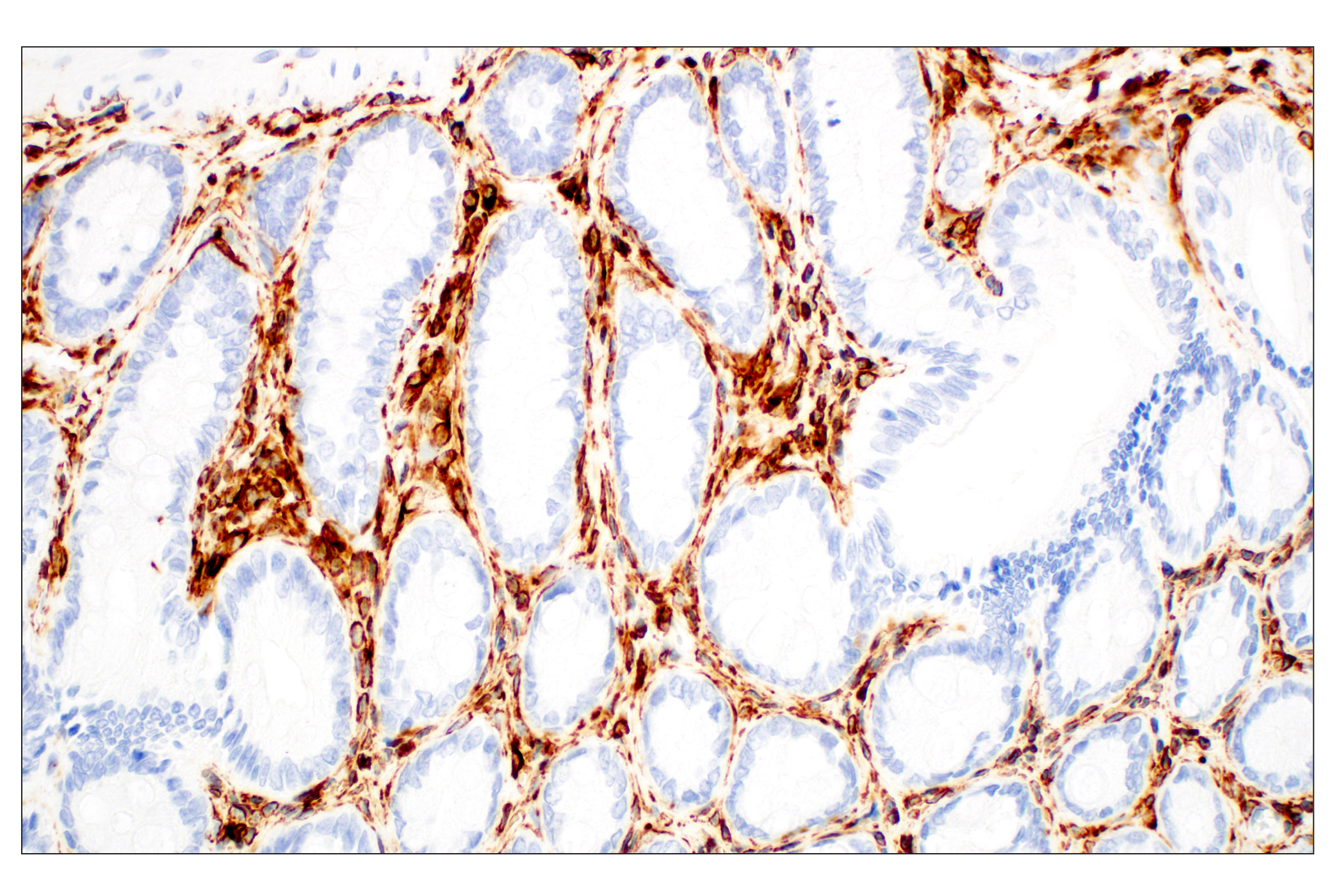
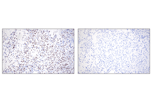
Epithelial-mesenchymal transition (EMT) is an essential process during development whereby epithelial cells acquire mesenchymal, fibroblast-like properties and display reduced intracellular adhesion and increased motility. This is a critical feature of normal embryonic development, which is also utilized by malignant epithelial tumors to spread beyond their origin (1-3). This tightly regulated process is associated with a number of cellular and molecular events. EMT depends on a reduction in expression of cell adhesion molecules. Cadherins mediate calcium-dependent cell-cell adhesion and play critical roles in normal tissue development (4). E-cadherin is considered an active suppressor of invasion and growth of many epithelial cancers (4-6). Recent studies indicate that cancer cells have upregulated N-cadherin in addition to loss of E-cadherin. This change in cadherin expression is called the "cadherin switch" and downregulation of E-cadherin is one of the hallmarks of EMT (1). Tight junctions, or zonula occludens, form a continuous barrier to fluids across the epithelium and endothelium. They function in regulation of paracellular permeability and in the maintenance of cell polarity, blocking the movement of transmembrane proteins between the apical and the basolateral cell surfaces. Tight junctions are composed of claudin and occludin proteins, which join the junctions to the cytoskeleton (7,8). Zona occludens (ZO) proteins (e.g., ZO-1) are peripheral membrane adaptor proteins that link junctional transmembrane proteins such as occludin and claudin to the actin cytoskeleton (9). ZO proteins are required for tight junction formation and function (10,11); mutations in ZO-1 and claudin induce EMT (12). Vimentin is an intermediate filament of mesenchymal origin and is present at early developmental stages. Vimentin's dynamic structural changes and spatial re-organization in response to extracellular stimuli helps to coordinate various signaling pathways (13). Slug (SNAI2) is a widely expressed transcriptional repressor and member of the Snail family of zinc finger transcription factors (14,15). Similar to the related Snail protein, Slug binds to the E-cadherin promoter region to repress transcription during development (16). The binding of Slug to integrin promoter sequences represses integrin expression and results in reduced cell adhesion (17). ZEB family proteins (e.g., ZEB1) are zinc finger and homeobox domain containing transcription factors, whose targets of regulation include E-cadherin (1). TWIST1 is a basic helix-loop-helix (b-HLH) transcription factor that functions as a master regulator of embryonic morphogenesis, and plays essential roles in mesenchymal differentiation (18,19). TWIST is upregulated in various human tumors and has been suggested to be a driver of EMT and metastasis (20-21).
- Aigner, K. et al. (2007) Oncogene 26, 6979-88.
- Peinado, H. et al. (2007) Nat Rev Cancer 7, 415-28.
- Moreno-Bueno, G. et al. (2008) Oncogene 27, 6958-69.
- Wheelock, M.J. and Johnson, K.R. (2003) Annu Rev Cell Dev Biol 19, 207-35.
- Christofori, G. (2003) EMBO J 22, 2318-23.
- Hazan, R.B. et al. (2004) Ann N Y Acad Sci 1014, 155-63.
- Shin, K. et al. (2006) Annu Rev Cell Dev Biol 22, 207-35.
- Oliveira, S.S. and Morgado-Díaz, J.A. (2007) Cell Mol Life Sci 64, 17-28.
- Matter, K. and Balda, M.S. (2007) J Cell Sci 120, 1505-11.
- Hernandez, S. et al. (2007) Exp Cell Res 313, 1533-47.
- Umeda, K. et al. (2006) Cell 126, 741-54.
- Reichert, M. et al. (2000) J Biol Chem 275, 9492-500.
- Helfand, B.T. et al. (2004) J Cell Sci 117, 133-41.
- Inukai, T. et al. (1999) Mol Cell 4, 343-52.
- Barrallo-Gimeno, A. and Nieto, M.A. (2005) Development 132, 3151-61.
- Bolós, V. et al. (2003) J Cell Sci 116, 499-511.
- Turner, F.E. et al. (2006) J Biol Chem 281, 21321-31.
- Lee, M.S. et al. (1999) J Cell Biochem 75, 566-77.
- Chen, Z.F. and Behringer, R.R. (1995) Genes Dev 9, 686-99.
- Yang, J. et al. (2004) Cell 117, 927-39.
- Watanabe, O. et al. (2005) Anticancer Res 24, 3851-6.
Background References
Trademarks and Patents
使用に関する制限
法的な権限を与えられたCSTの担当者が署名した書面によって別途明示的に合意された場合を除き、 CST、その関連会社または代理店が提供する製品には以下の条件が適用されます。お客様が定める条件でここに定められた条件に含まれるものを超えるもの、 または、ここに定められた条件と異なるものは、法的な権限を与えられたCSTの担当者が別途書面にて受諾した場合を除き、拒絶され、 いかなる効力も効果も有しません。
研究専用 (For Research Use Only) またはこれに類似する表示がされた製品は、 いかなる目的についても FDA または外国もしくは国内のその他の規制機関により承認、認可または許可を受けていません。 お客様は製品を診断もしくは治療目的で使用してはならず、また、製品に表示された内容に違反する方法で使用してはなりません。 CST が販売または使用許諾する製品は、エンドユーザーであるお客様に対し、使途を研究および開発のみに限定して提供されるものです。 診断、予防もしくは治療目的で製品を使用することまたは製品を再販売 (単独であるか他の製品等の一部であるかを問いません) もしくはその他の商業的利用の目的で購入することについては、CST から別途許諾を得る必要があります。 お客様は以下の事項を遵守しなければなりません。(a) CST の製品 (単独であるか他の資材と一緒であるかを問いません) を販売、使用許諾、貸与、寄付もしくはその他の態様で第三者に譲渡したり使用させたりしてはなりません。また、商用の製品を製造するために CST の製品を使用してはなりません。(b) 複製、改変、リバースエンジニアリング、逆コンパイル、 分解または他の方法により製品の構造または技術を解明しようとしてはなりません。また、 CST の製品またはサービスと競合する製品またはサービスを開発する目的で CST の製品を使用してはなりません。(c) CST の製品の商標、商号、ロゴ、特許または著作権に関する通知または表示を除去したり改変したりしてはなりません。(d) CST の製品をCST 製品販売条件(CST’s Product Terms of Sale) および該当する書面のみに従って使用しなければなりません。(e) CST の製品に関連してお客様が使用する第三者の製品またはサービスに関する使用許諾条件、 サービス提供条件またはこれに類する合意事項を遵守しなければなりません。