WB, IP, IHC-P
H M R
Endogenous
95
Rabbit IgG
#Q12929
2059
Product Information
Product Usage Information
| Application | Dilution |
|---|---|
| Western Blotting | 1:1000 |
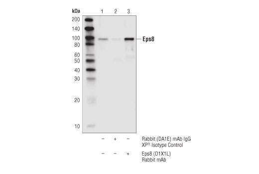
| Immunoprecipitation | 1:50 |
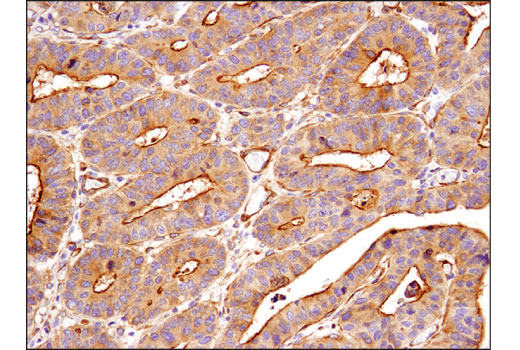
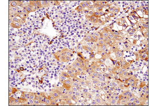
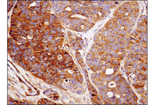
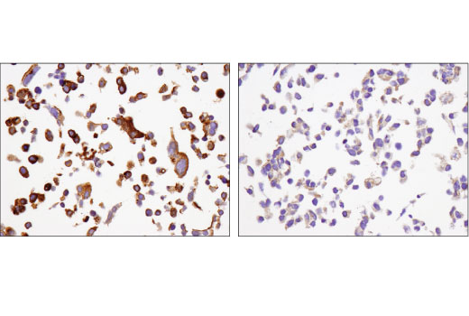
| Immunohistochemistry (Paraffin) | 1:100 |
Storage
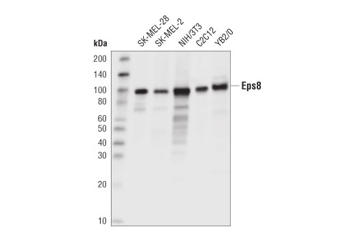
Specificity / Sensitivity
Species Reactivity:
Human, Mouse, Rat
Source / Purification
Monoclonal antibody is produced by immunizing animals with a synthetic peptide corresponding to residues surrounding Arg250 of human Eps8 protein.
Background
Epidermal growth factor receptor pathway substrate 8 (Eps8) is an adaptor protein and can be phosphorylated by several receptor tyrosine kinases including EGFR and Src (1,2). Eps8 is composed of an N-terminal PTB domain, followed by an SH3 domain and a C-terminal effector domain. Eps8 controls actin-based motility by capping the barbed end of actin and bundling actin subunits through its C-terminal effector domain (3,4). The C-terminal α hexlical structure of Eps8 interacts directly with actin to exert these capping and bundling functions (5). The actin capping activity requires the release of Eps8 autoinhibitory binding through SH3 domain interaction with an adaptor molecule, such as Abi-1 (6). This SH3 domain of Eps8 also binds to RN-tre to regulate the down stream Rab5-mediated endocytosis pathway (6). Eps8 functions by binding several receptor tyrosine kinases, such as EGFR or FGFR, to enhance receptor mediated mitogenic Rac signaling and Rab5 endocytosis (6,7). The effector region of Eps8 is necessary for this process. By association with Abi-1 and forming the Eps8/Abi-1/Sos-1 complex, Eps8 couples initial growth factor stimulation to actin motility and the Rac activation pathway (8,9). Eps8 has been shown to be important in the cellular function of filopodial protrusions, cell migration, microvilli formation, and focal adhesion (10-13). Research studies have demonstrated that through its involvement in actin related cellular functions, Eps8 plays a role in cancer cell growth, survival, motility, and invasiveness (14-18).
- Fazioli, F. et al. (1993) EMBO J 12, 3799-808.
- Gallo, R. et al. (1997) Oncogene 15, 1929-36.
- Disanza, A. et al. (2004) Nat Cell Biol 6, 1180-8.
- Disanza, A. et al. (2006) Nat Cell Biol 8, 1337-47.
- Hertzog, M. et al. (2010) PLoS Biol 8, e1000387.
- Lanzetti, L. et al. (2000) Nature 408, 374-7.
- Auciello, G. et al. (2013) J Cell Sci 126, 613-24.
- Scita, G. et al. (1999) Nature 401, 290-3.
- Innocenti, M. et al. (2003) J Cell Biol 160, 17-23.
- Welsch, T. et al. (2007) Cancer Lett 255, 205-18.
- Frittoli, E. et al. (2011) Immunity 35, 388-99.
- Zwaenepoel, I. et al. (2012) Mol Biol Cell 23, 1080-94.
- Maa, M.C. et al. (2007) J Biol Chem 282, 19399-409.
- Xu, M. et al. (2009) Endocrinology 150, 2064-71.
- Chen, Y.J. et al. (2008) Mol Cancer Ther 7, 1376-85.
- Yap, L.F. et al. (2009) Oncogene 28, 2524-34.
- Chen, H. et al. (2010) Cancer Res 70, 9979-90.
- Funato, Y. et al. (2004) Cancer Res 64, 5237-44.
Species Reactivity
Species reactivity is determined by testing in at least one approved application (e.g., western blot).
Western Blot Buffer
IMPORTANT: For western blots, incubate membrane with diluted primary antibody in 5% w/v BSA, 1X TBS, 0.1% Tween® 20 at 4°C with gentle shaking, overnight.
Applications Key
WB: Western Blotting IP: Immunoprecipitation IHC-P: Immunohistochemistry (Paraffin)
Cross-Reactivity Key
H: human M: mouse R: rat Hm: hamster Mk: monkey Vir: virus Mi: mink C: chicken Dm: D. melanogaster X: Xenopus Z: zebrafish B: bovine Dg: dog Pg: pig Sc: S. cerevisiae Ce: C. elegans Hr: horse GP: Guinea Pig Rab: rabbit All: all species expected
Trademarks and Patents
使用に関する制限
法的な権限を与えられたCSTの担当者が署名した書面によって別途明示的に合意された場合を除き、 CST、その関連会社または代理店が提供する製品には以下の条件が適用されます。お客様が定める条件でここに定められた条件に含まれるものを超えるもの、 または、ここに定められた条件と異なるものは、法的な権限を与えられたCSTの担当者が別途書面にて受諾した場合を除き、拒絶され、 いかなる効力も効果も有しません。
研究専用 (For Research Use Only) またはこれに類似する表示がされた製品は、 いかなる目的についても FDA または外国もしくは国内のその他の規制機関により承認、認可または許可を受けていません。 お客様は製品を診断もしくは治療目的で使用してはならず、また、製品に表示された内容に違反する方法で使用してはなりません。 CST が販売または使用許諾する製品は、エンドユーザーであるお客様に対し、使途を研究および開発のみに限定して提供されるものです。 診断、予防もしくは治療目的で製品を使用することまたは製品を再販売 (単独であるか他の製品等の一部であるかを問いません) もしくはその他の商業的利用の目的で購入することについては、CST から別途許諾を得る必要があります。 お客様は以下の事項を遵守しなければなりません。(a) CST の製品 (単独であるか他の資材と一緒であるかを問いません) を販売、使用許諾、貸与、寄付もしくはその他の態様で第三者に譲渡したり使用させたりしてはなりません。また、商用の製品を製造するために CST の製品を使用してはなりません。(b) 複製、改変、リバースエンジニアリング、逆コンパイル、 分解または他の方法により製品の構造または技術を解明しようとしてはなりません。また、 CST の製品またはサービスと競合する製品またはサービスを開発する目的で CST の製品を使用してはなりません。(c) CST の製品の商標、商号、ロゴ、特許または著作権に関する通知または表示を除去したり改変したりしてはなりません。(d) CST の製品をCST 製品販売条件(CST’s Product Terms of Sale) および該当する書面のみに従って使用しなければなりません。(e) CST の製品に関連してお客様が使用する第三者の製品またはサービスに関する使用許諾条件、 サービス提供条件またはこれに類する合意事項を遵守しなければなりません。