| Product Includes | Product # | Quantity | Mol. Wt | Isotype/Source |
|---|---|---|---|---|
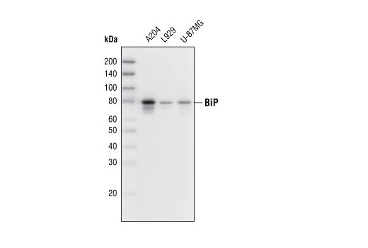
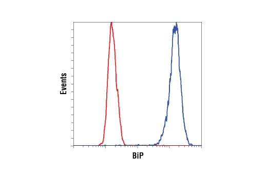
| BiP (C50B12) Rabbit mAb | 3177 | 20 µl | 78 kDa | Rabbit IgG |
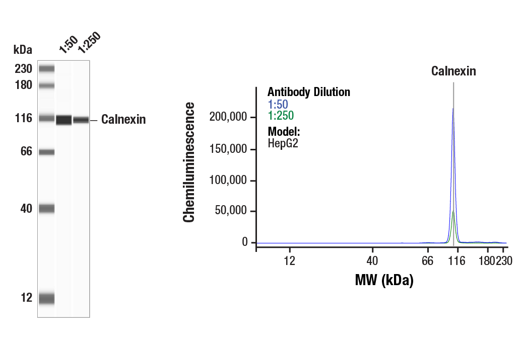
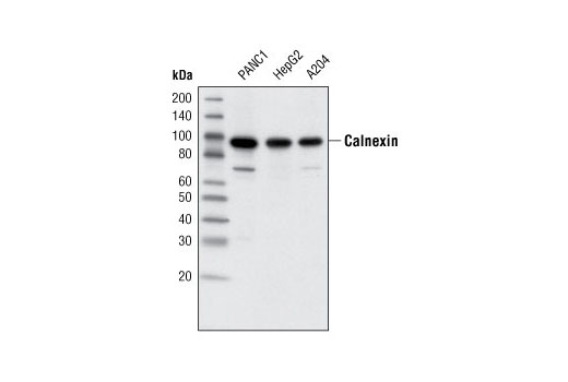
| Calnexin (C5C9) Rabbit mAb | 2679 | 20 µl | 90 kDa | Rabbit IgG |
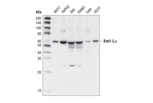
| Ero1-Lα Antibody | 3264 | 20 µl | 60 kDa | Rabbit |
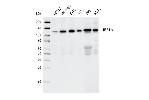
| IRE1α (14C10) Rabbit mAb | 3294 | 20 µl | 130 kDa | Rabbit IgG |
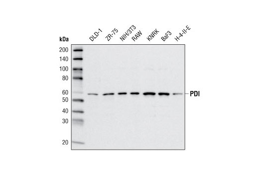
| PDI (C81H6) Rabbit mAb | 3501 | 20 µl | 57 kDa | Rabbit |
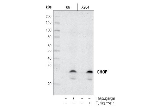
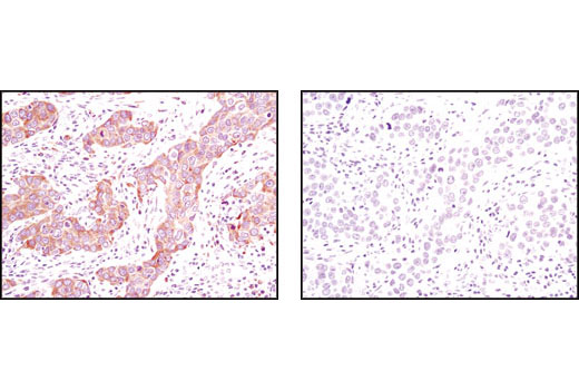
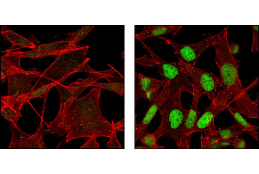
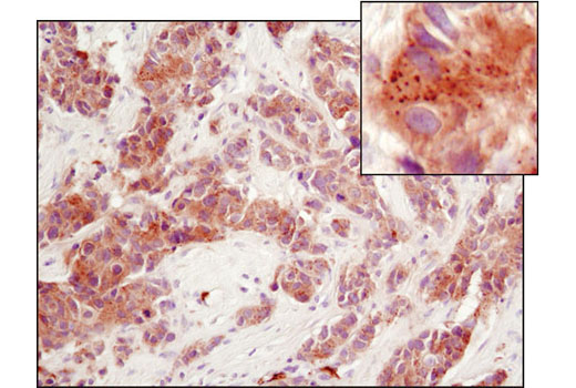
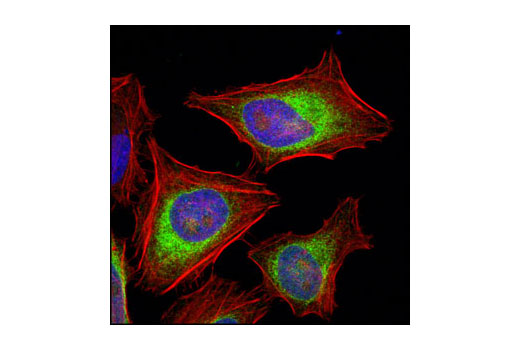
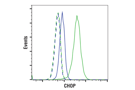
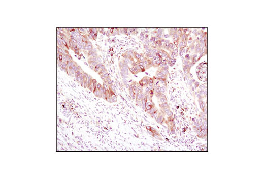
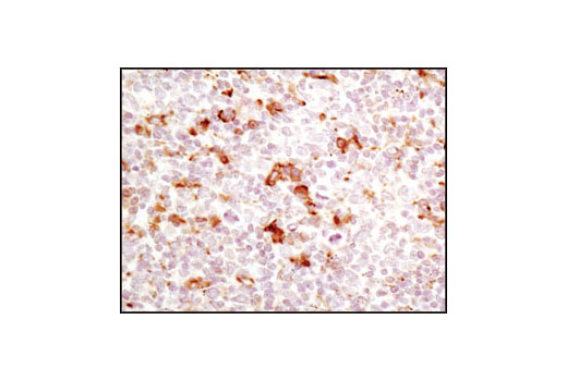
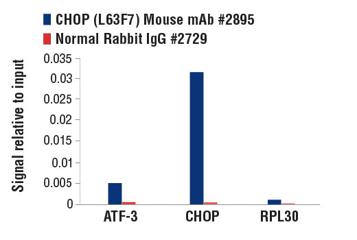
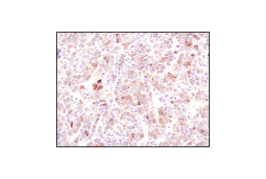
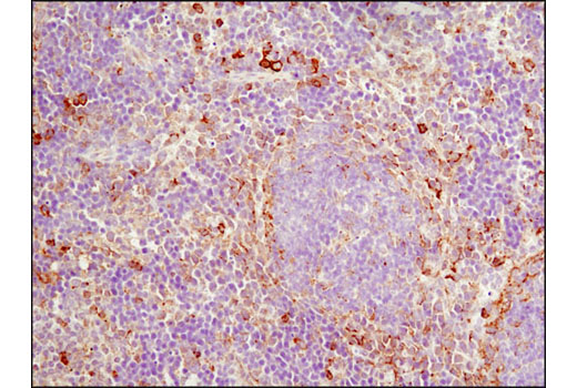
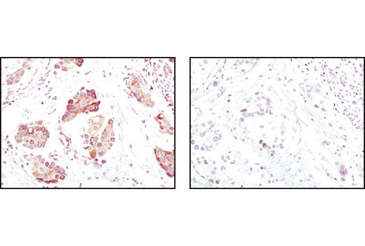
| CHOP (L63F7) Mouse mAb | 2895 | 20 µl | 27 kDa | Mouse IgG2a |
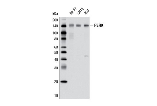
| PERK (D11A8) Rabbit mAb | 5683 | 20 µl | 140 kDa | Rabbit IgG |
| Anti-rabbit IgG, HRP-linked Antibody | 7074 | 100 µl | Goat | |
| Anti-mouse IgG, HRP-linked Antibody | 7076 | 100 µl | Horse |
Please visit cellsignal.com for individual component applications, species cross-reactivity, dilutions, protocols, and additional product information.
Description
The ER Stress Sampler Kit contains reagents to investigate ER stress within the cell. The kit contains enough primary and secondary antibodies to perform two Western blot experiments per primary antibody.
Storage
Background
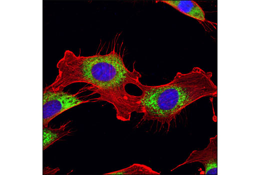
Secretory and transmembrane proteins are synthesized on polysomes and translocate into the endoplasmic reticulum (ER) where they are often modified by the formation of disulfide bonds, amino-linked glycosylation and folding. The ER contains a pool of molecular chaperone proteins including calnexin, BiP and protein disulfide isomerase (PDI). Calnexin is an ER membrane, calcium-binding protein that retains newly synthesized glycoproteins inside the ER to ensure proper folding and quality control (1,2). Irregular protein folding within the ER increases BiP synthesis, which binds misfolded proteins to prevent them from forming aggregates and to assist them to refold properly (3).
PDI catalyzes the formation and isomerization of disulfide bonds required for a protein to reach its native state (4). Studies have found that the resident ER protein endoplasmic oxidoreductin-1 (Ero1) provides oxidizing potential to the ER in Saccharomyces cerevisiae (5). Ero1-Lα is an ER membrane-associated N-glycoprotein that promotes oxidative protein folding (6). Disruptions of ER homeostasis leads to the accumulation of unfolded proteins. The ER has developed an adaptive mechanism called the unfolded protein response (UPR) to counteract compromised protein folding (7). This is regulated by proteins such as the membrane-bound transcription factor protease site 2 (MBTPS2) and the serine/threonine kinase IRE1 (8-12). The PERK eIF2α kinase is an ER resident transmembrane protein that couples ER stress signals to translation inhibition. ER stress increases PERK activity, which phosphorylates eIF2α to reduce protein translation. PERK activation during ER stress correlates with autophosphorylation of its cytoplasmic kinase domain (13,14). Phosphorylation of PERK at Thr980 can serve as a marker for its activation status.
During ER stress, the level of CHOP expression is elevated and CHOP functions to mediate programmed cell death (15).
- Bergeron, J.J. et al. (1994) Trends Biochem. Sci. 19, 124-128.
- Williams, D.B. (2006) J. Cell Sci. 119, 615-623.
- Kohno, K. et al. (1993) Mol. Cell. Biol. 13, 877-890.
- Ellgaard, L. and Ruddock, L.W. (2005) EMBO Rep. 6, 28-32.
- Frand, A.R. and Kaiser, C.A. (1998) Mol. Cell 1, 161-170.
- Cabibbo, A. et al. (2000) J. Biol. Chem. 275, 4827-4833.
- Kaufman, R.J. et al. (2002) Nat. Rev. Mol. Cell Biol. 3, 411-421.
- Nikawa, J. and Yamashita, S. (1992) Mol. Microbiol. 6, 1441-1446.
- Cox, J.S. et al. (1993) Cell 73, 1197-1206.
- Mori, K. et al. (1993) Cell 74, 743-756.
- Lee, K. et al. (2002) Genes Dev. 16, 452-466.
- Shen, J. and Prywes, R. (2004) J. Biol. Chem. 279, 43046-43051.
- Harding, H.P. et al. (1999) Nature 397, 271-274.
- Shi, Y. et al. (1998) Mol. Cell. Biol. 18, 7499-7509.
- Zinszner, H. et al. (1998) Genes Dev 12, 982-95.
Background References
Trademarks and Patents
使用に関する制限
法的な権限を与えられたCSTの担当者が署名した書面によって別途明示的に合意された場合を除き、 CST、その関連会社または代理店が提供する製品には以下の条件が適用されます。お客様が定める条件でここに定められた条件に含まれるものを超えるもの、 または、ここに定められた条件と異なるものは、法的な権限を与えられたCSTの担当者が別途書面にて受諾した場合を除き、拒絶され、 いかなる効力も効果も有しません。
研究専用 (For Research Use Only) またはこれに類似する表示がされた製品は、 いかなる目的についても FDA または外国もしくは国内のその他の規制機関により承認、認可または許可を受けていません。 お客様は製品を診断もしくは治療目的で使用してはならず、また、製品に表示された内容に違反する方法で使用してはなりません。 CST が販売または使用許諾する製品は、エンドユーザーであるお客様に対し、使途を研究および開発のみに限定して提供されるものです。 診断、予防もしくは治療目的で製品を使用することまたは製品を再販売 (単独であるか他の製品等の一部であるかを問いません) もしくはその他の商業的利用の目的で購入することについては、CST から別途許諾を得る必要があります。 お客様は以下の事項を遵守しなければなりません。(a) CST の製品 (単独であるか他の資材と一緒であるかを問いません) を販売、使用許諾、貸与、寄付もしくはその他の態様で第三者に譲渡したり使用させたりしてはなりません。また、商用の製品を製造するために CST の製品を使用してはなりません。(b) 複製、改変、リバースエンジニアリング、逆コンパイル、 分解または他の方法により製品の構造または技術を解明しようとしてはなりません。また、 CST の製品またはサービスと競合する製品またはサービスを開発する目的で CST の製品を使用してはなりません。(c) CST の製品の商標、商号、ロゴ、特許または著作権に関する通知または表示を除去したり改変したりしてはなりません。(d) CST の製品をCST 製品販売条件(CST’s Product Terms of Sale) および該当する書面のみに従って使用しなければなりません。(e) CST の製品に関連してお客様が使用する第三者の製品またはサービスに関する使用許諾条件、 サービス提供条件またはこれに類する合意事項を遵守しなければなりません。