| Product Includes | Product # | Quantity | Mol. Wt | Isotype/Source |
|---|---|---|---|---|
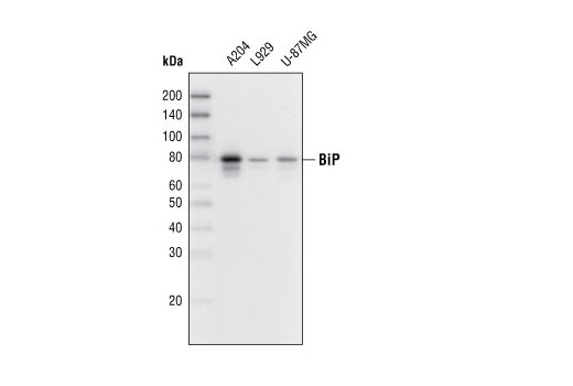
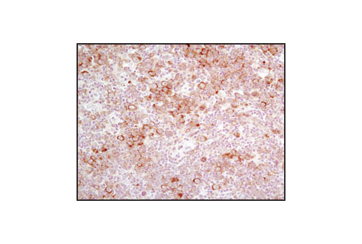
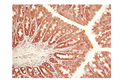
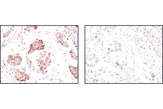
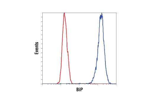
| BiP (C50B12) Rabbit mAb | 3177 | 20 µl | 78 kDa | Rabbit IgG |
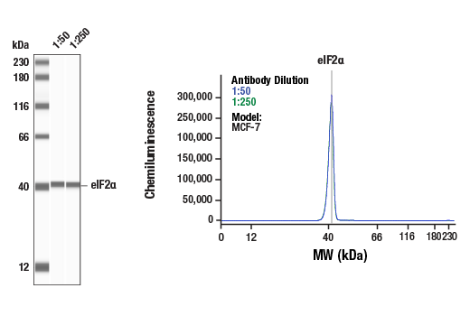
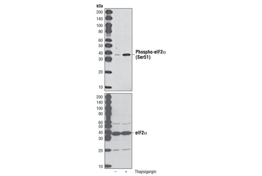
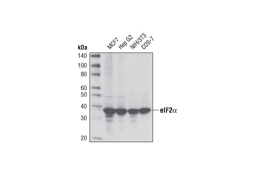
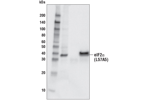
| eIF2α (D7D3) XP® Rabbit mAb | 5324 | 20 µl | 38 kDa | Rabbit IgG |
| Phospho-eIF2α (Ser51) (D9G8) XP® Rabbit mAb | 3398 | 20 µl | 38 kDa | Rabbit IgG |
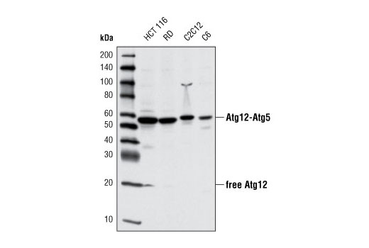
| Atg12 (D88H11) Rabbit mAb | 4180 | 20 µl | 16, 55 kDa | Rabbit IgG |
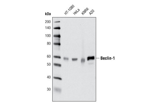
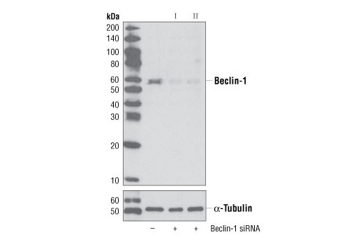
| Beclin-1 (D40C5) Rabbit mAb | 3495 | 20 µl | 60 kDa | Rabbit IgG |
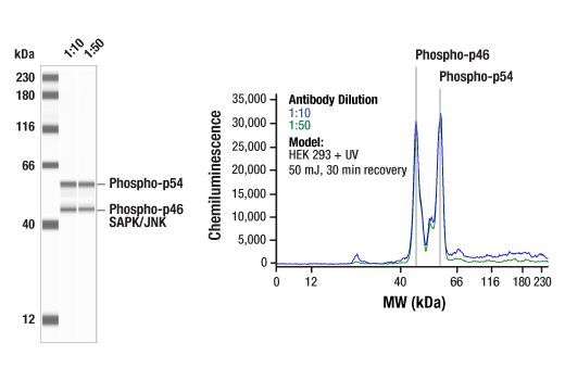
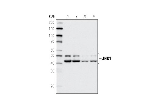
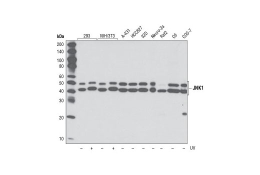
| JNK1 (2C6) Mouse mAb | 3708 | 20 µl | 46, 54 kDa | Mouse IgG1 |
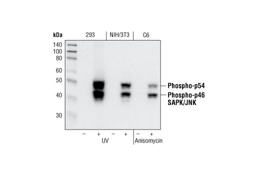
| Phospho-SAPK/JNK (Thr183/Tyr185) (81E11) Rabbit mAb | 4668 | 20 µl | 46, 54 kDa | Rabbit IgG |
| Anti-rabbit IgG, HRP-linked Antibody | 7074 | 100 µl | Goat | |
| Anti-mouse IgG, HRP-linked Antibody | 7076 | 100 µl | Horse |
Please visit cellsignal.com for individual component applications, species cross-reactivity, dilutions, protocols, and additional product information.
Description
The ER Stress-induced Antibody Sampler Kit contains reagents to investigate ER stress-induced signaling within the cell. The kit contains enough primary antibodies to perform four western blot experiments per primary antibody.
Storage
Background
The endoplasmic reticulum (ER) is an organelle with essential biosynthetic and signaling functions in eukaryotic cells (1). Post synthesis of secretory and transmembrane proteins on polysomes, proteins are translocated into the ER where they are often modified by disulfide bond formation, amino-linked glycosylation, and folding. Different physiological and pathological conditions can disturb proper protein folding in the ER causing ER stress (1). ER stress activates an intracellular signaling transduction pathway called unfolded protein response (UPR) and autophagy to avoid cell death (2). The main role of UPR is to improve the protein load on the ER by shutting down protein translation and gene transcription to enhance ER's folding capacity (2). On the other hand, autophagy is a catabolic process for the autophagosomic-lysosomal degradation of bulk cytoplasmc contents (3,4). One of the chaperones aiding in proper protein folding is Binding immunoglobulin Protein (BiP) (5,6). BiP works by binding to misfolded proteins to prevent them from forming aggregates and assists in proper refolding (7). The molecular machinery of autophagy was largely discovered in yeast and referred to as autophagy-related (Atg) genes. Formation of the autophagosome involves a ubiquitin-like conjugation system in which Atg12 is covalently bound to Atg5 and targeted to autophagosome vesicles (8-10). One of the proteins critical to autophagy process is Beclin-1, the mammalian orthologue of the yeast autophagy protein Apg6/Vps30 (11). Beclin-1 can complement defects in yeast autophagy caused by loss of Apg6 and can also stimulate autophagy when overexpressed in mammalian cells (12). Mammalian Beclin-1 was originally isolated in a yeast two-hybrid screen for Bcl-2 interacting proteins and has been shown to interact with Bcl-2 and Bcl-xL, but not with Bax or Bak (13). Phosphorylation of the eukaryotic initiation factor 2 (eIF2) α subunit is a well-documented mechanism to downregulate protein synthesis under a variety of stress conditions. eIF2 binds GTP and Met-tRNAi and transfers Met-tRNA to the 40S subunit to form the 43S preinitiation complex (14,15). Kinases that are activated by viral infection (PKR) can phosphorylate the α subunit of eIF2 (16,17). Induction of PKR by IFN-γ and TNF-α induces potent phosphorylation of eIF2α at Ser51 (18,19). There are three SAPK/JNK genes each of which undergoes alternative splicing, resulting in numerous isoforms (20). The IRE1, a transmembrane serine/threonine kinase (21,22), through its kinase activity activates SAPK/JNK in the early stage of ER stress in order to induce autophagosome formation (23).
- Verfaillie, T. et al. (2010) Int J Cell Biol 2010, 930509.
- Ogata, M. et al. (2006) Mol Cell Biol 26, 9220-31.
- Reggiori, F. and Klionsky, D.J. (2002) Eukaryot Cell 1, 11-21.
- Codogno, P. and Meijer, A.J. (2005) Cell Death Differ 12 Suppl 2, 1509-18.
- Wabl, M. and Steinberg, C. (1982) Proc Natl Acad Sci U S A 79, 6976-8.
- Haas, I.G. and Wabl, M. (2002) Nature 306, 387-9.
- Kohno, K. et al. (1993) Mol Cell Biol 13, 877-90.
- Mizushima, N. et al. (1998) J Biol Chem 273, 33889-92.
- Mizushima, N. et al. (1998) Nature 395, 395-8.
- Suzuki, K. et al. (2001) EMBO J 20, 5971-81.
- Kametaka, S. et al. (1998) J Biol Chem 273, 22284-91.
- Liang, X.H. et al. (1999) Nature 402, 672-6.
- Liang, X.H. et al. (1998) J Virol 72, 8586-96.
- Kimball, S.R. (1999) Int J Biochem Cell Biol 31, 25-9.
- de Haro, C. et al. (1996) FASEB J 10, 1378-87.
- Kaufman, R.J. (1999) Genes Dev 13, 1211-33.
- Sheikh, M.S. and Fornace, A.J. (1999) Oncogene 18, 6121-8.
- Cheshire, J.L. et al. (1999) J Biol Chem 274, 4801-6.
- Zamanian-Daryoush, M. et al. (2000) Mol Cell Biol 20, 1278-90.
- Kyriakis, J.M. and Avruch, J. (2001) Physiol Rev 81, 807-69.
- Nikawa, J. and Yamashita, S. (1992) Mol Microbiol 6, 1441-6.
- Cox, J.S. et al. (1993) Cell 73, 1197-206.
- Urano, F. et al. (2000) Science 287, 664-6.
Background References
Trademarks and Patents
使用に関する制限
法的な権限を与えられたCSTの担当者が署名した書面によって別途明示的に合意された場合を除き、 CST、その関連会社または代理店が提供する製品には以下の条件が適用されます。お客様が定める条件でここに定められた条件に含まれるものを超えるもの、 または、ここに定められた条件と異なるものは、法的な権限を与えられたCSTの担当者が別途書面にて受諾した場合を除き、拒絶され、 いかなる効力も効果も有しません。
研究専用 (For Research Use Only) またはこれに類似する表示がされた製品は、 いかなる目的についても FDA または外国もしくは国内のその他の規制機関により承認、認可または許可を受けていません。 お客様は製品を診断もしくは治療目的で使用してはならず、また、製品に表示された内容に違反する方法で使用してはなりません。 CST が販売または使用許諾する製品は、エンドユーザーであるお客様に対し、使途を研究および開発のみに限定して提供されるものです。 診断、予防もしくは治療目的で製品を使用することまたは製品を再販売 (単独であるか他の製品等の一部であるかを問いません) もしくはその他の商業的利用の目的で購入することについては、CST から別途許諾を得る必要があります。 お客様は以下の事項を遵守しなければなりません。(a) CST の製品 (単独であるか他の資材と一緒であるかを問いません) を販売、使用許諾、貸与、寄付もしくはその他の態様で第三者に譲渡したり使用させたりしてはなりません。また、商用の製品を製造するために CST の製品を使用してはなりません。(b) 複製、改変、リバースエンジニアリング、逆コンパイル、 分解または他の方法により製品の構造または技術を解明しようとしてはなりません。また、 CST の製品またはサービスと競合する製品またはサービスを開発する目的で CST の製品を使用してはなりません。(c) CST の製品の商標、商号、ロゴ、特許または著作権に関する通知または表示を除去したり改変したりしてはなりません。(d) CST の製品をCST 製品販売条件(CST’s Product Terms of Sale) および該当する書面のみに従って使用しなければなりません。(e) CST の製品に関連してお客様が使用する第三者の製品またはサービスに関する使用許諾条件、 サービス提供条件またはこれに類する合意事項を遵守しなければなりません。