| Product Includes | Product # | Quantity | Mol. Wt | Isotype/Source |
|---|---|---|---|---|
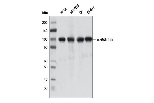
| α-Actinin (D6F6) XP® Rabbit mAb | 6487 | 20 µl | 100 kDa | Rabbit IgG |
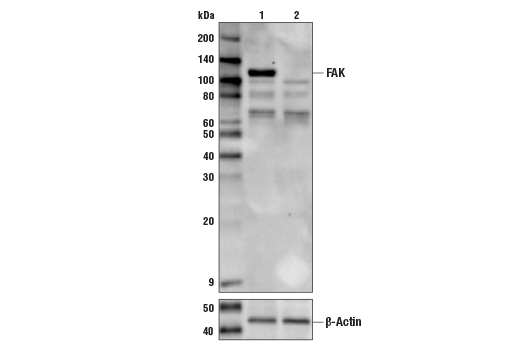
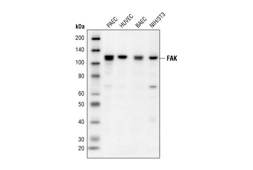
| FAK Antibody | 3285 | 20 µl | 125 kDa | Rabbit |
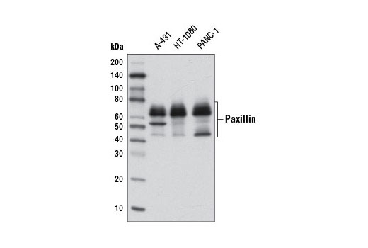
| Paxillin (D9G12) Rabbit mAb | 12065 | 20 µl | 54, 62, 68 kDa | Rabbit IgG |
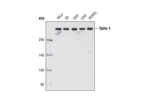
| Talin-1 (C45F1) Rabbit mAb | 4021 | 20 µl | 270 kDa | Rabbit IgG |
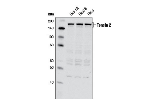
| Tensin 2 Antibody | 11990 | 20 µl | 145-155 kDa | Rabbit |
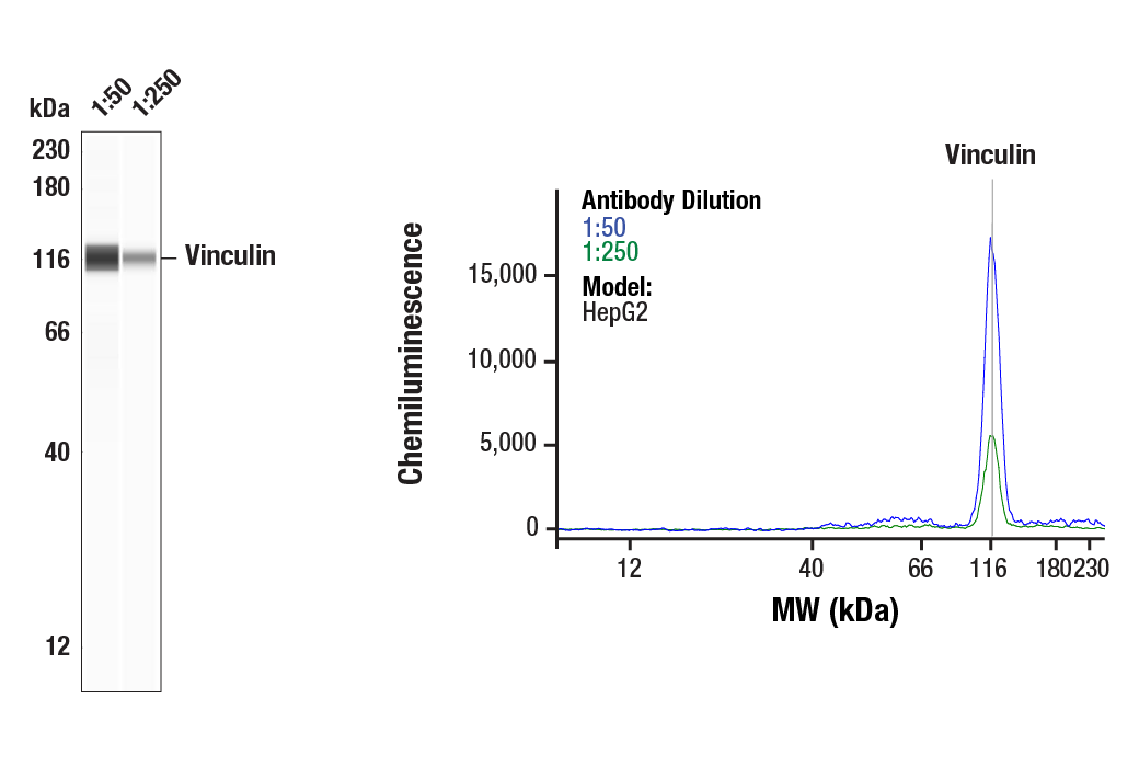
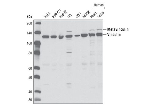
| Vinculin Antibody | 4650 | 20 µl | 124 kDa | Rabbit |
| Anti-rabbit IgG, HRP-linked Antibody | 7074 | 100 µl | Goat |
Please visit cellsignal.com for individual component applications, species cross-reactivity, dilutions, protocols, and additional product information.
Description
The Focal Adhesion Protein Antibody Sampler Kit provides an economical means to evaluate proteins involved in focal adhesions. The kit includes enough antibody to perform two western blot experiments per primary antibody.
Storage
Background
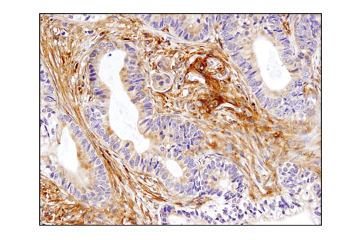
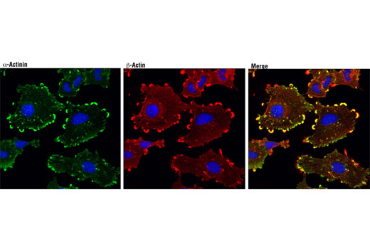
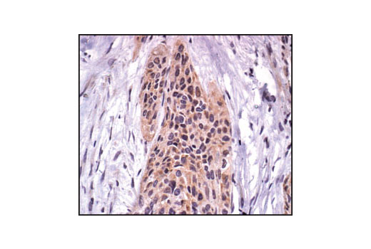
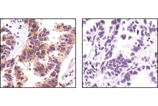
Focal adhesions connect the cytoskeleton with the extracellular matrix (ECM), a complex structure of secreted macromolecules that surrounds mammalian organs and tissues. Integrins clustered on the extracellular side of focal adhesions relay signals from the ECM to intracellular protein complexes that signal the actin cytoskeleton to regulate tension for cell motility. Internal signals converge on focal adhesions to regulate integrin receptor affinity and avidity. Signaling through focal adhesions regulates cell adhesion, migration, proliferation, apoptosis, and gene expression, and impacts cellular processes such as development, wound healing, immune response, invasion, metastasis and angiogenesis (reviewed in 1-3). Focal adhesion kinase (FAK) is a widely expressed cytoplasmic protein tyrosine kinase involved in integrin-mediated signal transduction. Integrin clustering triggers FAK recruitment to the focal adhesion complex (4). Talin is a large, multidomain focal adhesion protein that interacts with the intracellular domains of integrins and other focal adhesion proteins. Talin is involved in the formation of focal adhesions and in linking focal adhesions to the actin cytoskeleton (5). Paxillin is a key component of integrin signaling that localizes primarily to focal adhesion sites in the extracellular matrix (6). Tyrosine phosphorylation of paxillin is required for integrin-mediated cytoskeletal reorganization (7). Paxillin is phosphorylated by FAK at Tyr118 (8,9). Vinculin is a cytoskeletal protein involved in regulation of focal adhesions and embryonic development (10-13). Active vinculin translocates to focal adhesions where it may be involved in anchoring F-actin to the membrane and regulating cell migration. Vinculin binds a number of proteins, including talin, α-actinin and paxillin (11,13). Tensin 2 localizes to focal adhesions of various tissues and exhibits highest expression in heart, kidney, and liver (14,15). Tensin 2 belongs to a family of cytoskeletal proteins that include Tensin 1-3 and Cten, which couple integrins to the actin cytoskeleton (16). Tensin family proteins play an important role in signal transduction, cell proliferation, and motility (17-20). α-actinin is a member of the spectrin family of cytoskeletal proteins that was first recognized as an actin cross-linking protein, but also interacts with a large number of cytoskeletal signaling proteins, including those involved in cellular adhesion, migration, and immune cell targeting (21).
- Burridge, K. et al. (1988) Annu Rev Cell Biol 4, 487-525.
- Calderwood, D.A. et al. (2000) J Biol Chem 275, 22607-10.
- ffrench-Constant, C. and Colognato, H. (2004) Trends Cell Biol 14, 678-86.
- Parsons, J.T. et al. (2000) Oncogene 19, 5606-13.
- Nayal, A. et al. (2004) Curr Opin Cell Biol 16, 94-8.
- Turner, C.E. (2000) J Cell Sci 113 Pt 23, 4139-40.
- Burridge, K. et al. (1992) J Cell Biol 119, 893-903.
- Bellis, S.L. et al. (1995) J Biol Chem 270, 17437-41.
- Bellis, S.L. et al. (1997) Biochem J 325 ( Pt 2), 375-81.
- Izard, T. et al. (2004) Nature 427, 171-5.
- Humphries, J.D. et al. (2007) J Cell Biol 179, 1043-57.
- Witt, S. et al. (2004) J Biol Chem 279, 31533-43.
- Xu, W. et al. (1998) Development 125, 327-37.
- Clark, K. et al. (2010) J Cell Biochem 109, 808-17.
- Hafizi, S. et al. (2002) Biochem Biophys Res Commun 299, 793-800.
- Lo, S.H. et al. (1994) Bioessays 16, 817-23.
- Lo, S.H. et al. (1994) J Cell Biol 125, 1067-75.
- Chen, H. and Lo, S.H. (2003) Biochem J 370, 1039-45.
- Katz, M. et al. (2007) Nat Cell Biol 9, 961-9.
- Chen, H. et al. (2002) Proc Natl Acad Sci U S A 99, 733-8.
- Otey, C.A. and Carpen, O. (2004) Cell Motil Cytoskeleton 58, 104-11.
Background References
Trademarks and Patents
使用に関する制限
法的な権限を与えられたCSTの担当者が署名した書面によって別途明示的に合意された場合を除き、 CST、その関連会社または代理店が提供する製品には以下の条件が適用されます。お客様が定める条件でここに定められた条件に含まれるものを超えるもの、 または、ここに定められた条件と異なるものは、法的な権限を与えられたCSTの担当者が別途書面にて受諾した場合を除き、拒絶され、 いかなる効力も効果も有しません。
研究専用 (For Research Use Only) またはこれに類似する表示がされた製品は、 いかなる目的についても FDA または外国もしくは国内のその他の規制機関により承認、認可または許可を受けていません。 お客様は製品を診断もしくは治療目的で使用してはならず、また、製品に表示された内容に違反する方法で使用してはなりません。 CST が販売または使用許諾する製品は、エンドユーザーであるお客様に対し、使途を研究および開発のみに限定して提供されるものです。 診断、予防もしくは治療目的で製品を使用することまたは製品を再販売 (単独であるか他の製品等の一部であるかを問いません) もしくはその他の商業的利用の目的で購入することについては、CST から別途許諾を得る必要があります。 お客様は以下の事項を遵守しなければなりません。(a) CST の製品 (単独であるか他の資材と一緒であるかを問いません) を販売、使用許諾、貸与、寄付もしくはその他の態様で第三者に譲渡したり使用させたりしてはなりません。また、商用の製品を製造するために CST の製品を使用してはなりません。(b) 複製、改変、リバースエンジニアリング、逆コンパイル、 分解または他の方法により製品の構造または技術を解明しようとしてはなりません。また、 CST の製品またはサービスと競合する製品またはサービスを開発する目的で CST の製品を使用してはなりません。(c) CST の製品の商標、商号、ロゴ、特許または著作権に関する通知または表示を除去したり改変したりしてはなりません。(d) CST の製品をCST 製品販売条件(CST’s Product Terms of Sale) および該当する書面のみに従って使用しなければなりません。(e) CST の製品に関連してお客様が使用する第三者の製品またはサービスに関する使用許諾条件、 サービス提供条件またはこれに類する合意事項を遵守しなければなりません。