| Product Includes | Product # | Quantity | Mol. Wt | Isotype/Source |
|---|---|---|---|---|
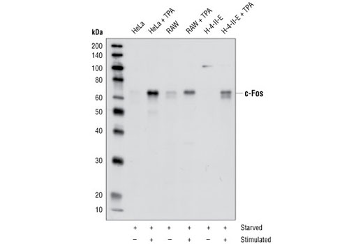
| c-Fos Antibody | 4384 | 20 µl | 62 kDa | Rabbit |
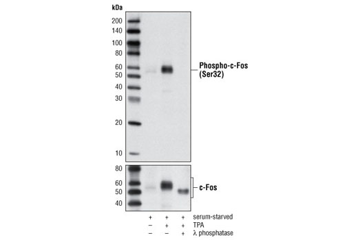
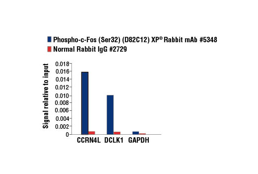
| Phospho-c-Fos (Ser32) (D82C12) XP® Rabbit mAb | 5348 | 20 µl | 62 kDa | Rabbit IgG |
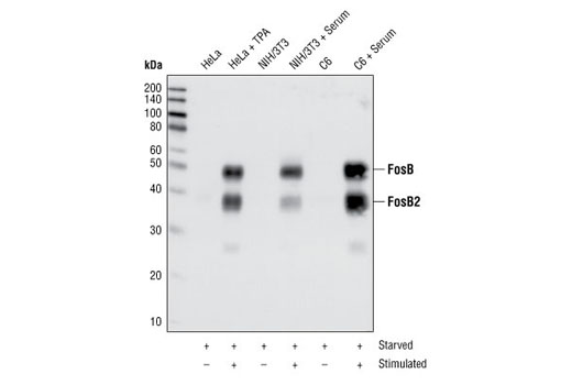
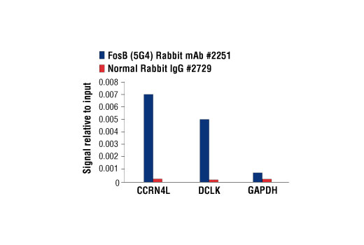
| FosB (5G4) Rabbit mAb | 2251 | 20 µl | 38 FosB2 48 FosB kDa | Rabbit IgG |
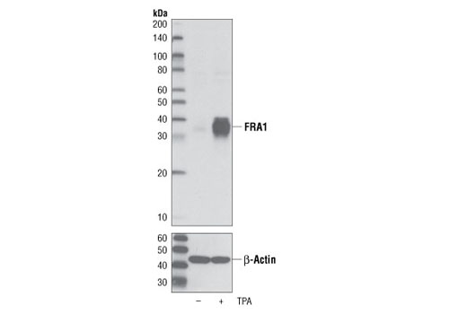
| FRA1 (D80B4) Rabbit mAb | 5281 | 20 µl | 40 kDa | Rabbit IgG |
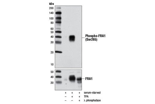
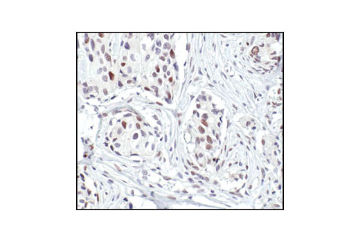
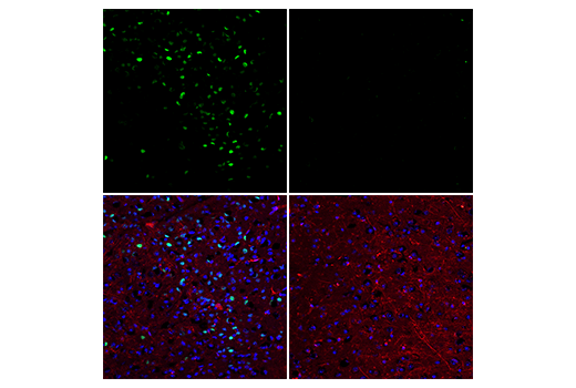
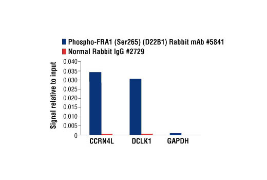
| Phospho-FRA1 (Ser265) (D22B1) Rabbit mAb | 5841 | 20 µl | 40 kDa | Rabbit IgG |
| Anti-rabbit IgG, HRP-linked Antibody | 7074 | 100 µl | Goat |
Please visit cellsignal.com for individual component applications, species cross-reactivity, dilutions, protocols, and additional product information.
Description
The Fos Family Antibody Sampler Kit provides an economical means to evaluate the Fos family of transcription factors. The kit includes enough antibody to perform two western blot experiments with each primary antibody.
Storage
Background
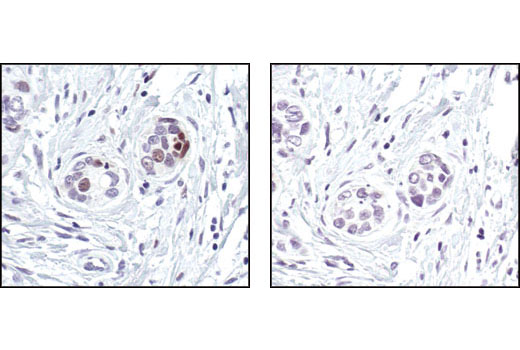
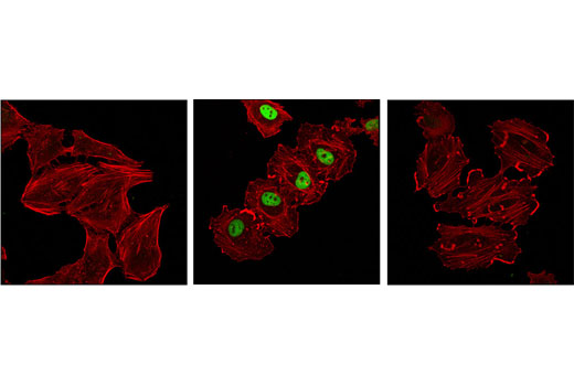
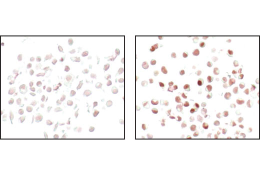
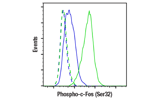
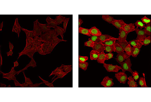
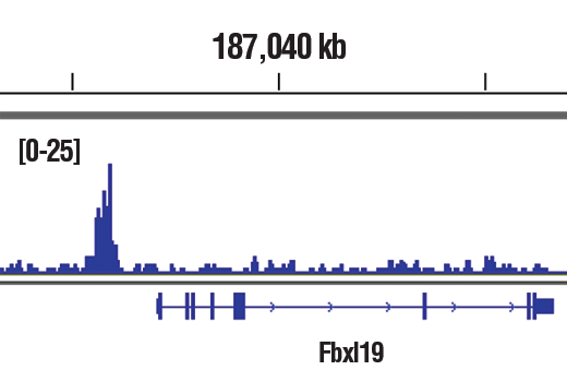
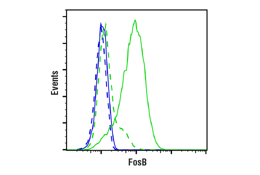
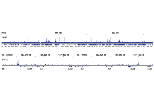
The Fos family of nuclear oncogenes includes c-Fos, FosB, Fos-related antigen 1 (FRA1), and Fos-related antigen 2 (FRA2) (1). While most Fos proteins exist as a single isoform, the FosB protein exists as two isoforms: full-length FosB and a shorter form, FosB2 (Delta FosB), which lacks the carboxy-terminal 101 amino acids (1-3). The expression of Fos proteins is rapidly and transiently induced by a variety of extracellular stimuli, including growth factors, cytokines, neurotransmitters, polypeptide hormones, and stress. Fos proteins dimerize with Jun proteins (c-Jun, JunB, and JunD) to form Activator Protein-1 (AP-1), a transcription factor that binds to TRE/AP-1 elements and activates transcription. Fos and Jun proteins contain the leucine-zipper motif that mediates dimerization and an adjacent basic domain that binds to DNA. The various Fos/Jun heterodimers differ in their ability to transactivate AP-1 dependent genes. In addition to increased expression, phosphorylation of Fos proteins by Erk kinases in response to extracellular stimuli may further increase transcriptional activity (4-6). Phosphorylation of c-Fos at Ser32 and Thr232 by Erk5 increases protein stability and nuclear localization (5). Phosphorylation of FRA1 at Ser252 and Ser265 by Erk1/2 increases protein stability and leads to overexpression of FRA1 in cancer cells (6). Following growth factor stimulation, expression of FosB and c-Fos in quiescent fibroblasts is immediate, but very short-lived, with protein levels dissipating after several hours (7). FRA1 and FRA2 expression persists longer, and appreciable levels can be detected in asynchronously growing cells (8). Deregulated expression of c-Fos, FosB, or FRA2 can result in neoplastic cellular transformation; however, Delta FosB lacks the ability to transform cells (2,3).
- Tulchinsky, E. (2000) Histol Histopathol 15, 921-8.
- Dobrazanski, P. et al. (1991) Mol Cell Biol 11, 5470-8.
- Nakabeppu, Y. and Nathans, D. (1991) Cell 64, 751-9.
- Rosenberger, S.F. et al. (1999) J Biol Chem 274, 1124-30.
- Sasaki, T. et al. (2006) Mol Cell 24, 63-75.
- Basbous, J. et al. (2007) Mol Cell Biol 27, 3936-50.
- Kovary, K. and Bravo, R. (1991) Mol Cell Biol 11, 2451-9.
- Kovary, K. and Bravo, R. (1992) Mol Cell Biol 12, 5015-23.
Background References
Trademarks and Patents
使用に関する制限
法的な権限を与えられたCSTの担当者が署名した書面によって別途明示的に合意された場合を除き、 CST、その関連会社または代理店が提供する製品には以下の条件が適用されます。お客様が定める条件でここに定められた条件に含まれるものを超えるもの、 または、ここに定められた条件と異なるものは、法的な権限を与えられたCSTの担当者が別途書面にて受諾した場合を除き、拒絶され、 いかなる効力も効果も有しません。
研究専用 (For Research Use Only) またはこれに類似する表示がされた製品は、 いかなる目的についても FDA または外国もしくは国内のその他の規制機関により承認、認可または許可を受けていません。 お客様は製品を診断もしくは治療目的で使用してはならず、また、製品に表示された内容に違反する方法で使用してはなりません。 CST が販売または使用許諾する製品は、エンドユーザーであるお客様に対し、使途を研究および開発のみに限定して提供されるものです。 診断、予防もしくは治療目的で製品を使用することまたは製品を再販売 (単独であるか他の製品等の一部であるかを問いません) もしくはその他の商業的利用の目的で購入することについては、CST から別途許諾を得る必要があります。 お客様は以下の事項を遵守しなければなりません。(a) CST の製品 (単独であるか他の資材と一緒であるかを問いません) を販売、使用許諾、貸与、寄付もしくはその他の態様で第三者に譲渡したり使用させたりしてはなりません。また、商用の製品を製造するために CST の製品を使用してはなりません。(b) 複製、改変、リバースエンジニアリング、逆コンパイル、 分解または他の方法により製品の構造または技術を解明しようとしてはなりません。また、 CST の製品またはサービスと競合する製品またはサービスを開発する目的で CST の製品を使用してはなりません。(c) CST の製品の商標、商号、ロゴ、特許または著作権に関する通知または表示を除去したり改変したりしてはなりません。(d) CST の製品をCST 製品販売条件(CST’s Product Terms of Sale) および該当する書面のみに従って使用しなければなりません。(e) CST の製品に関連してお客様が使用する第三者の製品またはサービスに関する使用許諾条件、 サービス提供条件またはこれに類する合意事項を遵守しなければなりません。