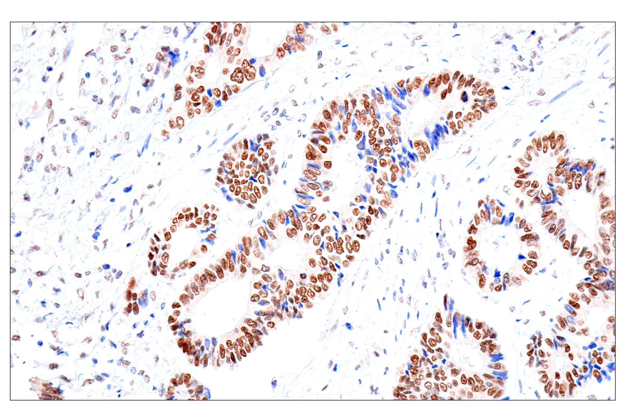
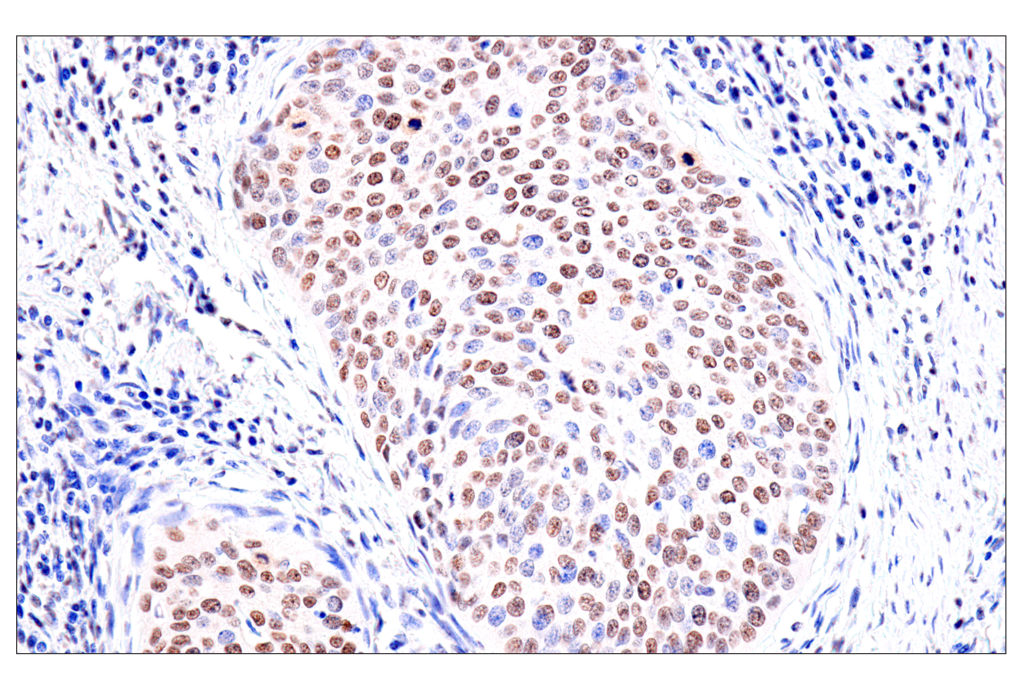
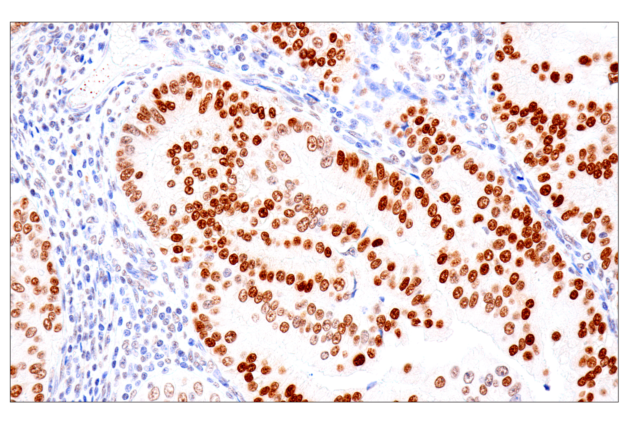
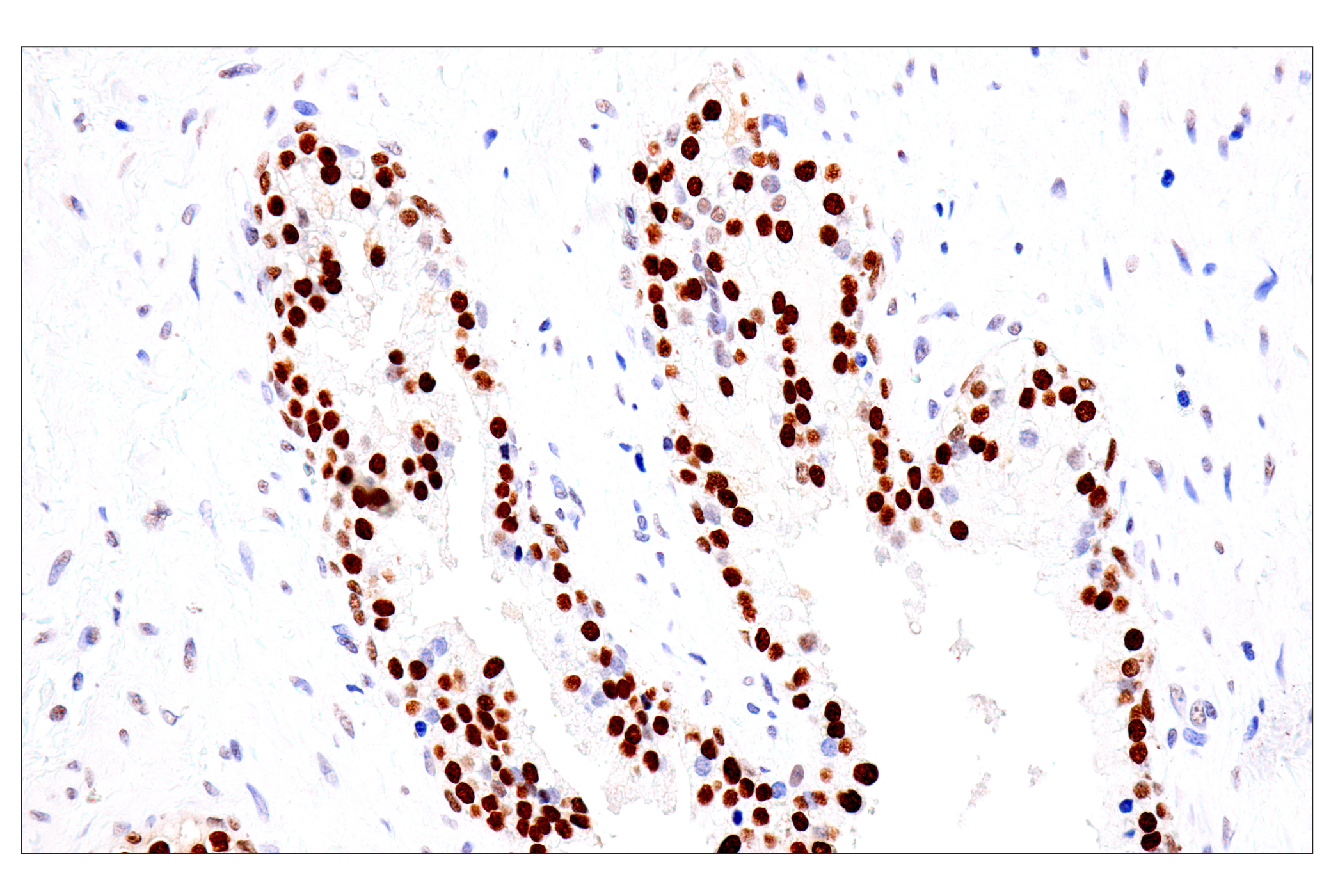
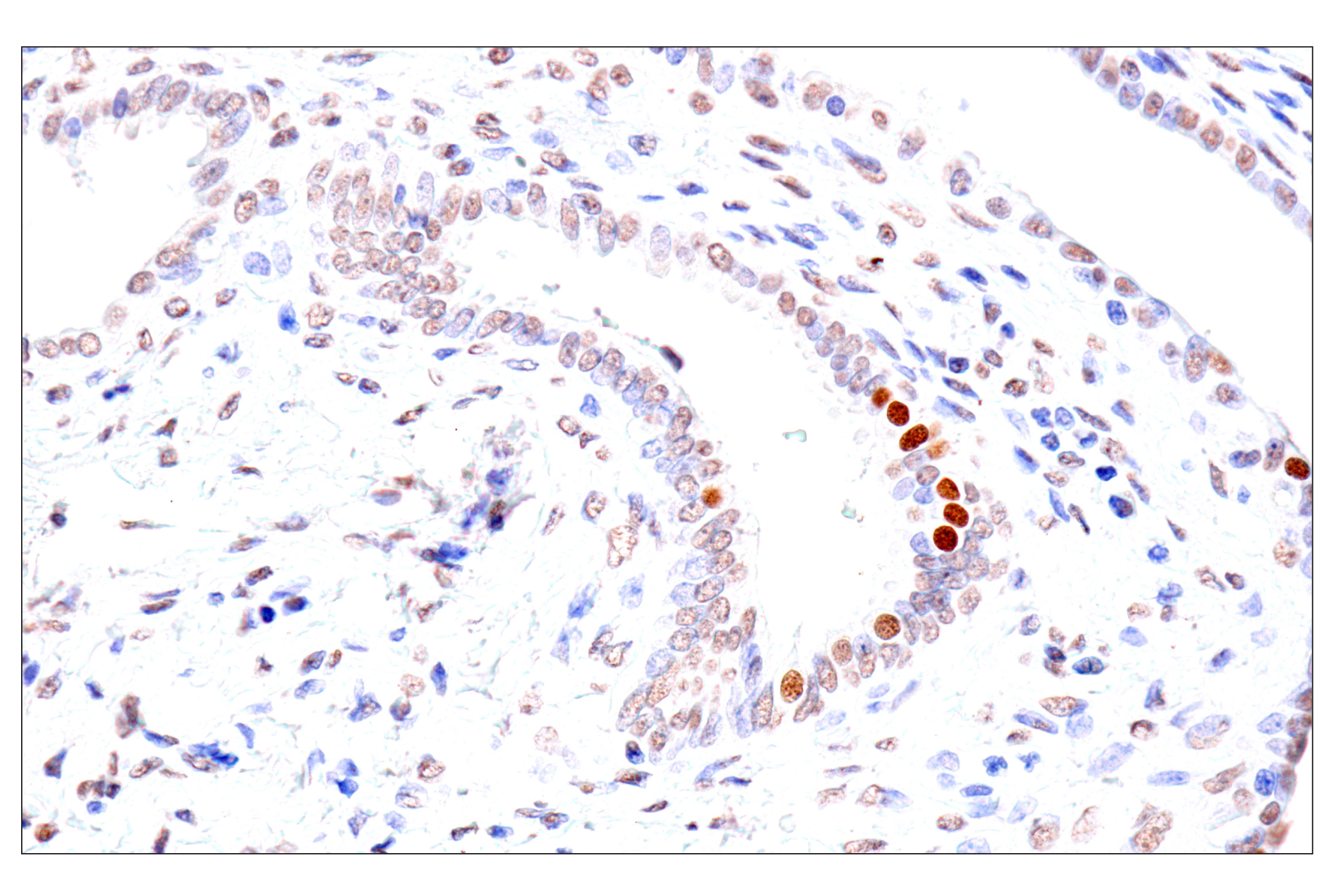
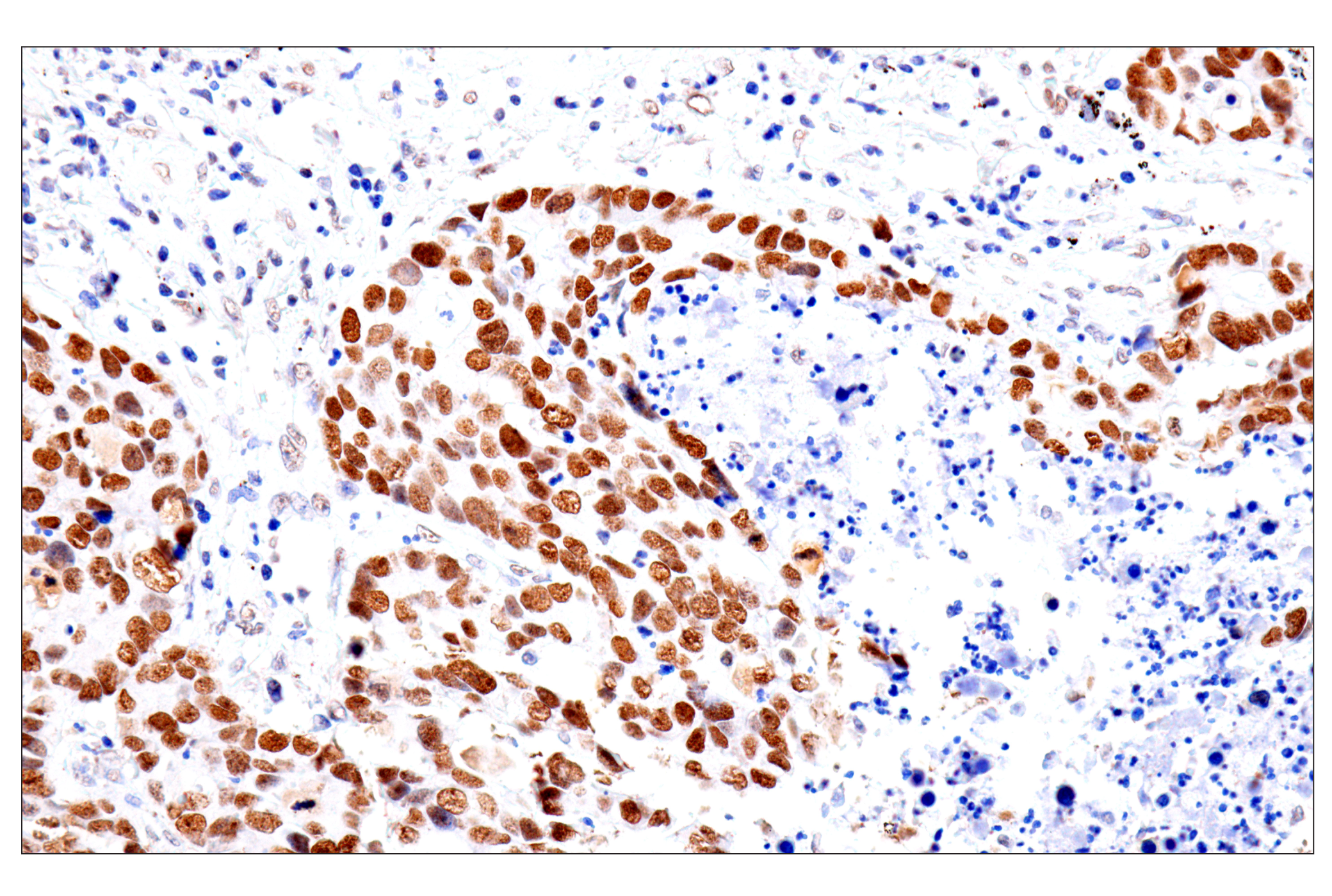
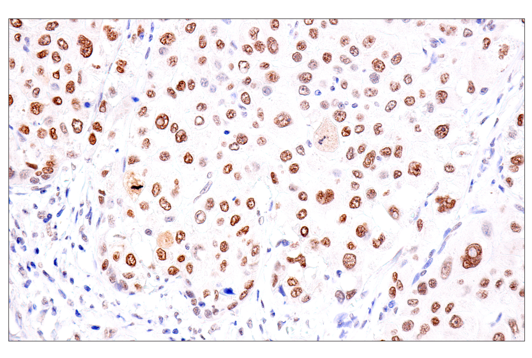
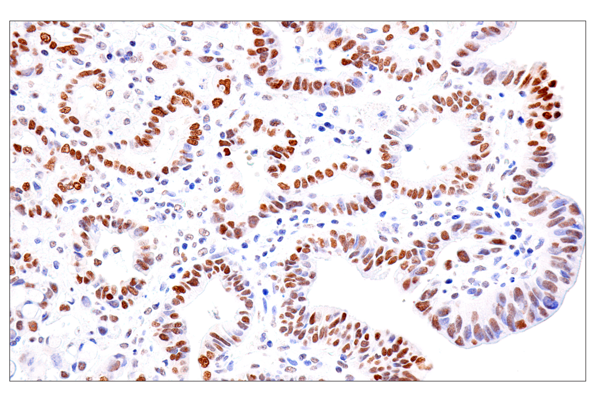
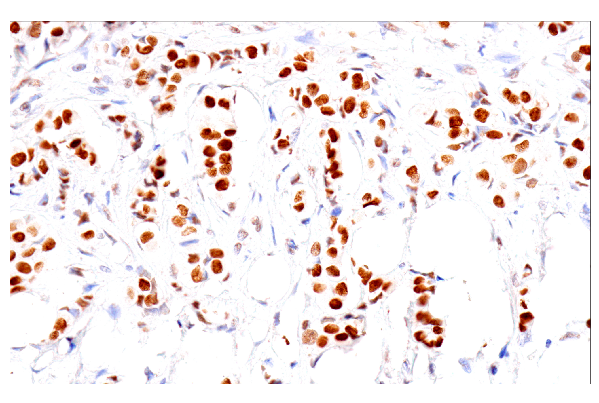
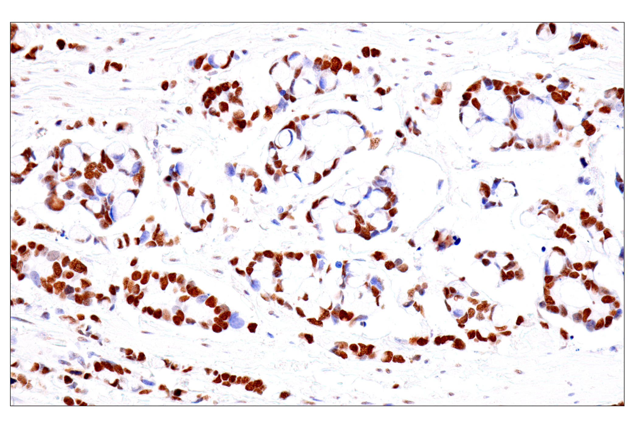
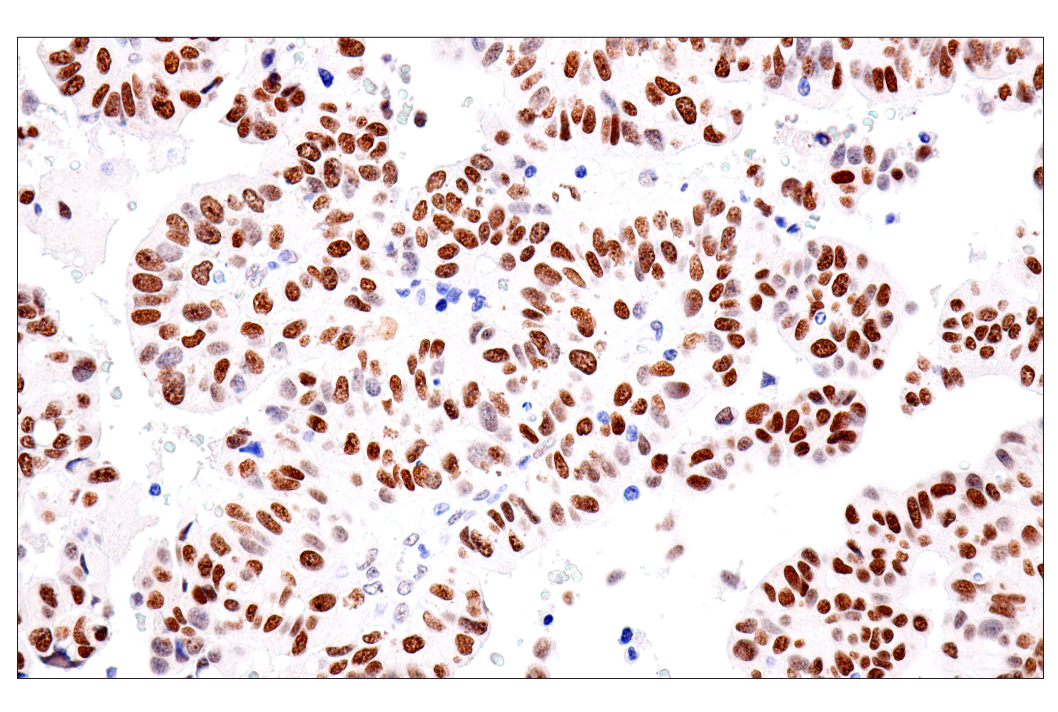
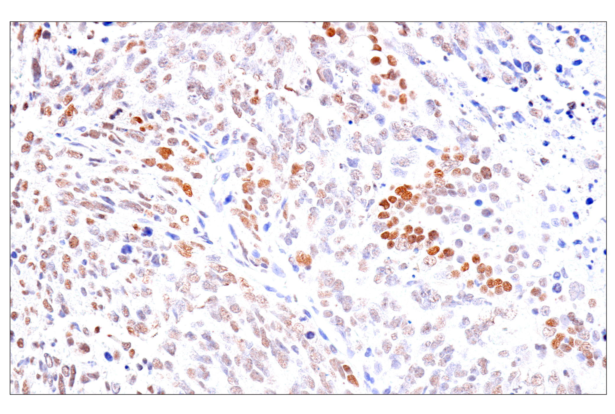
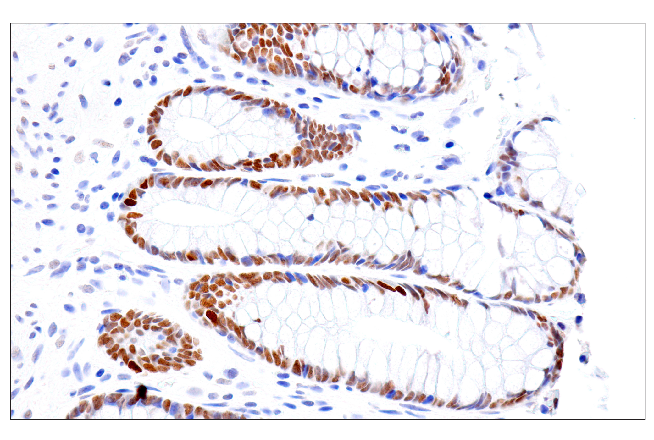
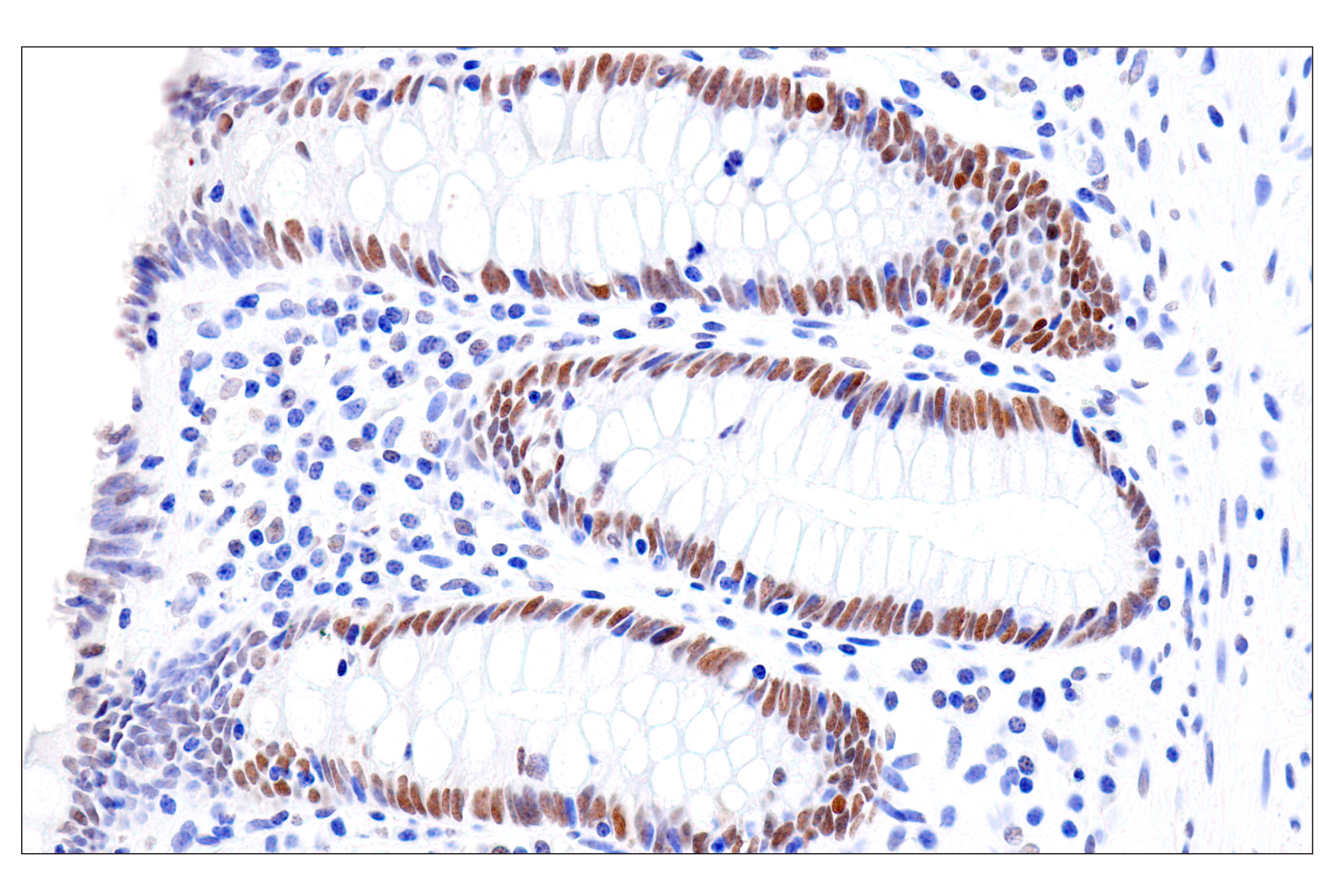
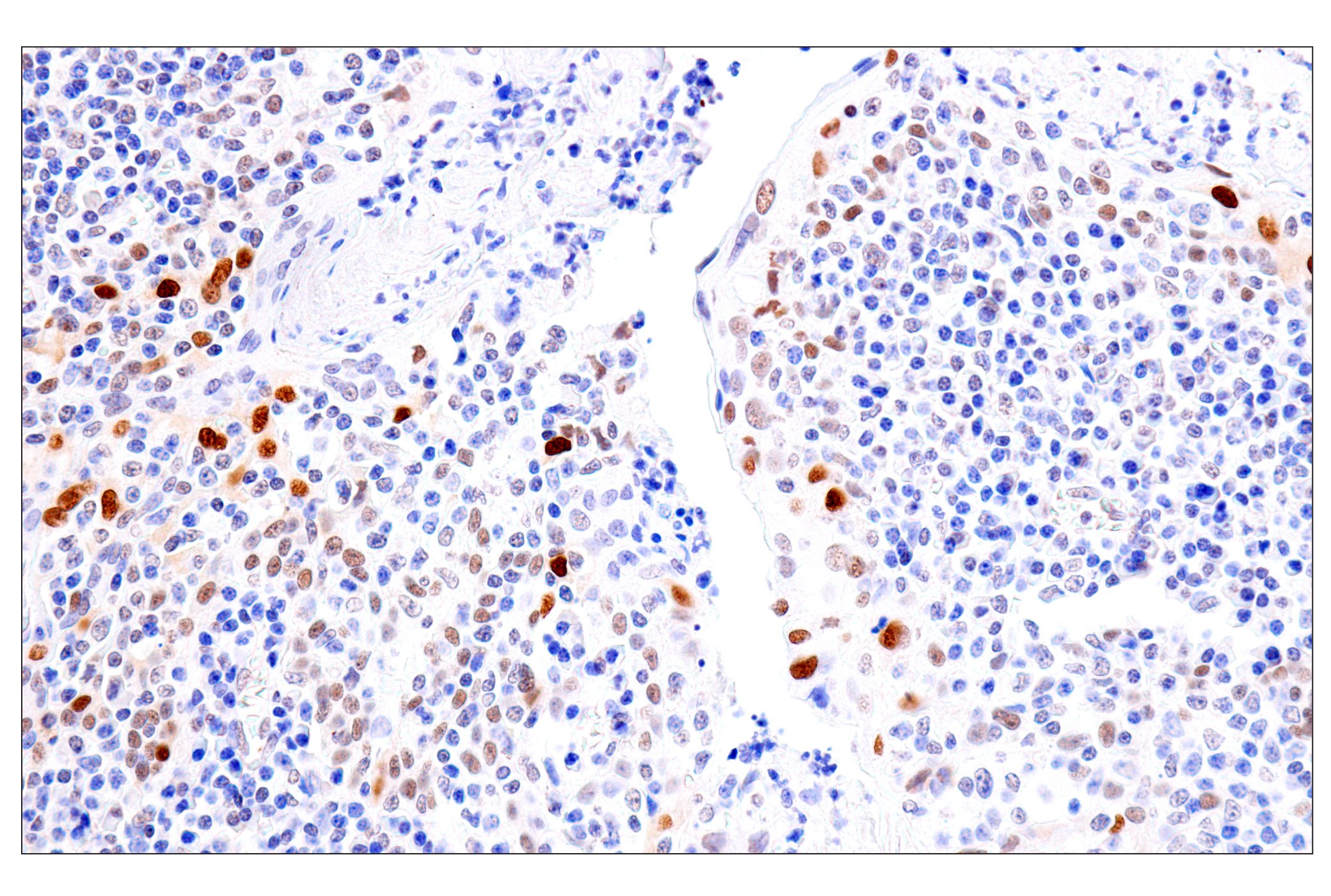
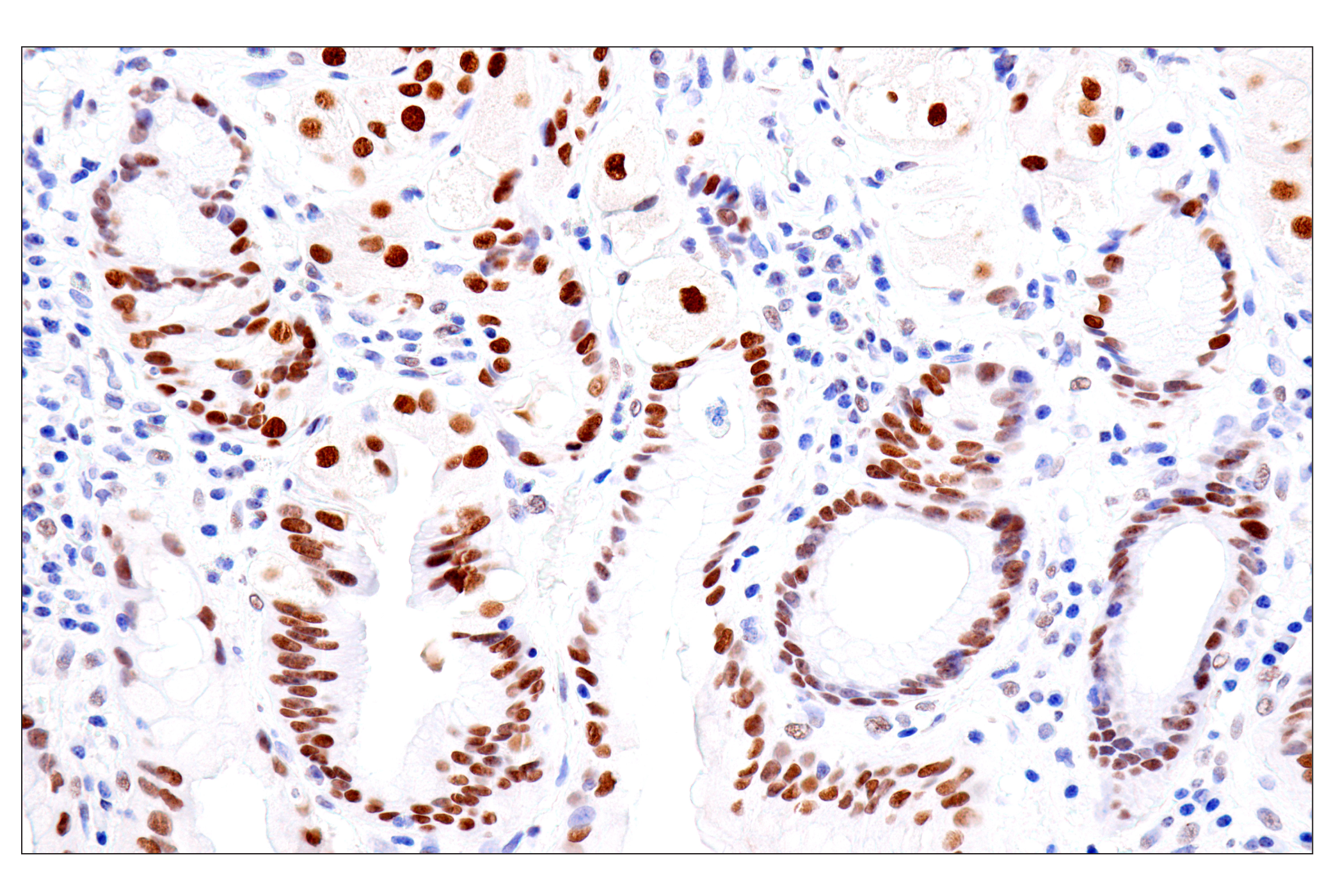
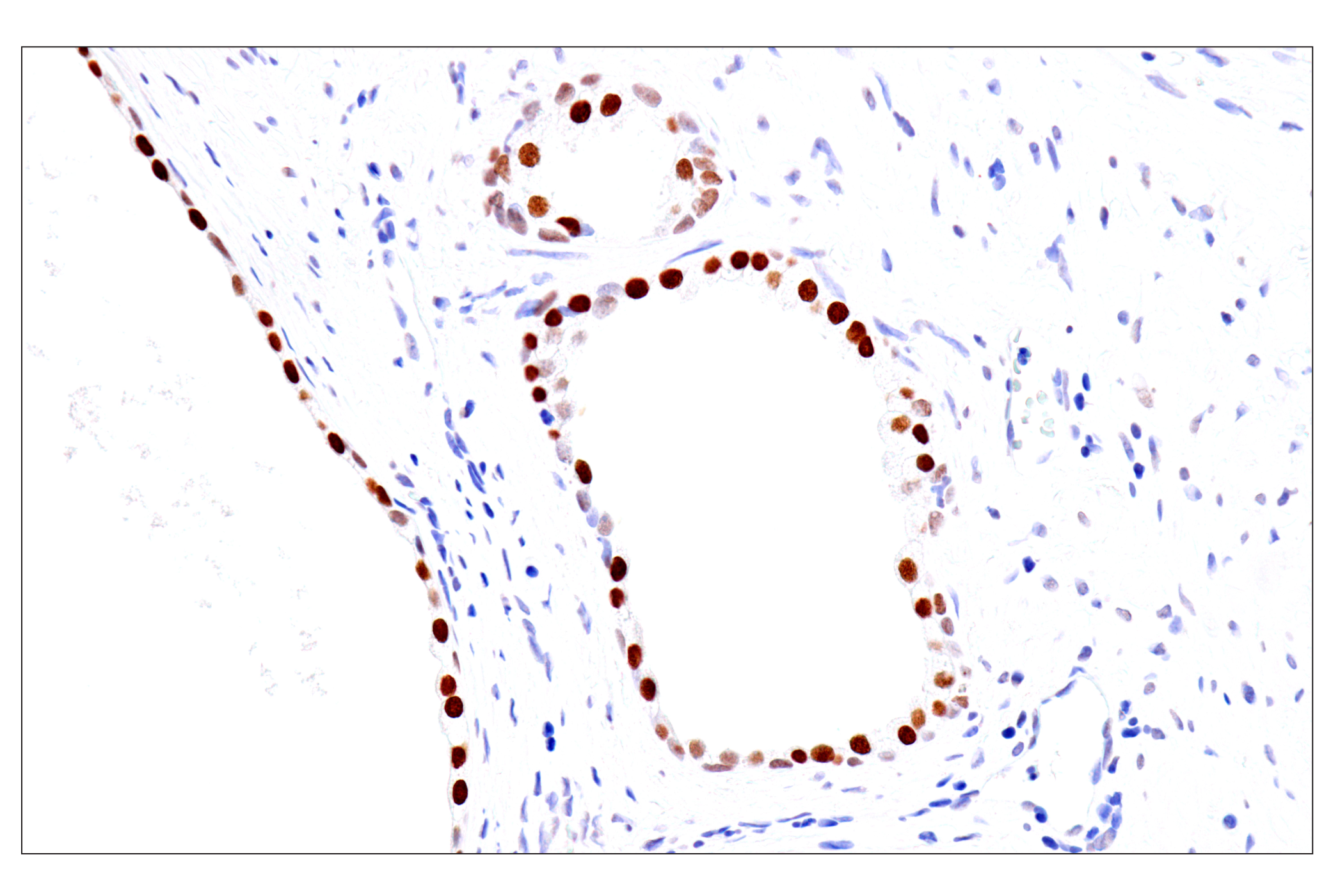
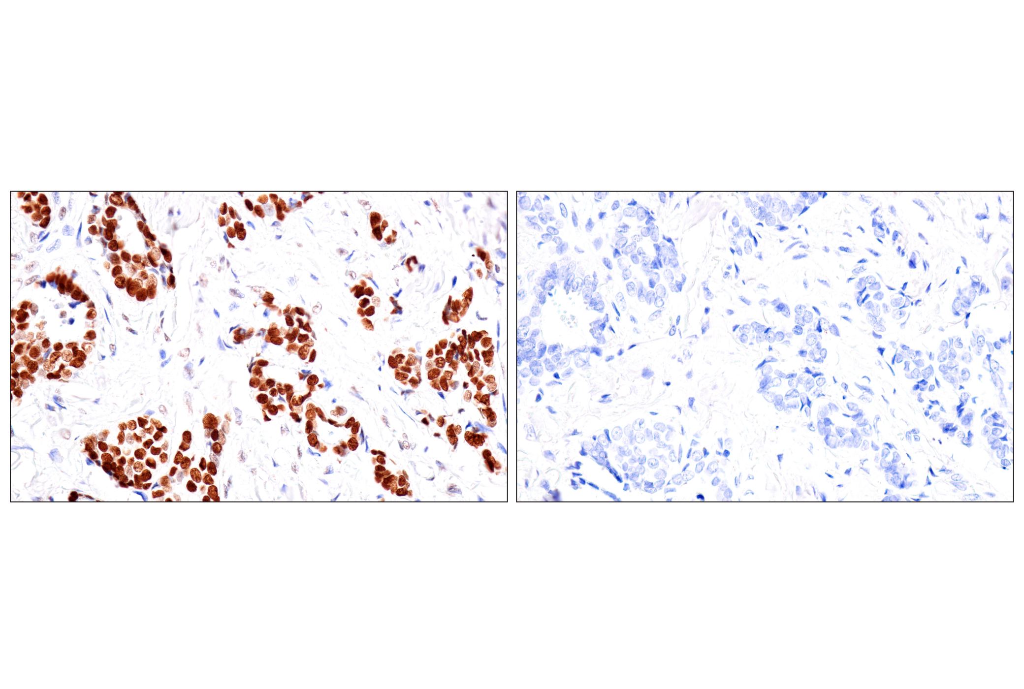
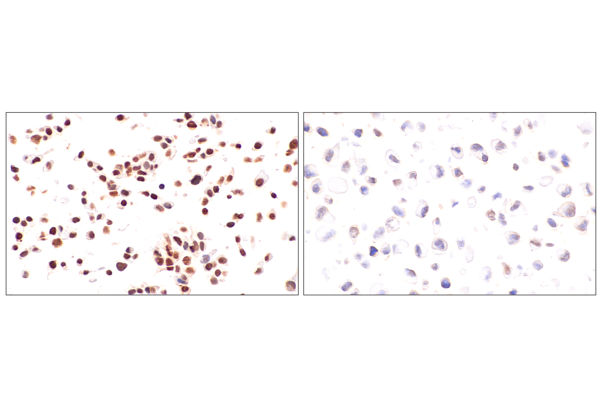
Revision 1

#59197Store at +4C

Cell Signaling Technology

Orders: 877-616-CELL (2355) [email protected]

Support: 877-678-TECH (8324)

Web: [email protected] cellsignal.com

3 Trask LaneDanversMassachusetts01923USA
For Research Use Only. Not for Use in Diagnostic Procedures.
Applications:

IHC-P

REACTIVITY:

H

SENSITIVITY:

Endogenous

MW (kDa):

Source/Isotype:

Mouse IgG1

UniProt ID:

#P55317

Entrez-Gene Id:

3169

Product Information

Product Usage Information

Application Dilution
Immunohistochemistry (Paraffin) 1:800 - 1:3200

Storage

Supplied in a Tris-based buffer with 1% BSA and less than 0.1% sodium azide. Stable for 24 months when stored at 4°C. Do not aliquot the antibody.

Specificity / Sensitivity

FoxA1/HNF3α (IHC578) Mouse mAb recognizes endogenous levels of total FoxA1/HNF3α protein.

Species Reactivity:

Human

Source / Purification

Monoclonal antibody is produced by immunizing animals with a recombinant protein corresponding to human FoxA1/HNF3α protein.

Background

Forkhead box protein A1 (FoxA1, HNF3α) is a transcription factor required for the development of endoderm-derived organs, such as liver, lung, and prostate (1). FoxA1 functions as a pioneer factor that is recruited primarily to the distant enhancers to change chromatin structure for transcription in a cell type-specific manner (2,3). The FoxA1 transcription factor is implicated in various diseases, playing a role in hormone-dependent disorders, such as breast and prostate cancers (4). The treatment of relapsing-remitting multiple sclerosis patients with IFN-β results in FoxA1-induced stimulation of a novel population of FoxA1(+) regulatory T cells, suggesting a possible immunosuppressive role for FoxA1 (5).

  1. Friedman, J.R. and Kaestner, K.H. (2006) Cell Mol Life Sci 63, 2317-28.
  2. Lupien, M. et al. (2008) Cell 132, 958-70.
  3. Wang, A. et al. (2015) Cell Stem Cell 16, 386-99.
  4. Augello, M.A. et al. (2011) EMBO J 30, 3885-94.
  5. Liu, Y. et al. (2014) Nat Med 20, 272-82.

Species Reactivity

Species reactivity is determined by testing in at least one approved application (e.g., western blot).

Applications Key

IHC-P: Immunohistochemistry (Paraffin)

Cross-Reactivity Key

H: human M: mouse R: rat Hm: hamster Mk: monkey Vir: virus Mi: mink C: chicken Dm: D. melanogaster X: Xenopus Z: zebrafish B: bovine Dg: dog Pg: pig Sc: S. cerevisiae Ce: C. elegans Hr: horse GP: Guinea Pig Rab: rabbit All: all species expected

Trademarks and Patents

Cell Signaling Technology is a trademark of Cell Signaling Technology, Inc.
All other trademarks are the property of their respective owners. Visit cellsignal.com/trademarks for more information.

使用に関する制限

法的な権限を与えられたCSTの担当者が署名した書面によって別途明示的に合意された場合を除き、 CST、その関連会社または代理店が提供する製品には以下の条件が適用されます。お客様が定める条件でここに定められた条件に含まれるものを超えるもの、 または、ここに定められた条件と異なるものは、法的な権限を与えられたCSTの担当者が別途書面にて受諾した場合を除き、拒絶され、 いかなる効力も効果も有しません。

研究専用 (For Research Use Only) またはこれに類似する表示がされた製品は、 いかなる目的についても FDA または外国もしくは国内のその他の規制機関により承認、認可または許可を受けていません。 お客様は製品を診断もしくは治療目的で使用してはならず、また、製品に表示された内容に違反する方法で使用してはなりません。 CST が販売または使用許諾する製品は、エンドユーザーであるお客様に対し、使途を研究および開発のみに限定して提供されるものです。 診断、予防もしくは治療目的で製品を使用することまたは製品を再販売 (単独であるか他の製品等の一部であるかを問いません) もしくはその他の商業的利用の目的で購入することについては、CST から別途許諾を得る必要があります。 お客様は以下の事項を遵守しなければなりません。(a) CST の製品 (単独であるか他の資材と一緒であるかを問いません) を販売、使用許諾、貸与、寄付もしくはその他の態様で第三者に譲渡したり使用させたりしてはなりません。また、商用の製品を製造するために CST の製品を使用してはなりません。(b) 複製、改変、リバースエンジニアリング、逆コンパイル、 分解または他の方法により製品の構造または技術を解明しようとしてはなりません。また、 CST の製品またはサービスと競合する製品またはサービスを開発する目的で CST の製品を使用してはなりません。(c) CST の製品の商標、商号、ロゴ、特許または著作権に関する通知または表示を除去したり改変したりしてはなりません。(d) CST の製品をCST 製品販売条件(CST’s Product Terms of Sale) および該当する書面のみに従って使用しなければなりません。(e) CST の製品に関連してお客様が使用する第三者の製品またはサービスに関する使用許諾条件、 サービス提供条件またはこれに類する合意事項を遵守しなければなりません。

Revision 1
#59197

FoxA1/HNF3α (IHC578) Mouse mAb

Immunohistochemistry Image 1: FoxA1/HNF3α (IHC578) Mouse mAb Expand Image

Immunohistochemical analysis of paraffin-embedded human colon adenocarcinoma using FoxA1/HNF3α (IHC578) Mouse mAb.

Immunohistochemistry Image 2: FoxA1/HNF3α (IHC578) Mouse mAb Expand Image

Immunohistochemical analysis of paraffin-embedded human squamous cell carcinoma of the cervix using FoxA1/HNF3α (IHC578) Mouse mAb.

Immunohistochemistry Image 3: FoxA1/HNF3α (IHC578) Mouse mAb Expand Image
Immunohistochemical analysis of paraffin-embedded human endometrioid adenocarcinoma using FoxA1/HNF3α (IHC578) Mouse mAb.
Immunohistochemistry Image 4: FoxA1/HNF3α (IHC578) Mouse mAb Expand Image
Immunohistochemical analysis of paraffin-embedded human prostate adenocarcinoma using FoxA1/HNF3α (IHC578) Mouse mAb.
Immunohistochemistry Image 5: FoxA1/HNF3α (IHC578) Mouse mAb Expand Image
Immunohistochemical analysis of paraffin-embedded human serous papillary carcinoma of the ovary using FoxA1/HNF3α (IHC578) Mouse mAb.
Immunohistochemistry Image 6: FoxA1/HNF3α (IHC578) Mouse mAb Expand Image
Immunohistochemical analysis of paraffin-embedded human bronchioalveolar adenocarcinoma using FoxA1/HNF3α (IHC578) Mouse mAb.
Immunohistochemistry Image 7: FoxA1/HNF3α (IHC578) Mouse mAb Expand Image
Immunohistochemical analysis of paraffin-embedded human hepatocellular carcinoma using FoxA1/HNF3α (IHC578) Mouse mAb.
Immunohistochemistry Image 8: FoxA1/HNF3α (IHC578) Mouse mAb Expand Image

Immunohistochemical analysis of paraffin-embedded human esophageal adenocarcinoma using FoxA1/HNF3α (IHC578) Mouse mAb.

Immunohistochemistry Image 9: FoxA1/HNF3α (IHC578) Mouse mAb Expand Image

Immunohistochemical analysis of paraffin-embedded human ductal carcinoma of the breast using FoxA1/HNF3α (IHC578) Mouse mAb.

Immunohistochemistry Image 10: FoxA1/HNF3α (IHC578) Mouse mAb Expand Image
Immunohistochemical analysis of paraffin-embedded human esophageal adenocarcinoma using FoxA1/HNF3α (IHC578) Mouse mAb.
Immunohistochemistry Image 11: FoxA1/HNF3α (IHC578) Mouse mAb Expand Image

Immunohistochemical analysis of paraffin-embedded human urothelial carcinoma using FoxA1/HNF3α (IHC578) Mouse mAb.

Immunohistochemistry Image 12: FoxA1/HNF3α (IHC578) Mouse mAb Expand Image
Immunohistochemical analysis of paraffin-embedded human carcinosarcoma of the thymus using FoxA1/HNF3α (IHC578) Mouse mAb.
Immunohistochemistry Image 13: FoxA1/HNF3α (IHC578) Mouse mAb Expand Image
Immunohistochemical analysis of paraffin-embedded normal human colon using FoxA1/HNF3α (IHC578) Mouse mAb.
Immunohistochemistry Image 14: FoxA1/HNF3α (IHC578) Mouse mAb Expand Image
Immunohistochemical analysis of paraffin-embedded human appendix using FoxA1/HNF3α (IHC578) Mouse mAb.
Immunohistochemistry Image 15: FoxA1/HNF3α (IHC578) Mouse mAb Expand Image
Immunohistochemical analysis of paraffin-embedded human tonsil using FoxA1/HNF3α (IHC578) Mouse mAb.
Immunohistochemistry Image 16: FoxA1/HNF3α (IHC578) Mouse mAb Expand Image
Immunohistochemical analysis of paraffin-embedded normal human stomach using FoxA1/HNF3α (IHC578) Mouse mAb.
Immunohistochemistry Image 17: FoxA1/HNF3α (IHC578) Mouse mAb Expand Image
Immunohistochemical analysis of paraffin-embedded normal human prostate using FoxA1/HNF3α (IHC578) Mouse mAb.
Immunohistochemistry Image 18: FoxA1/HNF3α (IHC578) Mouse mAb Expand Image
Immunohistochemical analysis of paraffin-embedded human ductal carcinoma of the breast using FoxA1/HNF3α (IHC578) Mouse mAb (left) compared to concentration-matched Mouse (G3A1) mAb IgG1 Isotype Control #5415 (right).
Immunohistochemistry Image 19: FoxA1/HNF3α (IHC578) Mouse mAb Expand Image
Immunohistochemical analysis of paraffin-embedded Hep G2 cell pellet (left, high-expressing) or SK-MEL-5 cell pellet (right, low-expressing) using FoxA1/HNF3α (IHC578) Mouse mAb.